Detailed analysis of the cell-inactivation mechanism by accelerated protons and light ions
Abstract
Published survival data for V79 cells irradiated by monoenergetic protons, helium-3, carbon, and oxygen ions and for CHO cells irradiated by carbon ions have been analyzed using the probabilistic two-stage model of cell inactivation. Three different classes of DNA damages formed by traversing particles have been distinguished, namely severe single-track damages which might lead to cell inactivation directly, less severe damages where cell inactivation is caused by their combinations, and damages of negligible severity that can be repaired easily. Probabilities of single ions to form these damages have been assessed in dependence on their linear energy transfer (LET) values.
Damage induction probabilities increase with atomic number and LET. While combined damages play crucial role at lower LET values, single-track damages dominate in high-LET regions. The yields of single-track lethal damages for protons have been compared with the Monte Carlo estimates of complex DNA lesions, indicating that lethal events correlate well with complex DNA double-strand breaks. The decrease in the single-track damage probability for protons of LET above approx. 30 keV/m, suggested by limited experimental evidence, is discussed, together with the consequent differences in the mechanisms of biological effects between protons and heavier ions. Applications of the results in hadrontherapy treatment planning are outlined.
1 Introduction
Hadron radiotherapy, based on irradiating tumours by beams of accelerated protons and other ions, is expected to significantly increase the cure rate of cancer in near future; compare e.g. (Mayer et al 2004, Krengli and Orecchia 2004, Brahme et al 2001, Debus et al 1998). The well-known rationale for using proton beams in radiotherapy lies in the characteristic pattern of their energy deposition to matter, the Bragg peak, leading to the possibility of achieving highly conformal dose distributions (Wilson 1946, Zurlo et al 2000, Cella et al 2001). Beams of light ions possess additional advantages: They are characterized by higher linear energy transfer (LET) values, leading to enhanced relative biological effectiveness (RBE) and diminishing oxygen enhancement ratio (OER), e.g. (Wambersie et al 2004). They are therefore expected to be especially suitable for curing tumours resistant to conventional radiotherapy; compare e.g. (Wambersie et al 2004, Schulz-Ertner et al 2002). Another advantage consists in the possibility of online dose monitoring by positron emission tomography, PET (Enghardt et al 2004). Furthermore, recent clinical studies indicate that unconventional fractionation schemes might be used for selected tumours, reducing the overall treatment time to a few sessions only (Tsujii et al 2004).
Several dedicated hadrontherapy facilities have been launched recently, compare (Sisterson 2005). In most of them only proton beams are available; however, in several centres, existing or being built, protons as well as light ions up to carbon or oxygen may be used. For the choice of an optimal ion in given clinical situations, analyses of energy loss, scattering and fragmentation phenomena for different ions and comparisons of their dose distributions are necessary (Brahme et al 2001, Krämer et al 2000, Schall et al 1996). These physical analyses have to be complemented then by biology-oriented studies, analyzing the mechanisms of biological effects for different ions using available data and adequate models.
However, existing hadrontherapy treatment planning approaches have been based on detailed description of the physical processes only, and have not addressed the biological phase of the underlying radiobiological mechanism in detail. E.g. the treatment planning procedure used at HIMAC (Chiba, Japan) is based on interpolating the coefficients of the linear-quadratic (LQ) fits to experimental survival data, and on the similarity in biological effects between carbon and neutron beams of similar LET values (Kanai et al 1997, Kanai et al 1999); compare also (Kagawa et al 2002). In the approach used in clinical hadrontherapy applications at GSI (Darmstadt, Germany), the local effect model (LEM), the effect of ion tracks on a microscopic scale is assumed to be equal to that of correspondingly high photon doses (compare (Krämer and Scholz 2000)); again, the underlying biological processes of damage induction and repair have not been taken into account explicitly. On the contrary, the present paper summarizes the results of a detailed biology-oriented analysis of cell inactivation data for protons, helium-3, carbon and oxygen ions. Experimental data gathered by several groups (Belli et al 1998, Perris et al 1986, Goodhead et al 1992, Folkard et al 1996, Weyrather et al 1999, Stoll et al 1995) have been analysed with the help of a detailed probabilistic radiobiological model (Kundrát et al 2005).
2 Probabilistic two-stage model of radiobiological effects
The processes running in a cell after irradiation (under conditions usual in radiotherapy) proceed in two distinct, subsequent phases. The first stage includes processes running immediately after the impact of individual ionizing particles, i.e. energy transfer events, radical formation and diffusion, chemical reactions and formation of DNA damages. The other stage is formed then by the subsequent cellular response which follows as a reaction to the effect of all ionizing particles having hit a cell nucleus or chromosomal system at a given dose; this stage includes the processes of damage repair or misrepair and further biological processes leading to cell survival or inactivation. Distinguishing these two phases represents the basis of the probabilistic two-stage model of biological effects of ionizing particles.
In the full scheme of the model, the following characteristics are taken into account: (i) the stochastic distribution of particle tracks over the irradiated cell population, (ii) the distribution of transferred energy, (iii) the probability of single traversing particles to induce DNA damages of different severity, (iv) saturation or synergetic combinations of individual damages, and (v) the effects of cellular repair systems. The model scheme, presented in (Kundrát et al 2005), allows to describe different classes of survival curves, including also low-dose hypersensitivity phenomena, on the basis of detailed characteristics of the mentioned processes, especially the effects of damage induction and repair processes. However, in the following comparison of the effects of different light ions that are expected to be used in hadrontherapy, a simplified model scheme will be used, taking into account only DNA damages that are practically unrepairable.
The cell inactivation probability at applied dose is given by
| (1) |
Here, the number () of primary particles (particle tracks) traversing individual cell nuclei is given by Poisson distribution
| (2) |
the average number per 1 Gy, , being proportional to the geometric cross-section of the nuclei, . The survival probability after particles have traversed the nucleus has been denoted by . It can be calculated by
| (3) |
where combinatorial numbers . Here and stand for the induction probability of different damages and for repair probabilities of their different combinations; (compare below). Three different classes of damage formed by individual particles have been distinguished:
- 1.
Severe damage formed by a single track that is capable of causing cell inactivation even if this is the only damage to the cell (”single-track” or ”single-particle induced” damage). The probability of inducing such a damage, per track, has been denoted by .
- 2.
Damages of lower severity, at least two of which must combine to be lethal (”combined” or ”two-track” damages). The probability of their formation (per track) has been denoted by .
- 3.
Lesions that do not represent any significant challenge to the cell and its repair systems. Such negligible lesions have not been included in Eq. (3).
The above classification of DNA damage is illustrated in Figure 1.

Eq. (3) represents the general formula for cell inactivation after a passage of particles. The effects of cell repair systems have been included in repair probabilities . In the present analysis, however, only unrepairable damages have been considered, for which . In other words, the probabilities and derived here represent the lowest estimates of damage induction at corresponding energy transfers (LET values), since unrepairable damages form, essentially, subsets of all (both repairable and unrepairable) damages in the above classification (Eq. (3), Figure 1).
Considering unrepairable damages only, cell inactivation follows, then, if at least one single-track or at least two combined-type damages have been formed by the particles traversing the nucleus. Indeed, for , Eq. (3) may be simplified to
| (4) |
(with and ), i.e., the cell survives if no single-track and not more than a single -type damage have been formed by the particles. 11 1 The corresponding Eqs. (9-15) in (Kundrát et al 2005) represented simplified versions of formulas derived from Eq. (3), as combined damages were taken into account in a simplified manner only: The synergetic effects were represented by terms only, yielding e.g. after neglecting the repair probabilities. By comparing this formula with Eq. (4) it is clear that the role of combined damages was slightly underestimated, especially in the region of higher particle numbers. I.e. the analyses based on precise formulas derived from Eq. (3) yield slightly higher combined-damage induction probabilities as compared to the simplified formulas used in (Kundrát et al 2005). This fact, however, does not affect the conclusions drawn in that paper, especially those concerning the role of single-particle and combined damage induction and repair processes with respect to the shapes of survival curves.
3 Analysis of experimental data
Survival data for V79 cells irradiated by protons at 0.57 – 7.4 MeV (Belli et al 1998, Folkard et al 1996, Perris et al 1986, Goodhead et al 1992), helium-3 at 3.4 – 6.9 MeV (Folkard et al 1996), carbon at 2.4 – 266 MeV/u (Weyrather et al 1999) and oxygen ions at 1.9 – 396 MeV/u (Stoll et al 1995) have been analysed using the model reviewed in the preceding section. The list of particle energies, LET values and ranges in water is given in Table 1.
| Energy | LET | Range in water | Reference | |
| [MeV/u] | [] | [mm] | ||
| p | 7.4 | 5.8 | 0.72 | Perris et al 1986 |
| 5.01 | 7.7 | 0.37 | Belli et al 1998 | |
| 3.66 | 10.1 | 0.21 | Folkard et al 1996 | |
| 3.20 | 11.0 | 0.17 | Belli et al 1998 | |
| 3.0 | 11.7 | 0.15 | Perris et al 1986 | |
| 1.83 | 17.8 | 0.065 | Folkard et al 1996 | |
| 1.41 | 20.0 | 0.043 | Belli et al 1998 | |
| 1.4 | 20.3 | 0.042 | Goodhead et al 1992 | |
| 1.16 | 23.0 | 0.040 | Goodhead et al 1992 | |
| 1.07 | 27.6 | 0.027 | Folkard et al 1996 | |
| 0.76 | 30.5 | 0.016 | Belli et al 1998 | |
| 0.64 | 34.6 | 0.012 | Belli et al 1998 | |
| 0.57 | 37.8 | 0.010 | Belli et al 1998 | |
| 2.30 | 58.9 | 0.074 | Folkard et al 1996 | |
| 1.39 | 88.3 | 0.033 | ||
| 1.13 | 105.8 | 0.024 | ||
| C | 266.4 | 13.7 | 140 | Weyrather et al 1999 |
| 190.7 | 16.8 | 80 | ||
| 76.9 | 32.4 | 15.9 | ||
| 18.0 | 103 | 1.2 | ||
| 11.0 | 153.5 | 0.5 | ||
| 5.4 | 275.1 | 0.15 | ||
| 4.2 | 339.1 | 0.1 | ||
| 2.4 | 482.7 | 0.045 | ||
| O | 396 | 18 | 202 | Stoll et al 1995 |
| 88 | 46 | 15.2 | ||
| 10.7 | 238 | 0.36 | ||
| 1.9 | 754 | 0.030 |
To facilitate a systematic analysis of the given data sets, the damage induction probabilities for a given ion have been considered in dependence on LET value instead of deriving their values for individual measured survival curves independently. These LET dependences have been represented by flexible test functions involving a low number of auxiliary parameters, and :
| (5) |
Parameter values have been determined using standard optimization methods; mainly the SIMPLEX and MIGRAD methods implemented in the MINUIT multivariate minimization tool (James 1994) have been applied to dedicated computer codes written in FORTRAN. Weighted least-square method has been used to construct the objective function, , as described previously (Kundrát et al 2005).
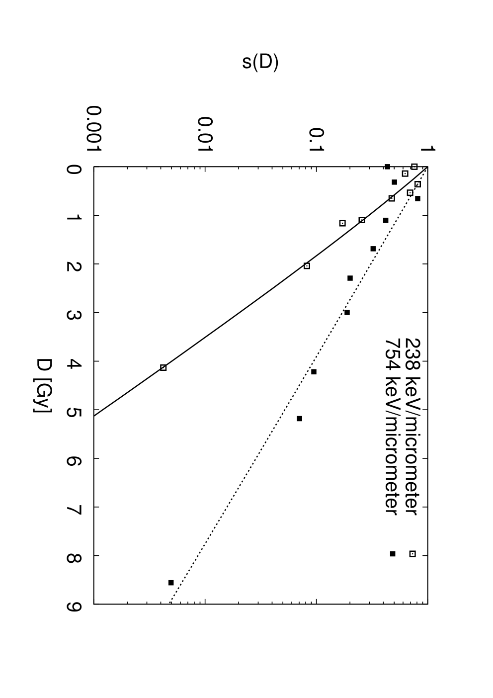
Model representations of survival curves for V79 cells irradiated by different ions are shown in Figures 2-5. In Figure 6, the probabilities of single-track and combined damage induction are plotted as functions of LET for different ions. Values of auxiliary model parameters are listed in Table 2.




| V79 | CHO-K1 | ||||
|---|---|---|---|---|---|
| p | 3He | C | O | C | |
| 0.045 | 0.25 | 0.41 | 0.80 | 0.50 | |
| [m/keV] | 0.036 | 0.015 | 0.0063 | 0.0042 | 0.0064 |
| 3.57 | 2.79 | 3.21 | 4.17 | 2.0 | |
| 0.017 | 0.0 | 1.0 | 0.39 | 0.26 | |
| [m/keV] | 0.13 | 0.0012 | 0.0093 | 0.0011 | |
| 2.81 | 1.0 | 1.44 | 0.69 |
A similar analysis has been performed for survival data of CHO-K1 cells irradiated by carbon ions, measured experimentally by Weyrather et al 1999. Model representation of survival curves and the derived damage induction probabilities are shown in Figure 7; the parameter values are given in Table 2.


The present results correspond to the geometrical cross section for V79 cells chosen uniquely for all data sets to be = 87.8 , based on the value reported in (Weyrather et al 1999), in order to enable comparisons between different ions. Values in the range of 50 – 135 , also reported in the literature (compare e.g. (Scholz and Kraft 1995, Belli et al 1998)), have yielded similar results (figures not shown here). For CHO cells, the value of geometrical cross section = 108 was taken, as reported in the given experiment (Weyrather et al 1999).
4 Discussion
4.1 Single-track damage induced by protons: Possible indications for differences in radiobiological mechanisms between protons and ions
Measured data for protons of LET values above approximately 30 keVm (Belli et al 1998) suggest that the single-track damage formation probability, , might decrease in this region; compare Figure 8 where more detailed fits to the data are shown, based on the parameterization
| (6) |
Whereas the value of the goodness-of-fits criterion is for the data of Belli et al 1998 in the joint fits to proton-induced survival shown in Figure 2, the corresponding value for the detailed fits shown in Figure 8 is . The goodness-of-fits to this data set can be, in fact, further improved if, in addition to a decreasing single-track damage probability, the combined-damages are allowed to dominate in the whole protons’ LET range and their repair is taken into account, compare (Kundrát et al 2005).
To verify the hypothesis of decreasing single-track damage probability for protons of high LET, further experimental studies are necessary, as the existing evidence is limited to two survival curves measured under a single experimental setup only. Even though this decrease corresponds to very limited proton ranges in tissues only (approximately 15 m or smaller) and will probably not manifest in clinical applications, this finding would be important for understanding the mechanisms underlying the radiobiological effects: For heavier ions, no such complex behaviour has been observed in the studied LET ranges, the damage induction probabilities exhibiting saturation characteristics only (Figure 6). It means that for heavier ions the RBE effects termed often as ”overkill” occur as a result of too high energy deposits to individual cells. For protons, on the other hand, this evidence suggests that they are already the effects of single particles on the level of DNA damage formation that get saturated and are responsible for the decrease of biological effectiveness observed experimentally (Belli et al 1998).


4.2 The influence of repair processes
As mentioned in the preceding paragraph, the effects of cellular repair processes might be important in representing the outcome of proton irradiation. Also the fits to 3He data in the high-dose region might be improved significantly by incorporating the repair probabilities (Figure 9). In this region, however, the experimental conditions are very challenging and, consequently, the data in this region have a limited reliability (Folkard et al 1996); therefore, the authors have interpreted the upward-bending of survival curves as artificial effects only. However, the results shown in Figure 9 demonstrate that similar upward bending might follow as a result of repair processes; compare also the general discussion in (Kundrát et al 2005). The damage induction probabilities are almost identical with those presented in Figure 6 (in both cases, the combined-damage probabilities have been neglected, ); the survival values at high doses being mainly influenced. Again, additional experimental data would be helpful in solving this issue.


The goodness-of-fits for heavier ions does not increase significantly when the repair processes are taken into account. For the sake of simplicity, only unrepairable damages have, therefore, been considered in the present work (Section 3). The induction probabilities for both single-track and combined damages have been estimated on the basis of survival data only. This slightly limits the reliability of estimating the roles of combined-track events, , in the higher-LET regions, whereas the unrepairable single-track damages, , have been assessed reliably in the whole studied LET regions. Somewhat higher values of damage induction probabilities would be obtained if the influence of repair processes were included. A corresponding concept, including a wider class of damages and their repair by the cells, has been used in analyzing radiobiological data for cell lines of different radiosensitivity; the results will be presented and discussed in detail elsewhere (Hromčíková et al 2005).
4.3 On the interpretation of the damage classification
Clustered DNA lesions of high complexity (locally multiply damaged sites, complex lesions) are thought to represent significant challenges to cellular repair systems, and to play crucial roles in cell inactivation by ionizing particles (Ward 1985, Goodhead 1994, Ottolenghi et al 1997). The existing estimates of their yields have been based mostly on Monte Carlo calculations involving track structure simulations and simplified models of DNA and/or chromatin structure. Due to their complexity and computational costs, these approaches have been performed for photons and low-energy particles only. On the other hand, the present approach, based on analyzing survival data, enables to derive the yields of lethal events also for high- high-energy ions (Figure 6). By comparing the present results with the mentioned Monte Carlo calculations, mechanistic interpretation of the damage classification used in the present work might be sought, and the fraction of lethal events among different classes of complex lesions can be estimated.
As the first step in this direction, the present results for lethal damage induced by protons in V79 cells have been compared to estimates of complex lesions calculated on the basis of a fast Monte Carlo damage simulation model proposed by Semenenko and Stewart 2004. In Figure 10, the LET-dependent yields of single-track lethal events, , are compared to the total number of double-strand breaks (DSBs) and DSBs composed of at least 4, 6, 8, and 10 elementary lesions (single-strand breaks (SSBs), base damages or abasic sites). This figure indicates that the present results are consistent with the trends predicted by Monte Carlo calculations. In Figure 11, the ratio between the number of lethal events and DSBs of different complexity is shown, again as a function of LET. Whereas only 0.05 – 0.5% of all DSBs formed (denoted by DSB total in Figures 10 and 11) are lethal, this portion increases to approximately 3 – 4% for DSBs composed of at least 8 elementary lesions (DSB 8+); this number being uniform (LET-independent), i.e. being consistent with the hypothesis of a uniform repair probability for these specific damages.


These results indicate that the single-track lethal damages, , might be related to clustered lesions of very high complexity, and correlate best with DSB 8+, containing at least 6 elementary DNA lesions in addition to 2 SSBs forming the DSB. Similarly, the combined damages, , might correspond to pairwise combinations of DSBs formed by different tracks, leading to large-scale chromosomal aberrations; compare e.g. (Sachs and Brenner 1993). Further damage induction and repair studies are, however, necessary to identify the biophysical nature of lethal events reliably. Furthermore, this comparison has been performed for protons only, given the limitations of existing Monte Carlo approaches. Efforts will therefore be made to extend these calculations to heavier ions and higher energies, using correspondingly simplified Monte Carlo-based models.
4.4 Applications in hadrontherapy treatment planning
The analyzed proton and helium-3 data (Belli et al 1998, Folkard et al 1996, Perris et al 1986, Goodhead et al 1992) concern irradiation by low-energy particles only, with ranges in tissue of approx. 10 m – 0.7 mm for protons and 24 – 74 m for 3He ions, respectively (Table 1), corresponding to track ends of these particles. On the other hand, data for carbon (Weyrather et al 1999) and oxygen ions (Stoll et al 1995) have been gathered over wide energy (and LET) ranges of these particles, corresponding to penetration depths of approx. 45 m to 140 mm for carbon ions and 30 m to 200 mm for oxygen ions, covering the majority of clinically relevant ranges. Figures 4 and 5 illustrate that the simplified probabilistic model enables to represent cell survival over such wide ranges in a systematic manner by using a low number of parameters only (the present analysis involves 6 parameters, and , plus the cross section of cell nuclei, ). This is a prerequisite if the model is to be used in treatment planning applications. In the present analysis, cell inactivation effects have been studied for monoenergetic particles as function of their LET value (corresponding to their energy). For treatment planning applications, an adequate physical model, describing the spectra of particle energy and LET values as function of penetration depths and including also energy-loss straggling, lateral scattering and fragmentation processes, has to be used together with the model of biological effects presented here. Such a physical model might be based on detailed Monte Carlo calculations (e.g. (Ziegler 2004)) or semi-phenomenological parameterization of measured experimental data (Krämer et al 2000, Chu et al 1993). Calculations of survival as function of penetration depth for several cell lines, using the radiobiological module presented here and a simplified description of physical processes based on SRIM-2003 calculations, have been already performed, showing good agreement with measured data; the results will be presented separately (Kundrát 2005). To enhance the predictive power of the model, relation between the damage induction probabilities and track structure characteristics of different ions will be sought, with the aim to help in identifying the optimal ion in different clinical situations. Additional systematic experimental data, especially for inactivation induced by light ions (helium to nitrogen), would be helpful in solving this issue, too.
5 Conclusion
Systematic analysis of published survival data for V79 cells irradiated by light ions has been performed using a simplified scheme of the probabilistic two-stage model. The probabilities of single ions to induce severe damage to DNA have been derived for different ions in dependence on their LET values. In the lower-LET regions, combined damages dominate, while survival at higher LET values is governed by single-particle damages (Figure 6). The present results give quantitative estimates of the increase in damage complexity with increasing atomic number and LET value. The analysis of survival data for CHO-K1 cell line shows then the differences in DNA damage induction for different cell lines.
The derived probabilities of inducing single-track lethal damages have been compared with the Monte Carlo estimates of the yields of complex DNA lesions. Good agreement between these two methodologically different approaches was demonstrated, indicating the biophysical interpretation of lethal events and the importance of damage complexity with respect to its biological consequences.
The results of this work will be used in proposing detailed biology-oriented approaches for hadrontherapy treatment planning. The results may also contribute to understanding the differences between the mechanisms of biological effects of different ions.
References
References
- Belli et al (1998) Belli M, Cera F, Cherubini R, Dalla Vecchia M, Haque AMI, Ianzini F, Moschini G, Sapora O, Simone G, Tabocchini MA, Tiveron P 1998 RBE-LET relationships for cell inactivation and mutation induced by low energy protons in V79 cells: further results at the LNL facility. Int. J. Radiat. Biol. 74 501–509
- Berger et al (2000) Berger MJ, Coursey JS, and Zucker MA 2000 ESTAR, PSTAR, and ASTAR: Computer Programs for Calculating Stopping-Power and Range Tables for Electrons, Protons, and Helium Ions (version 1.2.2). Available: http://physics.nist.gov/Star. National Institute of Standards and Technology, Gaithersburg, MD. Originally published as: Berger MJ, NISTIR 4999, National Institute of Standards and Technology, Gaithersburg, MD (1993).
- Brahme et al (2001) Brahme A, Lewensohn R, Ringborg U, Amaldi U, Gerardi F, Rossi S 2001 Design of a centre for biologically optimised light ion therapy in Stockholm. Nucl. Instrum. Meth. B 184 569-588.
- Cella et al (2001) Cella L, Lomax A, Miralbell R 2001 Potential role of intensity modulated proton beams in prostate cancer radiotherapy. Int. J. Radiat. Oncol. Biol. Phys. 49 217-223.
- Chu et al (1993) Chu WT, Ludewigt BA, Renner TR 1993 Instrumentation for treatment of cancer using proton and light-ion beams Rev. Sci. Instrum. 64 2055-2122
- Debus et al (1998) Debus J, Gross KD, Pavlovic M (Eds) 1998 Proposal for a Dedicated Ion Beam Facility for Cancer Therapy. GSI, Darmstadt.
- Enghardt et al (2004) Enghardt W, Parodi K, Crespo P, Fiedler F, Pawelke J, Ponisch F 2004 Dose quantification from in-beam positron emission tomography Radiother. Oncol. 73 Suppl. 2, S96-S98.
- Folkard et al (1996) Folkard M, Prise K M, Vojnovic B, Newman H C, Roper M J, Michael B D 1996 Inactivation of V79 cells by low-energy protons, deuterons and helium-3 ions Int. J. Radiat. Biol. 69 729–738
- Goodhead et al (1992) Goodhead DT, Belli M, Mill AJ, Bance DA, Allen LA, Hall SC, Ianzani F, Simone G, Stevens DL, Stretch A, Tabocchini MA, Wilkinson RE 1992 Direct comparison between protons and alpha-particles of the same LET. I. Irradiation methods and inactivation of asynchronous V79, HeLa and C3H 10T1/2 cells Int. J. Radiat. Biol. 61 611–624
- Goodhead (1994) Goodhead DT 1994 Initial events in the cellular effects of ionizing-radiations - clustered damage in DNA Int. J. Radiat. Biol. 65 7-17
- Hromčíková et al (2005) Hromčíková H, Kundrát P, Lokajíček M 2005 Analysis of damage induction and repair processes after carbon irradiation in different cell lines (in preparation)
- James (1994) James F 1994 MINUIT Minimization package - Reference Manual CERN Program Library Long Writeup D506, CERN Geneva
- Kagawa et al (2002) Kagawa K, Murakami M, Hishikawa Y, Abe M, Akagi T, Yanou T, Kagiya G, Furusawa Y, Ando K, Nojima K, Aoki M, Kanai T 2002 Preclinical biological assessment of proton and carbon ion beams at Hyogo Ion Beam Medical Center. Int. J. Radiat. Oncol. Biol. Phys. 54 928–938
- Kanai et al (1997) Kanai T, Furusawa Y, Fukutsu K, Itsukaichi H, Eguchi-Kasai K, Ohara H 1997 Irradiation of Mixed Beam and Design of Spread-out Bragg Peak for Heavy-Ion Radiotherapy. Radiat. Res. 147 78–85
- Kanai et al (1999) Kanai T, Endo M, Minohara S, Miyahara N, Koyama-Ito H, Tomura H, Matsufuji N, Futami Y, Fukumura A, Hiraoka T, Furusawa Y, Ando K, Suzuki M, Soga F, Kawachi K 1999 Biophysical characteristics of HIMAC clinical irradiation system for heavy-ion radiation therapy. Int. J. Radiat. Oncol. Biol. Phys. 44 201–210
- Krämer et al (2000) Krämer M, Jäkel O, Haberer T, Kraft G, Schardt D, Weber U 2000 Treatment planning for heavy-ion radiotherapy: physical beam model and dose optimization. Phys. Med. Biol. 45 3299-3317.
- Krämer and Scholz (2000) Krämer M, Scholz M 2000 Treatment planning for heavy-ion radiotherapy: calculation and optimization of biologically effective dose Phys. Med. Biol. 45 3319-3330
- Krengli and Orecchia (2004) Krengli M, Orecchia R 2004 Medical aspects of the National Centre for Oncological Hadrontherapy (CNAO - Centro Nazionale Adroterapia Oncologica) in Italy. Radiother. Oncol. 73 Suppl. 2, S21-S23.
- Kundrát et al (2005) Kundrát P, Lokajíček M, Hromčíková H 2005 Probabilistic two-stage model of cell inactivation by ionizing particles Phys. Med. Biol. 50 1433-1447
- Kundrát (2005) Kundrát P 2005 Towards biology-oriented hadrontherapy treatment planning (in preparation)
- Mayer et al (2004) Mayer R, Mock U, Jager R, Potter R, Vutuc C, Eiter H, Krugmann K, Hammer J, Hirn B, Hawliczek R, Knocke-Abulesz TH, Lukas P, Nechville E, Pakisch B, Papauschek M, Wolfgang R, Rhomberg W, Sabitzer H, Schratter-Sehn A, Felix S, Wedrich I, Auberger T 2004 Epidemiological aspects of hadron therapy: A prospective nationwide study of the Austrian project MedAustron and the Austrian Society of Radiooncology (OEGRO). Radiother. Oncol. 73 Suppl. 2, S24-S28.
- Ottolenghi et al (1997) Ottolenghi A, Merzagora M, Paretzke HG 1997 DNA complex lesions induced by protons and alpha-particles: Track structure characteristics determining linear energy transfer and particle type dependence Radiat. Environ. Biophys. 36 97-103
- Perris et al (1986) Perris A, Pialoglou P, Katsanos AA, Sideris EG 1986 Biological effectiveness of low-energy protons. 1. Survival of Chinese-hamster cells Int. J. Radiat. Biol. 50 1093–1101
- Sachs and Brenner (1993) Sachs RK, Brenner DJ 1993 Effect of LET on chromosomal aberration yields. 1. Do long-lived, exchange-prone double-strand breaks play a role Int. J. Radiat. Biol. 64 677-688
- Schall et al (1996) Schall I, Schardt D, Geissel H, Irnich H, Kankeleit E, Kraft G, Magel A, Mohar MF, Munzenberg G, Nickel F, Scheidenberger C, Schwab W 1996 Charge-changing nuclear reactions of relativistic light-ion beams () passing through thick absorbers Nucl. Instrum. Meth. B 117 221-234
- Scholz and Kraft (1995) Scholz M, Kraft G 1995 Track structure and the calculation of biological effects of heavy charged-particles Adv. Space Res. 18 5-14
- Schulz-Ertner et al (2002) Schulz-Ertner D, Haberer T, Jakel O, Thilmann C, Kramer M, Enghardt W, Kraft G, Wannenmacher M, Debus JR 2002 Radiotherapy for chordomas and low-grade chondrosarcomas of the skull base with carbon ions Int. J. Radiat. Oncol. Biol. Phys. 53 36-42.
- Semenenko and Stewart (2004) Semenenko VA, Stewart RD 2004 A fast Monte Carlo algorithm to simulate the spectrum of DNA damages formed by ionizing radiation Radiat. Res. 161 451-457
- Sisterson (2005) Sisterson J (Ed) (2005) Particles Newsletter 36. http://ptcog.web.psi.ch/archive.html
- Stoll et al (1995) Stoll U, Schmidt A, Schneider E, Kiefer J 1995 Killing and mutation of Chinese-hamster V79 cells exposed to accelerated oxygen and neon ions. Radiat. Res. 142 288-294
- Tsujii et al (2004) Tsujii H, Mizoe J, Kamada T, Baba M, Kato S, Kato H, Tsuji H, Yamada S, Yasuda S, Ohno T, Yanagi T, Hasegawa A, Sugawara T, Ezawa H, Kandatsu S, Yoshikawa K, Kishimoto R, Miyamoto T 2004 Overview of clinical experiences on carbon ion radiotherapy at NIRS. Radiother. Oncol. 73 Suppl. 2, S41-S49
- Wambersie et al (2004) Wambersie A, Hendry J, Gueulette J, Gahbauer R, Potter D, Gregoire V 2004 Radiobiological rationale and patient selection for high-LET radiation in cancer therapy Radiother. Oncol. 73 Suppl. 2, S1-S14.
- Ward (1985) Ward JF 1985 Biochemistry of DNA lesions Radiat. Res. 104 S103-S111
- Weyrather et al (1999) Weyrather WK, Ritter S, Scholz M, Kraft G 1999 RBE for carbon track-segment irradiation in cell lines of differing repair capacity Int. J. Radiat. Biol. 75 1357–1364
- Wilson (1946) Wilson RR 1946 Radiological use of fast protons. Radiology 47 487-491
- Ziegler (2004) Ziegler JF 2004 SRIM-2003 Nucl. Instrum. Meth. B 219 1027-1036
- Zurlo et al (2000) Zurlo A, Lomax A, Hoess A, Bortfeld T, Russo M, Goitein G, Valentini V, Marucci L, Capparella R, Loasses A 2000 The role of proton therapy in the treatment of large irradiation volumes: A comparative planning study of pancreatic and biliary tumors. Int. J. Radiat. Oncol. Biol. Phys. 48 277-288.