2 Shanghai Academy of Artificial Intelligence for Science, Shanghai, China.
3 Human Phenome Institute, Fudan University, Shanghai, China.
4 Shanghai Universal Medical Imaging Diagnostic Center, Shanghai, China.
* Corresponding authors: 11email: {qiyuan,cheng_yuan,huzixin}@fudan.edu.cn
Universality Reconsidered: Rethinking the Validation of Foundation Models for General-Purpose 3D Medical Segmentation
Abstract
Foundation models have emerged as a transformative paradigm in 3D medical imaging, with the promise of unified quantitative analysis across diverse targets and imaging modalities. Yet the prevailing conception of universality remains incomplete. Current models are predominantly developed and evaluated on datasets largely concentrated around a limited set of imaging modalities and anatomical regions. In this Perspective, we evaluate representative 3D segmentation foundation models using paired whole-body structural and functional imaging data. Our analysis reveals a substantial gap between benchmark-reported performance and real-world generalization, with marked degradation on previously unseen data and particularly severe failures on functional imaging modalities. These findings suggest that current foundation models remain far from achieving true universality. We argue that progress requires not only scaling models and datasets, but also a reconsideration of how universality is defined and validated, extending evaluation beyond regional structural benchmarks toward whole-body structural and functional imaging. Our observations highlight the need to distinguish benchmark success from genuine clinical generalization. Bridging this gap will be essential for translating foundation models from controlled evaluation settings to real-world medical practice.
Medical imaging is a cornerstone of modern healthcare, playing an indispensable role in disease diagnosis, treatment planning, and patient monitoring [21]. Among the diverse tasks in medical image analysis, volumetric segmentation of 3D medical imaging occupies a central position as it underpins quantitative measurements of organ volumes, tumor burden, and treatment response that are essential for clinical decision-making [9]. Manual segmentation has long been the gold standard for delineating anatomical structures and lesions. However, this procedure is highly time-consuming, labor-intensive, and requires significant domain expertise. Semi- or fully-automatic segmentation methods can significantly reduce the time and manual effort required, improve consistency across results, and enable the rapid analysis of large-scale datasets.
Deep learning models have shown strong potential in medical image segmentation by extracting intricate features from massive annotated datasets and achieve accurate segmentation across a diverse range of tasks [2, 16, 13, 7]. Despite these advances, a prominent bottleneck of existing models remains their strict task-specificity. These models are typically designed and trained for specific predefined tasks and often suffer from severe performance degradation when exposed to new tasks or out-of-distribution data. Consequently, their capacity to generalize to other clinical scenarios is highly constrained, necessitating the development of distinct models for each specific imaging modality or target organ, which poses a major obstacle to broad clinical translation given that routine clinical workflows demand the robust segmentation of highly heterogeneous structures across a wide array of imaging modalities [15].
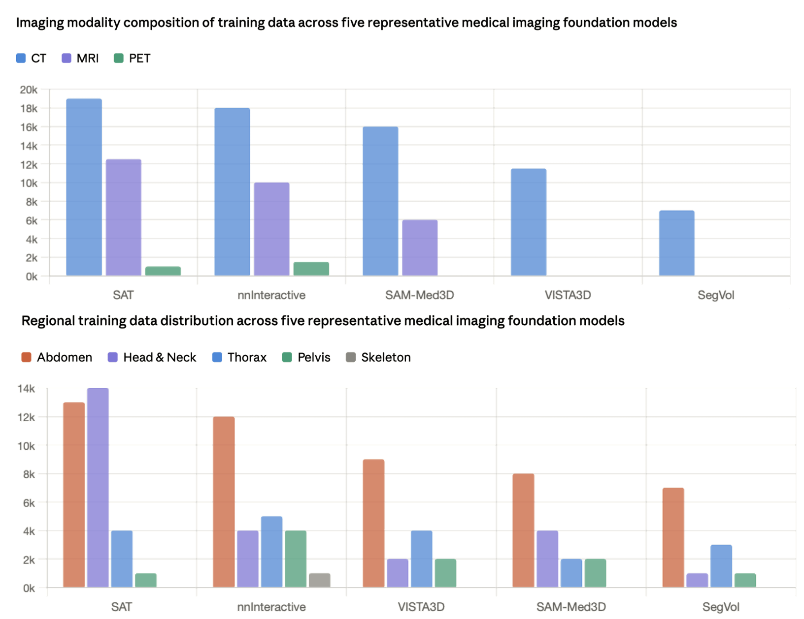

Recent advancement of artificial intelligence has been defined by the emergence of foundation models (FMs), which are large-scale, pre-trained models capable of performing multi-task segmentation within a unified framework [3, 24]. The field of medical image analysis is undergoing a paradigm shift from task-specific models to general-purpose foundation models. Inspired by the success of the Segment Anything Model (SAM) in computer vision [12], the community has witnessed a proliferation of efforts to adapt this paradigm to medical image segmentation for quantitative image analysis [17, 26]. However, direct adaptation of natural-image-based models to the medical domain faces significant structural hurdles. A primary limitation is that many existing adaptations operate via 2D slice-wise segmentation [14, 27], when adapted to 3D medical images, the volumes are segmented slice-by-slice and reconstructed through post-processing. This strategy inherently overlooks the critical volumetric contextual information and inter-slice spatial correlations that are fundamental to radiological interpretation [25]. To address the shortcomings of 2D models, a series of 3D general-purpose medical foundation models have emerged [6, 8, 11, 22, 28] to leverage intrinsic volumetric features directly and achieve remarkable generalization capabilities across a wide array of 3D segmentation tasks. While these 3D medical foundation models claim to provide a unified solution for diverse clinical tasks, their development and validation remain largely restricted. As shown in Figure 1, we quantify this severe imbalance by tallying the total 3D training volumes adopted during model construction across leading universal segmentation backbones. As visualized, CT and MRI scans constitute the overwhelming majority of training corpora for every benchmarked model, while functional PET data occupies only a negligible fraction of all training samples. Beyond modality imbalance, the regional distribution of training data reveals an equally pronounced bias. This skewed data distribution inherently embeds strong modality and regional bias into model weights, limiting the inherent capacity of foundation models to generalize toward underrepresented functional modalities at the training stage.
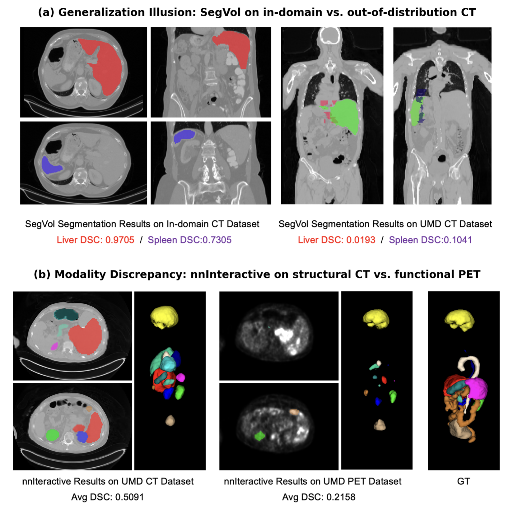
Compounding this training-set limitation, conventional evaluation pipelines further obscure true cross-modality robustness through flawed benchmark design. Standard validation protocols split assessments across disjoint, task-heterogeneous cohorts, pairing distinct imaging modalities with unrelated anatomical tasks. Under this setup, observed performance gaps cannot be attributed purely to modality differences variations in task difficulty, organ morphology and imaging contrast are fully confounded with modality effects. Consequently, performance metrics become fundamentally confounded, as it remains impossible to discern whether a performance gap arises from the underlying imaging physics or the intrinsic geometric difficulty of the anatomical target, as shown in Figure 2(a). Together, biased training data and confounded heterogeneous benchmarks create systemic blind spots: existing model development and validation workflows fail to reliably characterize the genuine generalization ability in diverse medical imaging applications.
A significant limitation in current evaluations of general-purpose medical segmentation models is the reliance on heterogeneous datasets, where different modalities often correspond to disparate anatomical regions or clinical tasks. Here, we constructed a benchmark of 490 paired PET/CT and 464 paired PET/MRI for whole-body segmentation, enabling a direct comparison of model performance across co-registered modalities, as shown in Figure 2(b). This paired design, with multi-modal scans acquired simultaneously from the same patient cohort, ensures that segmentation results for identical organs can be used to objectively compare performance differences across modalities. By utilizing intra-subject controlled comparisons, we effectively isolate imaging modality as the primary independent variable, enabling a rigorous quantification of the inherent modality discrepancy. Furthermore, our benchmark serves as a pristine testbed to evaluate the true zero-shot generalization performance of these models on previously unseen data. Our systematic evaluation reveals a stark discrepancy between literature-reported benchmarks and real-world performance, with substantial degradation in effectiveness and, in some cases, complete failure.
Our findings expose the systemic limitations of current foundation models and underscore the urgent necessity to bridge the gap between idealized benchmarking and comprehensive clinical utility. The promise of whole-body, modality-agnostic universality, we argue, remains substantially unaddressed, constrained by the fundamental bias originating from existing training and validation protocols. We conceptualize universality along two orthogonal axes. (1) modality universality: the capacity to process and reason across distinct 3D imaging modalities, spanning both structural and functional domains. (2) regional universality: the ability to perform competently across the full topographic extent of the human body, rather than within a restricted set of organ systems.

| Model | Setting | Reported Performance∗ | UMD Evaluation Performance | |||||
|---|---|---|---|---|---|---|---|---|
| CT | MRI | PET | CT | PET | MRI | PET | ||
| SAM-Med3D-turbo | 1 point prompt | 0.790 | 0.754 | / | 0.404 | 0.410 | 0.433 | 0.397 |
| SegVol | 1 point prompt | 0.793 | / | / | 0.107 | 0.075 | 0.134 | 0.148 |
| nnInteractive | 1 point prompt | 0.55 | 0.55 | / | 0.481 | 0.342 | 0.511 | 0.217 |
| VISTA3D | class id | 0.711 | / | / | 0.071 | 0.005 | 0.003 | 0.006 |
| SAT | text prompt | 75.6 | 83.8 | 63.4 | 0.018 | 0.003 | 0.006 | 0.000 |
| nnUNet | 10 Training Samples | - | - | - | 0.686 | 0.605 | 0.652 | 0.560 |
Benchmark Design
To bridge the gap and uncover modality discrepancy, we present UMD, a segmentation benchmark dataset by retrospectively collecting 490 whole-body PET/CT and 464 whole-body PET/MRI scans, with approximately 675,000 2D slices. For each case, the structural (CT or MRI) and functional (PET) volumes were acquired from the same subject during a single diagnostic session, ensuring intrinsic spatial and anatomical consistency. This intra-subject paired nature guarantees that the underlying morphology, scale, and orientation of the target organs remain constant across modalities. Patients fasted for at least 6 hours with blood glucose levels confirmed at mmol/L prior to receiving an intravenous injection of 18F-FDG. The scanning field was from the mid-thigh level to the top of the head with the subject in the supine position. All paired multimodal scans cover consistent whole-body anatomical ranges, enabling direct intra-subject comparison between structural anatomical contrasts and metabolic signals.
We provide voxel-wise fine-grained annotations for 13 diverse organs, including the liver, left kidney, right kidney, brain, heart, spleen, aorta, lung, colon, urinary bladder, pancreas, esophagus, and stomach, which supports a comprehensive and systematic evaluation of general-purpose segmentation models across varied anatomical complexities and imaging modalities. To construct reliable segmentation ground truth, we adopted a two-stage annotation pipeline. Initial coarse segmentation masks were automatically generated via pre-trained segmentation tools [23, 5], and all preliminary annotations were subsequently reviewed, corrected, and refined by radiologists using the LIFEx [18] platform. By providing paired, co-registered ground-truth labels for both structural and functional imaging volumes, our protocol enables a head-to-head comparison between different modalities, free from confounding factors such as inter-subject anatomical differences. Furthermore, as an originally collected dataset rather than a recompilation of existing public data sources, we eliminate the risk of data leakage, guaranteeing the validity and authenticity of the evaluation results. As a result, the evaluation performance can be reliably attributed to modality-specific generalization capabilities of these models.
Evaluation Details
To comprehensively evaluate the robustness of current 3D medical foundation models, we conducted a systematic comparison of five representative state-of-the-art general-purpose segmentation foundation models, including SAM-Med3D-turbo [22], SegVol [6], SAT-Pro [28], VISTA3D [8], and nnInteractive [11]. These models represent the cutting edge of general-purpose segmentation, utilizing diverse strategies such as large-scale pre-training on volumetric data, prompt-based interaction, and universal feature encoders. Notably, each of these frameworks has claimed to achieve superior zero-shot generalization performance across a wide array of 3D medical imaging tasks and anatomical structures.
To ensure a fair and rigorous evaluation, we utilized the official, latest, and largest-scale pre-trained weights and codebases for all comparing foundation models. All experiments were conducted on NVIDIA A100 GPUs with 80GB of memory. Following the standardized evaluation protocols associated with each model, we implemented two primary interaction strategies to facilitate zero-shot segmentation. For VISTA3D and SAT, we provided the specific textual category or class ID corresponding to each of the target organs. For other point-based models, we utilized simulated point prompts generated from the ground truth masks to mimic ideal user interaction.
Results and Analysis
Among five representative state-of-the-art models tested, all models exhibit performance degradation. Comparing literature-reported performance with actual performance on our benchmark reveals systematic overestimation across all models, as shown in Figure 3(a) and Table 1. Critically, even within CT alone, most models exhibit substantial performance gaps between their reported benchmark scores and their actual efficacy on previously unseen data from different acquisition protocols. SAM-Med3D-turbo, which reports a CT Dice score of 0.790, achieves only 0.404 on the benchmark CT data. SegVol drops from a reported 0.793 to 0.107, and VISTA3D collapses from 0.711 to 0.071, suggesting that current models may rely heavily on distribution-specific patterns rather than fully generalizable anatomical representations.
The transition to functional PET imaging further amplifies these failures. Semantic-guided models such as VISTA3D and SAT suffer near-complete failure on PET data, with Dice scores plummeting to near zero across nearly all organs, indicating that their learned representations are not grounded in anatomical understanding but rather in distribution-specific patterns. Point-based interactive models such as SAM-Med3D-turbo and nnInteractive maintain marginal continuity through spatial prompt localization, yet their performance remains far below clinical acceptability, particularly for organs with intricate geometries or low contrast, while even the best-performing model nnInteractive achieves only 0.342 on PET compared to 0.481 on CT. Comparison in Figure 3(b) confirms that the modality discrepancy is systemic rather than organ-specific. The few exceptions, where PET segmentation achieves non-trivial scores, are confined to organs with extreme radiotracer uptake such as the bladder, where the high signal intensity creates sufficient contrast for modality-agnostic spatial heuristics to succeed, with the signal sufficiently distinctive to be captured. These findings indicate that current foundation models remain strongly modality-dependent, having not yet developed the capacity to bridge the gap between anatomical density and metabolic activity. The benchmarks on which these models are evaluated and celebrated are themselves structurally biased, creating a self-reinforcing cycle where models optimized for a narrow data regime are validated on data drawn from that same regime.
Given the inherent differences in imaging characteristics across different modalities, we disentangle whether the aforementioned performance gap arises from the intrinsic segmentation difficulty of each modality itself, or from the distributional bias of these foundation models. We train nnU-Nets [10] of each modality as a reference of state-of-the-art task-specific models for comparison. As shown in Table. 1, most models exhibit a significant performance gap compared to their reported benchmarks, indicating that current 3D foundation models are overfitted to their validation data distributions and lack the robustness required for diverse clinical scenarios. Specifically, semantic guided models like SAT and VISTA3D undergo a near total collapse upon evaluation on unseen data, revealing that their purported generalization is restricted to specific morphological patterns.This persistent performance gap across all frameworks confirms that structural proficiency does not inherently translate into functional understanding, highlighting a critical limitation in the current foundation model paradigm.
Qualitative analysis further corroborates these quantitative findings and provides intuitive insight into the nature of model failure. Figure 4(a) illustrates the generalization illusion through SegVol. On its in-domain CT cases, the model achieves near-perfect segmentation of the liver (DSC: 0.97) and spleen (DSC: 0.73). However, when applied to previously unseen CT data acquired under different protocols, performance collapses catastrophically (liver DSC: 0.02, spleen DSC: 0.10). The segmentation masks are fragmented, spatially incoherent, and bear little resemblance to the target anatomy, revealing that the model’s in-domain competence derives from memorization of dataset-specific intensity patterns rather than genuine anatomical understanding. Figure 4(b) demonstrates the modality discrepancy through nnInteractive, the best-performing model in our evaluation. While achieving moderate multi-organ segmentation on structural CT (average DSC: 0.51), the same model produces severely degraded and anatomically implausible predictions on the co-registered PET scan of the same patient (average DSC: 0.22). This controlled comparison provides direct visual evidence that current foundation models are predominantly anchored to structural intensity profiles and face substantial difficulty transferring their learned representations to the metabolic signal distributions characteristic of functional imaging.
These empirical evidences underscore a critical methodological insight for the evaluation of foundation models: performance on aggregated, in-distribution benchmarks is a necessary but profoundly insufficient condition for claiming generality. Without controlled, out-of-distribution evaluation that systematically probes model behavior across the structural-functional divide, the field risks perpetuating what we term the generalization illusion: a state in which high reported metrics may create unwarranted confidence in model robustness while deployment on underrepresented modalities reveals significant and previously uncharacterized performance gaps. Moving beyond this illusion requires both the development of new evaluation paradigms and a fundamental reconceptualization of what constitutes a valid test of universality.
Universality Reconsidered
The performance gaps revealed by our paired evaluation are not isolated failures of individual architectures. Rather, they expose a deeper issue concerning how universality itself is currently defined and validated in medical imaging foundation models. Contemporary benchmarks often equate strong performance across multiple datasets with general-purpose capability. However, our findings suggest that such conclusions may be premature because the underlying training and evaluation ecosystems remain systematically biased along two fundamental dimensions: modality discrepancy and regional discrepancy.
Modality discrepancy: the under-representation of functional imaging. Current foundation models are overwhelmingly developed on structural imaging modalities, particularly CT and MRI, while functional imaging remains severely underrepresented. This imbalance reflects practical challenges in acquiring and annotating PET data, but it also creates a fundamental blind spot in model development. The discrepancy is not merely a matter of missing data. Structural and functional imaging encode fundamentally different representations of human biology. CT and MRI primarily capture anatomical morphology, whereas PET reflects metabolic and physiological activity. Consequently, models trained predominantly on structural signals learn priors centered on boundaries, textures, and geometric organization. These priors transfer poorly to functional imaging, where clinically meaningful information is often diffuse and heterogeneous. As the clinical significance of functional imaging is increasing towards more precise characterization of pathological processes, integrating metabolic and physiological information with structural anatomy is becoming indispensable [1, 19]. Our evaluation demonstrates that this limitation persists even when segmentation targets are held constant. The substantial degradation observed across all evaluated models indicates that current foundation models have not yet achieved modality universality, despite strong performance on conventional structural-imaging benchmarks.
Regional discrepancy: the fragmentation of whole-body understanding. A similar limitation emerges in anatomical coverage. Existing training datasets are heavily concentrated in a limited number of body regions, most notably the abdomen and brain. Although recent initiatives have expanded anatomical diversity, the majority of foundation models continue to learn from regionally fragmented supervision. This fragmentation becomes particularly problematic in whole-body imaging applications. Many clinically important tasks, especially in oncology, require understanding relationships across multiple organs and organ systems simultaneously rather than interpreting isolated anatomical structures. Whole-body PET imaging is a representative example, where disease burden, metastatic spread, and treatment response often manifest as distributed biological processes spanning the entire body [20]. In contrast, anatomical segmentation tasks are routinely framed as localized problems to segment a specific organ, evaluated in isolation from its systemic context. As a result, models trained primarily on localized segmentation tasks may perform adequately within familiar anatomical regions while struggling to generalize across the broader anatomical landscape encountered in routine clinical practice.
Importantly, modality discrepancy and regional discrepancy do not operate independently. Instead, they reinforce one another. Whole-body PET represents a simultaneous extrapolation along both dimensions: a modality that is largely absent from current training corpora and an imaging paradigm that inherently requires global anatomical reasoning. This observation motivates a broader perspective on universality. Rather than viewing generalization as a single property, we argue that universality should be considered along at least two orthogonal axes:
-
•
Modality universality: robustness across structural and functional imaging paradigms.
-
•
Anatomical universality: competence across the full spatial extent of the human body.
Current foundation models have made substantial progress toward general-purpose segmentation within structurally biased training distributions. However, the evidence presented here suggests that genuine whole-body multimodal universality remains an open challenge. Recognizing this distinction is essential for designing future benchmarks and foundation models that more faithfully reflect the complexity of real-world clinical imaging.
Outlook
Over the past several years, remarkable progress has been achieved in developing general-purpose segmentation foundation models. These systems have demonstrated impressive capabilities across diverse anatomical targets and have substantially advanced the field toward unified medical image analysis. However, our findings suggest that strong performance across existing benchmarks should not be conflated with genuine universality. Current evaluations predominantly assess models within a restricted subspace of medical imaging, largely centered on structural modalities and region-specific tasks. As a result, important dimensions of clinical reality remain insufficiently represented. Truly general-purpose foundation models must bridge two critical gaps: the transition from structural to functional imaging, and the transition from regional understanding to whole-body reasoning. These dimensions are increasingly important as clinical imaging evolves toward integrated anatomical-metabolic assessment and system-level disease characterization.
Consequently, future progress should be driven not only by larger models or larger datasets, but also by a redefinition of evaluation itself. In the era of deep learning, evaluation benchmarks serve as the compass for the research community, shaping how progress is defined. When the benchmarks themselves reflect the modality and regional discrepancies described above, evaluation becomes a reflection of bias rather than a test of genuine capability. Benchmarks should evolve from heterogeneous collections of isolated tasks toward unified grounded testbeds that explicitly measure robustness across modalities and across the full spatial extent of the human body. Such evaluations are essential for distinguishing genuine generalization from benchmark-specific optimization. Recent efforts such as Touchstone [4] have begun to establish standardized evaluation protocols for abdominal CT segmentation, representing an important step toward more rigorous and reproducible evaluation. We view this as a promising beginning. An important next step may be to extend such benchmarking frameworks beyond regional structural imaging tasks toward whole-body multimodal evaluation settings that better reflect the diversity of real-world clinical imaging.
Ultimately, the goal is not merely to develop models capable of segmenting more organs, but to build foundation models that can interpret the human body as an integrated biological system. Achieving this vision will require closer convergence between structural and functional imaging, between local and global understanding, and between benchmark performance and real-world clinical utility. We therefore advocate a shift from the pursuit of general-purpose segmentation toward the broader objective of whole-body multimodal universality. While current foundation models represent an important milestone on this trajectory, the evidence presented here suggests that the path toward true universality remains only partially explored. In this sense, universality should not be viewed as an established achievement, but as an evolving objective that must be continually re-examined as models expand to increasingly diverse imaging conditions and clinical scenarios. Such a perspective, we believe, is essential for truly reconsidering what universality means in medical imaging foundation models.
Data availability
Our dataset and evaluation pipeline will be publicly accessible at https://github.com/YichiZhang98/UMD.
References
- [1] (2022) Multiplexed imaging in oncology. Nature Biomedical Engineering 6 (5), pp. 527–540. Cited by: Universality Reconsidered.
- [2] (2022) The medical segmentation decathlon. Nature communications 13 (1), pp. 4128. Cited by: Universality Reconsidered: Rethinking the Validation of Foundation Models for General-Purpose 3D Medical Segmentation.
- [3] (2025) Foundation models defining a new era in vision: a survey and outlook. IEEE Transactions on Pattern Analysis and Machine Intelligence 47 (4), pp. 2245–2264. Cited by: Universality Reconsidered: Rethinking the Validation of Foundation Models for General-Purpose 3D Medical Segmentation.
- [4] (2024) Touchstone benchmark: are we on the right way for evaluating ai algorithms for medical segmentation?. Advances in Neural Information Processing Systems 37, pp. 15184–15201. Cited by: Outlook.
- [5] (2024) TotalSegmentator mri: sequence-independent segmentation of 59 anatomical structures in mr images. External Links: 2405.19492 Cited by: Benchmark Design.
- [6] (2024) Segvol: universal and interactive volumetric medical image segmentation. Advances in neural Information Processing Systems 37, pp. 110746–110783.. Cited by: Evaluation Details, Universality Reconsidered: Rethinking the Validation of Foundation Models for General-Purpose 3D Medical Segmentation.
- [7] (2024) Results from the autopet challenge on fully automated lesion segmentation in oncologic pet/ct imaging. Nature Machine Intelligence 6 (11), pp. 1396–1405. Cited by: Universality Reconsidered: Rethinking the Validation of Foundation Models for General-Purpose 3D Medical Segmentation.
- [8] (2025) VISTA3D: a unified segmentation foundation model for 3d medical imaging. In Proceedings of the Computer Vision and Pattern Recognition Conference, pp. 20863–20873. Cited by: Evaluation Details, Universality Reconsidered: Rethinking the Validation of Foundation Models for General-Purpose 3D Medical Segmentation.
- [9] (2024) Reviewing 3d convolutional neural network approaches for medical image segmentation. Heliyon 10 (6). Cited by: Universality Reconsidered: Rethinking the Validation of Foundation Models for General-Purpose 3D Medical Segmentation.
- [10] (2021) NnU-net: a self-configuring method for deep learning-based biomedical image segmentation. Nature methods 18 (2), pp. 203–211. Cited by: Results and Analysis.
- [11] (2025) NnInteractive: redefining 3d promptable segmentation. arXiv preprint arXiv:2503.08373. Cited by: Evaluation Details, Universality Reconsidered: Rethinking the Validation of Foundation Models for General-Purpose 3D Medical Segmentation.
- [12] (2023) Segment anything. In Proceedings of the IEEE/CVF international conference on computer vision, pp. 4015–4026. Cited by: Universality Reconsidered: Rethinking the Validation of Foundation Models for General-Purpose 3D Medical Segmentation.
- [13] (2022) Deep learning methods for automatic evaluation of delayed enhancement-mri. the results of the emidec challenge. Medical Image Analysis 79, pp. 102428. Cited by: Universality Reconsidered: Rethinking the Validation of Foundation Models for General-Purpose 3D Medical Segmentation.
- [14] (2024) Segment anything in medical images. Nature Communications 15, pp. 1–9. Cited by: Universality Reconsidered: Rethinking the Validation of Foundation Models for General-Purpose 3D Medical Segmentation.
- [15] (2023) Towards foundation models of biological image segmentation. Nature Methods 20 (7), pp. 953–955. Cited by: Universality Reconsidered: Rethinking the Validation of Foundation Models for General-Purpose 3D Medical Segmentation.
- [16] (2022) AbdomenCT-1k: is abdominal organ segmentation a solved problem?. IEEE Transactions on Pattern Analysis and Machine Intelligence 44 (10), pp. 6695–6714. Cited by: Universality Reconsidered: Rethinking the Validation of Foundation Models for General-Purpose 3D Medical Segmentation.
- [17] (2023) Segment anything model for medical image analysis: an experimental study. Medical Image Analysis 89, pp. 102918. Cited by: Universality Reconsidered: Rethinking the Validation of Foundation Models for General-Purpose 3D Medical Segmentation.
- [18] (2018) LIFEx: a freeware for radiomic feature calculation in multimodality imaging to accelerate advances in the characterization of tumor heterogeneity. Cancer research 78 (16), pp. 4786–4789. Cited by: Benchmark Design.
- [19] (2022) Molecular imaging in oncology: current impact and future directions. CA: a cancer journal for clinicians 72 (4), pp. 333–352. Cited by: Universality Reconsidered.
- [20] (2023) Advances in pet imaging of cancer. Nature Reviews Cancer 23 (7), pp. 474–490. Cited by: Universality Reconsidered.
- [21] (2017) Deep learning in medical image analysis. Annual review of biomedical engineering 19, pp. 221–248. Cited by: Universality Reconsidered: Rethinking the Validation of Foundation Models for General-Purpose 3D Medical Segmentation.
- [22] (2025) SAM-med3d: a vision foundation model for general-purpose segmentation on volumetric medical images. IEEE Transactions on Neural Networks and Learning Systems. Cited by: Evaluation Details, Universality Reconsidered: Rethinking the Validation of Foundation Models for General-Purpose 3D Medical Segmentation.
- [23] (2023) TotalSegmentator: robust segmentation of 104 anatomic structures in ct images. Radiology: Artificial Intelligence 5 (5), pp. e230024. Cited by: Benchmark Design.
- [24] (2024) On the challenges and perspectives of foundation models for medical image analysis. Medical image analysis 91, pp. 102996. Cited by: Universality Reconsidered: Rethinking the Validation of Foundation Models for General-Purpose 3D Medical Segmentation.
- [25] (2022) Bridging 2d and 3d segmentation networks for computation-efficient volumetric medical image segmentation: an empirical study of 2.5 d solutions. Computerized Medical Imaging and Graphics, pp. 102088. Cited by: Universality Reconsidered: Rethinking the Validation of Foundation Models for General-Purpose 3D Medical Segmentation.
- [26] (2024) Segment anything model for medical image segmentation: current applications and future directions. Computers in Biology and Medicine, pp. 108238. Cited by: Universality Reconsidered: Rethinking the Validation of Foundation Models for General-Purpose 3D Medical Segmentation.
- [27] (2025) A foundation model for joint segmentation, detection and recognition of biomedical objects across nine modalities. Nature methods 22 (1), pp. 166–176. Cited by: Universality Reconsidered: Rethinking the Validation of Foundation Models for General-Purpose 3D Medical Segmentation.
- [28] (2025) Large-vocabulary segmentation for medical images with text prompts. NPJ Digital Medicine 8 (1), pp. 566. Cited by: Evaluation Details, Universality Reconsidered: Rethinking the Validation of Foundation Models for General-Purpose 3D Medical Segmentation.
| Model | SAM-Med3D-tb | SegVol | nnIteractive | VISTA3D | SAT |
|---|---|---|---|---|---|
| Liver | \cellcolorgray!15 0.7039(0.1155) | 0.0806(0.1982) | \cellcolorgray!40 0.4993(0.3319) | \cellcolorgray!40 0.9186(0.0378) | 0.0057(0.0205) |
| 0.6835(0.1100) | \cellcolorgray!40 0.1618(0.2539) | 0.3904(0.3769) | 0.0676(0.0475) | 0.0020(0.0451) | |
| Kidney-L | 0.4701(0.1300) | \cellcolorgray!40 0.1203(0.1967) | \cellcolorgray!40 0.7930(0.1636) | 0.0000(0.0000) | 0.0001(0.0018) |
| \cellcolorgray!40 0.5678(0.1411) | 0.0687(0.1329) | 0.4464(0.3436) | 0.0000(0.0000) | 0.0041(0.0638) | |
| Kidney-R | 0.4387(0.1491) | \cellcolorgray!40 0.1437(0.2279) | \cellcolorgray!40 0.7780(0.1753) | 0.0000(0.0001) | 0.0061(0.0780) |
| \cellcolorgray!40 0.5596(0.1843) | 0.0630(0.1178) | 0.4187(0.3413) | \cellcolorgray!15 0.0003(0.0020) | 0.0082(0.0900) | |
| Brain | 0.6760(0.2378) | \cellcolorgray!40 0.3024(0.3926) | 0.7940(0.1432) | 0.0000(0.0000) | 0.0020(0.0451) |
| 0.6609(0.3030) | 0.1399(0.2408) | \cellcolorgray!40 0.8796(0.1250) | 0.0000(0.0000) | 0.0020(0.0451) | |
| Heart | \cellcolorgray!40 0.5073(0.0992) | 0.0993(0.1863) | \cellcolorgray!40 0.5607(0.2908) | 0.0000(0.0000) | 0.0020(0.0451) |
| 0.4581(0.1323) | 0.1148(0.1994) | 0.2214(0.2641) | 0.0001(0.0013) | 0.0041(0.0638) | |
| Spleen | 0.5110(0.1483) | 0.1208(0.2065) | \cellcolorgray!40 0.6064(0.2937) | 0.0000(0.0000) | 0.0061(0.0780) |
| \cellcolorgray!40 0.5827(0.1295) | 0.1105(0.2000) | 0.4378(0.3516) | 0.0001(0.0014) | 0.0061(0.0780) | |
| Aorta | 0.1343(0.0588) | 0.0465(0.0900) | \cellcolorgray!40 0.2356(0.1325) | 0.0000(0.0000) | 0.0024(0.0452) |
| \cellcolorgray!40 0.1635(0.0600) | 0.0541(0.0894) | 0.0662(0.0936) | 0.0000(0.0000) | 0.0020(0.0451) | |
| Lung | \cellcolorgray!40 0.6243(0.0767) | \cellcolorgray!40 0.1426(0.2239) | \cellcolorgray!40 0.4805(0.2361) | 0.0002(0.0015) | \cellcolorgray!40 0.0429(0.0151) |
| 0.5307(0.1522) | 0.0236(0.0594) | 0.3345(0.2679) | 0.0002(0.0006) | 0.0020(0.0451) | |
| Colon | 0.1316(0.0838) | \cellcolorgray!15 0.0384(0.0769) | 0.0701(0.0631) | 0.0000(0.0000) | \cellcolorgray!40 0.1465(0.0727) |
| 0.1298(0.0652) | 0.0264(0.0519) | 0.0685(0.0792) | 0.0000(0.0000) | 0.0000(0.0000) | |
| Bladder | 0.3960(0.1774) | 0.0972(0.1704) | 0.5234(0.2486) | 0.0000(0.0000) | 0.0021(0.0451) |
| \cellcolorgray!40 0.5900(0.2343) | 0.1016(0.1950) | \cellcolorgray!40 0.6676(0.2430) | 0.0000(0.0000) | 0.0020(0.0451) | |
| Pancreas | 0.1233(0.0813) | \cellcolorgray!40 0.0743(0.1296) | \cellcolorgray!40 0.4007(0.1399) | 0.0000(0.0000) | 0.0115(0.0252) |
| \cellcolorgray!40 0.1589(0.0767) | 0.0299(0.0594) | 0.0844(0.0894) | 0.0002(0.0022) | 0.0041(0.0638) | |
| Esophagus | \cellcolorgray!40 0.0904(0.0545) | \cellcolorgray!15 0.0356(0.0690) | \cellcolorgray!40 0.1721(0.0867) | 0.0000(0.0000) | 0.0057(0.0472) |
| 0.0620(0.0321) | 0.0235(0.0416) | 0.0749(0.0662) | 0.0000(0.0000) | 0.0020(0.0451) | |
| Stomach | \cellcolorgray!40 0.4459(0.1482) | \cellcolorgray!15 0.0875(0.1680) | 0.3536(0.2567) | 0.0000(0.0000) | 0.0022(0.0036) |
| 0.1945(0.1477) | 0.0546(0.1127) | 0.3563(0.3034) | 0.0000(0.0000) | 0.0041(0.0638) | |
| Avg | 0.4035(0.0417) | 0.1071(0.1376) | 0.4813(0.0737) | 0.0706(0.0045) | 0.0181(0.0207) |
| 0.4101(0.5016) | 0.0747(0.0971) | 0.3416(0.0999) | 0.0053(0.0037) | 0.0033(0.0440) |
| Model | SAM-Med3D-tb | SegVol | nnInteractive | VISTA3D | SAT |
|---|---|---|---|---|---|
| Liver | \cellcolorgray!15 0.7719(0.0755) | 0.3986(0.2433) | \cellcolorgray!40 0.8209(0.2412) | 0.0384(0.0548) | 0.0000(0.0005) |
| 0.6259(0.2050) | 0.3731(0.2500) | 0.1225(0.2252) | \cellcolorgray!40 0.0749(0.0244) | 0.0000(0.0000) | |
| Kidney-L | 0.4857(0.1422) | 0.1090(0.1136) | \cellcolorgray!40 0.6346(0.2242) | 0.0000(0.0000) | 0.0000(0.0000) |
| 0.5666(0.1142) | 0.1047(0.0867) | 0.1679(0.2398) | 0.0000(0.0000) | 0.0000(0.0000) | |
| Kidney-R | 0.5521(0.1488) | 0.0917(0.1088) | \cellcolorgray!40 0.7040(0.1546) | 0.0000(0.0000) | 0.0000(0.0000) |
| 0.5708(0.1257) | 0.0884(0.0785) | 0.2225(0.2494) | \cellcolorgray!40 0.0057(0.0207) | 0.0000(0.0000) | |
| Brain | 0.5797(0.1767) | 0.2342(0.1919) | 0.6405(0.3937) | \cellcolorgray!15 0.0002(0.0010) | \cellcolorgray!40 0.0378(0.0072) |
| 0.7707(0.0746) | \cellcolorgray!15 0.2654(0.2306) | \cellcolorgray!40 0.7860(0.3120) | 0.0000(0.0000) | 0.0000(0.0000) | |
| Heart | 0.6799(0.0893) | 0.0987(0.1069) | \cellcolorgray!40 0.5633(0.3423) | 0.0000(0.0000) | 0.0000(0.0000) |
| 0.6685(0.1012) | \cellcolorgray!40 0.2382(0.2124) | 0.1430(0.2047) | 0.0000(0.0000) | 0.0000(0.0000) | |
| Spleen | \cellcolorgray!40 0.5609(0.1130) | 0.1564(0.1477) | \cellcolorgray!40 0.6719(0.2582) | 0.0000(0.0000) | 0.0000(0.0000) |
| 0.4353(0.1606) | 0.1655(0.1422) | 0.1368(0.2115) | 0.0000(0.0000) | 0.0000(0.0000) | |
| Aorta | 0.0709(0.0458) | 0.0866(0.0872) | \cellcolorgray!40 0.2009(0.1384) | 0.0000(0.0000) | 0.0000(0.0000) |
| 0.0837(0.0437) | \cellcolorgray!40 0.1186(0.0811) | 0.0465(0.0662) | 0.0000(0.0000) | 0.0000(0.0000) | |
| Lung | \cellcolorgray!40 0.6046(0.0938) | \cellcolorgray!40 0.2611(0.2298) | \cellcolorgray!40 0.5700(0.1768) | 0.0000(0.0000) | \cellcolorgray!40 0.0005(0.0022) |
| 0.1892(0.1183) | 0.0469(0.0718) | 0.2685(0.2484) | \cellcolorgray!40 0.0002(0.0007) | 0.0000(0.0000) | |
| Colon | 0.1091(0.0586) | 0.0478(0.0648) | \cellcolorgray!40 0.1115(0.1110) | 0.0000(0.0000) | \cellcolorgray!40 0.0346(0.0556) |
| \cellcolorgray!15 0.1225(0.0661) | 0.0416(0.0478) | 0.0532(0.0632) | 0.0000(0.0000) | 0.0000(0.0000) | |
| Bladder | 0.6176(0.1912) | 0.0452(0.0863) | 0.5908(0.2813) | 0.0000(0.0000) | 0.0000(0.0000) |
| \cellcolorgray!15 0.6548(0.2074) | \cellcolorgray!40 0.2613(0.2188) | 0.5999(0.2067) | 0.0000(0.0000) | 0.0000(0.0000) | |
| Pancreas | 0.1642(0.0834) | 0.0579(0.0625) | \cellcolorgray!40 0.2961(0.1992) | 0.0002(0.0025) | 0.0043(0.0655) |
| 0.1702(0.0831) | 0.0552(0.0559) | 0.0554(0.0732) | 0.0000(0.0000) | 0.0043(0.0655) | |
| Esophagus | 0.0352(0.0209) | \cellcolorgray!40 0.0837(0.0678) | \cellcolorgray!40 0.1177(0.0936) | 0.0000(0.0000) | 0.0000(0.0000) |
| 0.0335(0.0169) | 0.0435(0.0362) | 0.0360(0.0448) | 0.0000(0.0000) | 0.0000(0.0000) | |
| Stomach | \cellcolorgray!40 0.4073(0.1306) | 0.0701(0.0925) | \cellcolorgray!40 0.7182(0.2040) | 0.0000(0.0000) | 0.0000(0.0000) |
| 0.2888(0.1211) | \cellcolorgray!40 0.1204(0.0967) | 0.1825(0.1667) | 0.0000(0.0000) | 0.0000(0.0000) | |
| Avg | 0.4334(0.0395) | 0.1340(0.0411) | 0.5107(0.0859) | 0.0030(0.0042) | 0.0059(0.0065) |
| 0.3974(0.0552) | 0.1480(0.0427) | 0.2170(0.0766) | 0.0062(0.0027) | 0.0003(0.0050) |