Mechano-chemical modeling of glia initiated secondary injury of neurons under mechanical load
Abstract
Traumatic Brain Injury (TBI) is associated with an impact or concussion to the head with the injury being specifically characterized through pathological degradation at various biological length scales. To quantify the sequence of events following TBI, various mechanical modeling techniques have been proposed in the literature that seek to quantify neuronal-scale to tissue-scale metrics of brain damage. Broadly, the two categories of degradation encompass physiological deterioration of neurons and upregulation of chemical entities such as neurotransmitters which causes initiation of downstream pathophysiological effects. Despite the plethora of pathways which contribute to the downstream processes, in this work, we delineate and model a potential glia-initiated injury pathway that leads to secondary injury. The primary aim of this work is to demonstrate a continuum framework which models the multiphysics of mechano-chemical interactions underlying TBI. Using a coupled PDE (partial differential equation) formulation and FEM (finite element method) discretization, the framework highlights evolution of field variables which spatio-temporally resolve mechanical metrics and chemical species across neuronal clusters. The modeling domain encompasses microglia, neurons and the extracellular matrix. The continuum framework used to model the mechano-chemical interactions assumes a three dimensional viscoelastic network to capture the mechanical response underlying proteins constituting the neuron microstructure and advection-diffusion equations modeling spatio-temporal evolution of chemical species. We use this framework to numerically estimate different critical concentrations of chemical species originating as a result of mechanical strain distribution across the domain. In this work, we identified expression of critical biomarkers within the labyrinth of molecular pathways and construct a mathematical framework which captures the underlying multiphysics of mechano-chemical interactions. This framework is an attempt to quantify secondary injury and thus assist in developing targeted TBI treatments.
keywords:
Biomarkers , necroptosis , chemical injury , mechanochemistry , excitotoxicity , neurotransmitters , neuron clusters , pathophysiology1 Introduction
A broad classification of injuries caused due to an impact or concussion to the head is referred as Traumatic Brain Injury (TBI). The difficulty in TBI diagnoses originates from the heterogeneity of the brain, structural complexity of the neuronal microstructure and the complex interplay of proteins at different length and time scales [35]. TBI is generally manifested as primary and secondary, with primary referring to the immediate mechanical response of the brain and the secondary type culminating as a biochemical response [44]. The initiation of the secondary injury is triggered by a pathological grade of mechanical load and as the initial trauma unfolds, a cascade of complex cellular and molecular events follow, potentially exacerbating the damage inflicted during the primary injury. The injury progression involves upregulation of critical neurotransmitters, inflammasomes and prolonged activation of ionic channels which are crucial to maintaining physiological homeostasis within neurons and neuronal clusters [24]. Identifying the cascade of these molecular events in accelerating neurological degradation remains as a fundamental challenge in post injury diagnoses. In this work we aim to qualitatively and quantitatively demonstrate one of the many pathways initiated by glial cells , underlying TBI secondary injury.
Previous studies have highlighted key biomarkers relevant to TBI which trigger downstream pathological pathways [25]. Recent studies have highlighted activation of the transmembrane channel proteins Pannexin-1 (Panx1) [62] present in neurons and glial cells, due to an increase in mechanical strain [2], which are key mediators of ATP (Adenosine Triphosphate) release into the extracellular region, thereby initiating neurodegeneration [19]. An increase in extracellular ATP, which is widely known for its role in energy metabolism, activates trimeric purinergic receptors P2X which are ATP-sensitive ion channels present in the microglia [29]. Xing et al. [72] quantitatively highlighted the influence of increasing extracellular ATP concentration on P2X and P2Y receptor sensitivity and activation dynamics of the P2X7 receptor, whose expression was associated with inflammation [4, 60]. Further, the P2X7 receptor also assists in production and release of TNF- (Tumor Necrosis Factor), another major pro-inflammatory cytokine, responsible for triggering an inflammatory response [75, 8]. TNF- has been implicated in modulating glutamate transmission and excitotoxicity [50] in the brain and can increase the release of glutamate, promoting excitotoxicity by several mechanisms [54] such as enhancing expression of the NMDA receptor [33] and disrupting glutamate clearance mechanisms [53] in the brain which leads to neuronal damage [65]. Together they cause irregular signal transmission across neurons and uncontrolled inflammation leading to neurodegeneration [34, 66].
To address this complexity in spatio-temporally quantifying downstream neurodegenerative processes, we identify a clinically relevant ensemble of biomarkers constituting glia-induced secondary injury. Additionally, we demonstrate spatial localisation of these species and their downstream influence on associated elements within the pathway. Therefore, our objective is to quantify the gap between isolated molecular events at the cellular scale and neurodegeneration at the tissue scale using a qualitative narrative.
Our approach of quantifying the mechanotransduction process underlying TBI is structured into two components. Firstly, we construct a mechano-chemical formulation consisting of relevant chemical and mechanical fields which dictate the multi-physics of secondary injury. Secondly, we numerically develop geometrical domains resembling neurons, microglia and extracellular matrix (ECM) to spatially represent localisation and evolution of chemical species within a larger neuronal cluster domain. This enables visualisation of the interactions between mechanical and chemical fields and their spatio-temporal resolution within single and multiple neuron-microglia-ECM assembly. Underlying this multiphysics setup for approximating mechanics induced chemical injury, the mechanical response is modeled using a three dimensional viscoelastic network consisting of proteins which confer structural stability to the neuron-microglia-ECM assembly. The chemical response is modeled using advection-diffusion equations with chemical fields representing molecular species (ATP, TNF-, P2X7, glutamate) and geometrical localisation accounting for the spatial heterogeneity of these constituents across the domain. The finite element (FE) method is used to discretize the underlying coupled PDE formulation. Using appropriate sets of boundary conditions across the domain, we demonstrate spatio-temporal evolution of biomarkers and characterize mechano-chemical basis of injury thresholds.
To summarize we propose a multiphysics model of molecular mechanotransduction and capture the mechano-chemical dynamics arising out of secondary injury. In Section 2 we provide a detailed description of the chemical pathway proposed. In Section 3 we introduce the biomarkers and quantify them using PDEs. This is followed by the numerical formulation of the multiphysics setup. In Section 4 we discuss about specific boundary value problems (BVPs) similar to TBI conditions and demonstrate mechano-chemical interactions. Additionally we construct a computational injury curve and increase the phase spectrum for TBI diagnoses. We conclude with Section 5 with a brief discussion and directions for future possibilities in the computational treatment of TBI.
2 Chemical Pathway of Necroptosis
TBI is often addressed as a ”biphasic injury” having a primary and a secondary component [49]. While experimental and clinical studies have established treatment strategies for primary injury, a detailed understanding of secondary injury remains unclear primarily due to the existence of the plethora of degradation pathways [41]. This study aims to elucidate one such mechanism driving secondary injury, focusing on identifying, quantifying biomarkers, and spatio-temporally modeling the proposed pathway that leads to necroptosis [31].
Cell death, traditionally categorized into three primary types namely apoptosis, autophagy, and necrosis showcases distinct morphological and biochemical transformations, each exhibiting unique pathways during cellular degradation [30]. In the context of TBI, cellular damage is characterized through apoptosis and necrosis [21]. In the context of secondary injury, emerging research suggests the presence of a meticulously regulated form of cell death, termed as necroptosis [51, 31] which is characterized by controlled activation and programmed cell death unlike apoptosis [1] or necrosis [18]. In this communication, our objective is to intricately delineate and quantify a possible chemical pathway governing necroptosis.
Following an insult, a cascade of downstream molecular pathways initiate within the brain and here we refer them as biomarkers. Crucial to biomarker expression is the activation of microglia, the resident immune cells of the central nervous system which causes production and release of molecules such as NLRP3 inflammasome [52], pro-inflammatory cytokines such as interleukins [74], TNF- [70, 40] and neurotransmitters such as glutamate [27] most of which are expressed in varying degrees during a concussion as illustrated in Fig.1. The pathway begins with a concussion to the brain characterized by mechanical deformation within neurons and neuron clusters and terminates with glutamate excitotoxicity, a classic indicator suggestive of neuronal degeneration [5]. Initiating the molecular cascade upon concussion, mechanosensitive receptor channels in the microglia, identified as Pannexins, become activated as the first elemental response in the pathway [7]. Recent studies have demonstrated the influence of mechanical deformation in prolonged activation of pannexin channels which trigger excessive release of ATP [2, 71].
Excess extracellular ATP, beyond its normal physiological levels, can trigger several detrimental effects including neuronal excitotoxicity [15], inflammation [13], oxidative stress [17] and impaired synaptic function [67]. This surplus ATP present in the extracellular space can overstimulate purinergic receptors which are widely expressed in microglia [12]. Of particular interest is the P2X7 receptor, a distinct member of the purinergic receptor family of ligand-gated ion channels which plays multifaceted roles in various physiological and pathological processes [4]. Xing et al. [72] characterized P2X receptor responses across varying concentrations of ATP, examining both human and rodent receptors. Activation of these receptors, typically at high concentrations of ATP allows the influx of calcium and sodium ions and subsequent efflux of potassium ions [56, 73]. which causes hyperexcitability [22], altered synaptic activity [64] and neuronal degradation [47, 37]. A reduction in intracellular potassium concentration serves as a signal for NLRP3 inflammasome [47] activation in microglia, and consequently leads to the processing and release of pro-inflammatory cytokines into the extracellular region [10]. In addition to the molecular pathway under consideration, an increased post injury oxygen consumption in the brain leads to heightened production of reactive oxygen species (ROS) [20] which directly stimulates the production of TNF- [39].
TNF- is a pleiotropic pro-inflammatory cytokine which is primarily released by activated microglia and other immune cells in the brain during secondary injury [14]. Despite its beneficial in immune defence and tissue repair [63, 23], in this study we focus on the pathophysiological impacts of this cytokine and its influence on downstream molecules. TNF-’s detrimental impact within the molecular pathway manifests notably through its influence on glutamate excitotoxicity [65]. Glutamate, often revered as the ”commander” of the brain [43] orchestrates several pivotal functions crucial for neuronal signalling [58], synaptic plasticity [48], memory and cognition [55]. Neurons initiate the process of glutamate synthesis by transforming glutamine into glutamate and subsequently releasing it to astrocytes which capture the released amino acid, converting it back into glutamine before transporting it back to neurons. During pathophysiological conditions, TNF- downregulates the expression and function of glutamate transporters on astrocytes, such as excitatory amino acid transporters (EAATs). The interference of the inflammatory cytokine with glutamate uptake by astrocytes leads to increased extracellular glutamate levels, contributing to excitotoxicity and neuronal damage [54, 27].
Within glial cells, the activation of tumor necrosis factor receptor 1 (TNFR1) by TNF- elicits a dual effect. Firstly it stimulates the process of glutamate removal from astrocytes and secondly it impedes their ability to efficiently clear glutamate from the synaptic cleft. Simultaneously, TNF- decreases the surface presence of inhibitory gamma aminobutyric acid (GABA)-A receptors, further tipping the balance towards increased glutamate [57]. The consequences of glutamate excitotoxicity commences at the molecular level, where dysregulated glutamate signaling disrupts neuronal homeostasis, triggers necroptosis [6, 38] and also contributes to chronic neurodegeneration, culminating in conditions such as Alzheimer’s disease [68], Parkinson’s disease [32] and Huntington’s disease [3]. Incorporating the qualitative insights from the proposed pathway we have developed a PDE based multiphysics (mechano-chemical) framework. We use our framework primarily to model neurons, neuron clusters and their microenvironment to visualise and resolve the spatio-temporal dynamics of the molecular biomarkers.

2.1 Reduced pathway considered for quantification of chemical species
Given the challenges and complexities in mathematically modeling the entire pathway in Fig.1, primarily due to insufficient quantifiable data for every element, we suggest a more succinct approach. Our proposal involves a reduced pathway consisting of fundamental chemical biomarkers within the cascade, supported by substantial experimental validation found in the literature Fig.2.
As illustrated in Fig.2 the cascade initiates with mechanical deformation, manifested as strain within neurons and neuron clusters. Mathematically, the imposed boundary condition is treated as deformation while the corresponding strain response is obtained from the viscoelastic constitutive network of the underlying neuronal microstructure. Using this strain field generated across the domain of interest (neurons or neuron clusters), we use it as an input for driving ATP generation. This assumption is hypothesized from the phenomena that mechanical deformation causes activation of Panx1 channels leading to ATP release from the microglia to the extracellular region.
Excess concentration of extracellular ATP causes activation of the purinergic P2X7 receptors situated in the microglia which facilitate formation of pro-inflammatory complexes. Recent findings [72] have quantified the effect of ATP on members of the P2X and P2Y receptor family which correlate the effect of ATP concentration with P2X7 receptor opening probability. In the modeling process, the purinergic receptor channel has been estimated numerically as a phase field. The consequences of this activation leads to production of enzymes, formation of inflammasomes and release of cytokines from the microglia such as TNF-.
The release of TNF- is regulated heavily by modulation of the P2X7 receptor. Studies have indicated increase in concentration of TNF- due to receptor activation. This inflammatory molecule is mathematically modeled as a diffusive chemical field and driven by a function which depends on the P2X7 phase field. While there are a number of crucial elements preceding the formation and release of TNF-, they have not been considered in the reduced pathway due to a paucity in quantifiable data relating intermediate molecules with any of the cascade components considered. Due to the increased extracellular concentration of TNF-, the microglial uptake of glutamate reduces in the glial cells and subsequently increases in the post synaptic region. In our reduced pathway, glutamate is also modeled as a diffusive chemical field localised in the neuronal synapses. Our modeling process is facilitated by data associating TNF with glutamate, where evolution of the latter is driven by a function relating the former.
We discuss the multiphysics of the functional dependencies of all of the above chemical species’ and derive reaction terms which demonstrate quantifiable relations between them. The idea is to present a novel mathematical framework incorporating a first-of-its-kind necroptotic pathway modeled with a rigorous numerical implementation using state-of-the-art scientific computing tools in FEM and demonstrate mechanical and chemical metrics underlying secondary injury. The computational framework is made available to the wider research community as an open source library [16].

3 Mathematical Formulation
The first step in the multiphysics formulation is identifying the biological species constituting the molecular mechanotransduction pathway and characterizing their diffusive dynamics, sources and chemo-mechanical interactions. We consider concentrations of the following chemical species as the primary fields: ATP (), TNF- (), Glutamate () and ionic channels represented as a phase field: P2X7 (). The idea behind a phase field assumption of ionic channels is the appoximation of gating probability (0-closed, 1-open) due to concentration sensitivity of certain chemical species. The evolution of these chemical fields are modeled using the following advection-diffusion equations:
| (1) | |||
| (2) | |||
| (3) | |||
| (4) |
where the source terms are expressed as and represents receptor sensitivity as a function of concentration of ATP. The diffusivity terms are expressed as where the subscript refers to individual species.
In order to model the mechanical response of the underlying neuronal microstructure, a viscoelastic network consisting of three dimensional springs and dampeners have been considered. The network illustrated in Fig.3, capable of mimicing the extracellular matrix is similar to the work of Wang et al.[69] who have characterised the mechanical properties of the elements constituting the network. The response of this structure is modeled using the conservation of linear momentum.
| (5) |

Variational Formulation
Casting the above governing equations in their variational (integral/weak) form. This formulation is used to solve these equations within a standard finite element method framework. We consider treatment of the chemical species dynamics followed by the mechanical equilibrium equations.
Find the primal concentration fields {} where,
| (6) |
where and the phase field,
| (7) |
finally displacement,
| (8) |
such that for all variations,
| (9) | |||
| (10) | |||
| (11) |
we have,
| (12) | |||
| (13) | |||
| (14) |
where are the variations in chemical concentrations and phase field respectively, is the variation in displacement and . is the problem domain, are the Dirichlet boundaries for the chemical concentration fields, phase field and displacement vector respectively. Similarly, and are the Newmann boundaries for the chemical fields and displacment respectively and n is the unit normal vector. In this formulation, we assume that there are traction boundary conditions t for the displacement boundaries and no chemical species flux at all the boundaries .
The mechanical and chemical properties which have been used from literature are summarized in Table 1.
Multiphysics of Reaction Terms
The equations representing evolution of the chemical species are driven by source terms which depend on evolution characteristics of other chemical fields. In our formulation, we have introduced four different chemical species and a displacement variable, which are related to each other sequentially. Based on the quantitative evidence from recent literature, the functional dependence of each primary field with others have been established.
ATP
As discussed earlier, when an injury manifests, it does so by activating pannexin channels embedded in the microglia which causes excess energy molecules, ATP, to efflux. While we have established a qualitative relationship between external mechanical deformation and spatial modulation of ATP concentration, there is a lack of quantitative evidence on specific metrics which relates the two phenomena. Therefore, we propose a linear relationship between the amount of mechanical strain and strain rate obtained from the underlying constitutive framework and the amount of ATP produced subsequently. The mathematical relation between the source term in Eq.1 driving ATP evolution and mechanical deformation is expressed as:
| (15) |
where and are fitting constants with units M and M-sec respectively. The functions and are plotted in Fig.4 below. The evolution profile is presented in Fig.5(A).

P2X7 Receptor
Xing et al.[72] demonstrated a critical concentration of ATP beyond which the purinergic P2X7 receptor activates leading to increased probability of inflammasome complex formation. Based on the quantitative data provided in their experiments, a tan hyperbolic function has been used to recreate the normalised response of the receptor. We have illustrated (Fig.5(B)) and compared the data with our proposed function as a mathematical source term for P2X7 receptor channel properties. The specific expression used in Eq.4 to quantify the phase field evolution representing the purinergic receptor is given as:
| (16) |
where and varies between M, which is considered keeping in view the typical ballpark of ATP produced during pathological conditions.
TNF-
Upon P2X7 receptor activation, a cascade of events unfolds, of which the pivotal step involves release of pro-inflammatory molecules namely TNF-. Based on the work of Barberà-Cremades et al. [8], a relationship between amount of this cytokine release upon P2X7 receptor activation over time has been shown. We compare the experimental findings with another tanh function tailored to incorporate contribution of the purinergic receptor as illustrated in Fig.5(B). In this comparison, TNF- accumulates as a function of time and saturates at a certain concentration. We propose a mathematical model which formulates a source term, as mentioned in Eq.2 by associating the P2X7 receptor based on the experimental findings and spatio-temporally resolving the maximum limits of this chemical species.
| (17) |
where and a critical concentration of ATP () is considered as a trigger for TNF- evolution. The reaction profile is shown in Fig.5(C).
Glutamate
Once the concentration of TNF- increases, glutamate uptake by nearby astrocytes decreases and post synaptic presence increases. This leads to excitotoxicity of the primary neurotransmitter. We have identified a study by Zou et al. [77] which presents some evidence of glutamate uptake reduction due to increase in cytokine concentration. Since there is no explicit data measuring excitotoxicity, we have hypothesized a metric which states that the reduction in uptake is inversely proportional to the increase in extracellular glutamate concentration. Using this principle we have quantified the increase in glutamate as a function of TNF-. The specific mathematical form representing the source term for Eq.3 is expressed as:
| (18) |
where is a fitting parameter. The reaction profile is highlighted in Fig.5(D).

4 Results
We proceed onto numerically implementing the multiphysics based variational formulation in a standard FE setting in two dimensions. In order to achieve visualisation of the chemical species’ evolution firstly we need to geometrically allocate regions within the given domain. These localised domains will signify neurons, microglia and the ECM and will correspond to presence of specific chemical fields in those regions. In order to construct a geometry which resembles a neuron-microglia-ECM assembly at single and multi-neuron length scale we subdivide a given domain into realistic shapes highlighted by the Gaussian point. This approach allow spatio-temporal visualisation of chemical species’ diffusion and interaction with the underlying mechanics in a realistic morphological setting, as observed in biological systems. Furthermore, simulation of boundary value problems enables a wide class of kinematics/mechanics driven mechano-chemical phenomena to be analysed.
Single neuron-microglia-ECM morphology
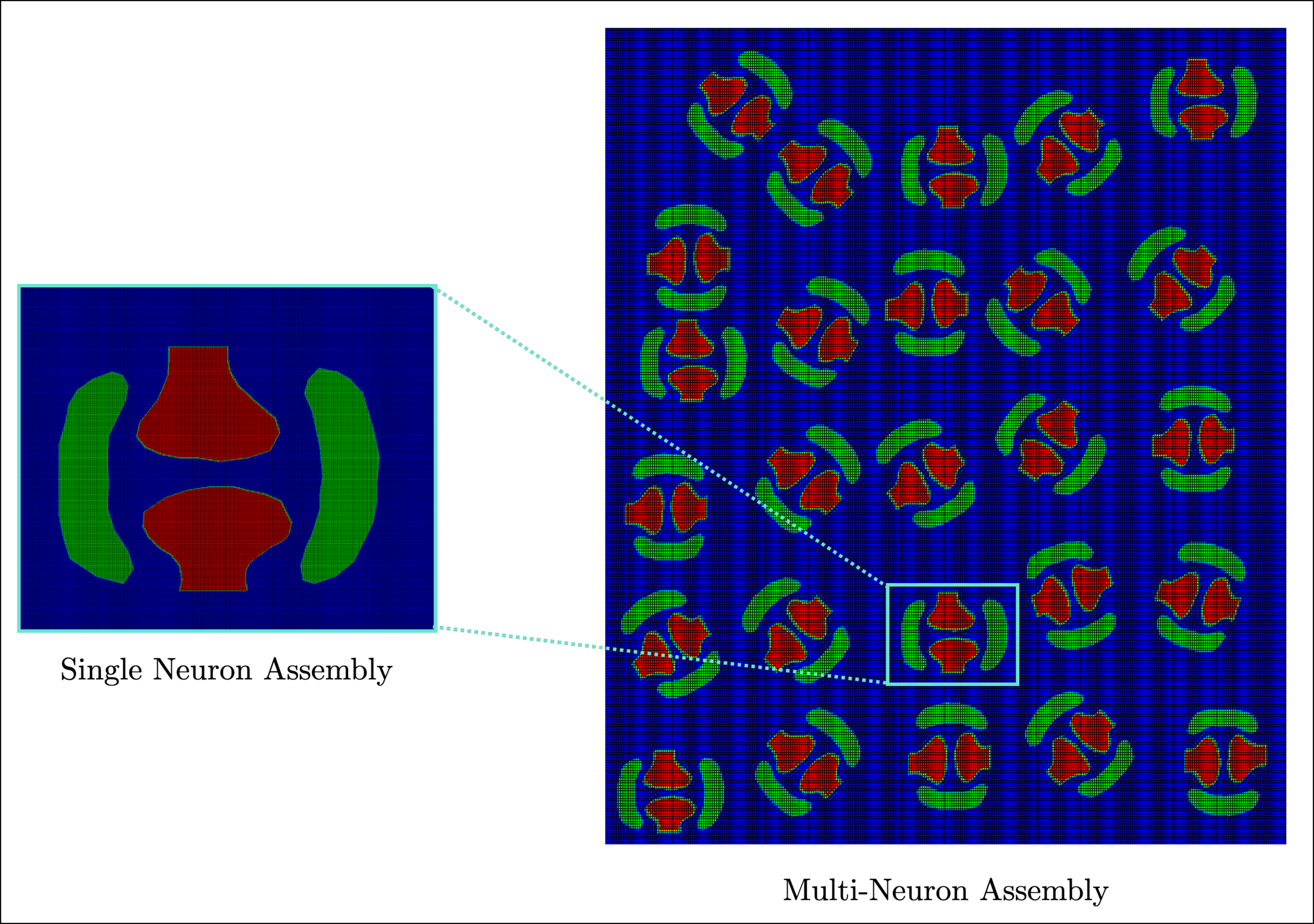
We have illustrated a morphological representation of single neuron-glia-ECM assembly (Fig.6). This consists of neuron synapses indicated by red, padded on both sides by microglia painted in green. The microglia is a pivotal region in numerical simulations, as it facilitates release of ATP to the ECM, houses the P2X7 receptor and modulates channel sensitivity and finally enables production of inflammatory cytokines. All the above mentioned chemical fields are spatially localised in the microglial region and the numerical framework allows diffusion across the ECM. For a single neuron-microglia-ECM assembly we observe the effects of various mechanical loading conditions on this domain through evolution of the underlying chemical and mechanical fields. Upon tensile loading, the biomarkers represented by chemical fields diffuse across the illustrated domain.

Multiple neuron-microglia-ECM geometry
In a more realistic setting, the length scale of the domain is increased to accommodate more number of neuron-glia-ECM assemblies which illustrate a better representation of neuron clusters (Fig.7). As we observe from this construction, more assemblies have been added at random orientations each having the characteristic spatial localisation as mentioned in the single neuron setup. For simplicity we have considered all neuron assemblies of the same type, meaning, the chemical and mechanical properties are the same for all assembly configurations. Primarily, this improvised structure allows us to visualise the spatio-temporal heterogeneity of chemical field evolution and capture essential chemical and mechanical injury metrics across the domain. Similar to the previous setup, we perform different BVPs on this domain and observe field evolution characteristics.
Simulations
For single and multiple neuron-microglia-ECM assembly, we demonstrate key results based on unaxial tension numerical simulations. For single assembly, as shown in Fig.8(A), the domain is fixed at one end and a displacement-type load is applied across a spectrum of strain and strain-values. In this simulation we highlight results obtained at a strain rate of upto strain. A fundamental metric of deformation based loading problems is strain; accordingly the axial strain field , obtained from the underlying mechanical network is plotted for the domain as given in Fig.8(B). Using a combination of strain and strain-rates, an array of results are presented in Fig.8(C). The strain field induces ATP to release from the microglia to the ECM. From left to right, field characteristics for ATP due to the above mentioned strain field is shown. As observed, ATP diffuses out of the microglia into the ECM demonstrating the heterogeneous nature of field distribution. The following sub-figure shows P2X7 characteristics which is operating at maximum potential as the critical concentration of ATP, needed to trigger the receptor, has already reached. To its right, TNF- field plot is shown, which depends on P2X7 receptor opening probability and diffuses out of the microglia. The final sub-plot highlights glutamate concentration due to the influence of TNF-. The neurotransmitter is localised at the synaptic region and diffuses into the ECM. While Fig.8(C) demonstrates full field profiles for each chemical entity, Fig.8(D) illustrates field evolution characteristics for increasing strain-rates from up to keeping the maximum strain fixed at 0.8 for each simulation. The following sub-plots (left to right) describe the nature of evolution of ATP (near the microglia), P2X7 (within the microglia), TNF- (near the microglia) and Glutamate (near the synapse). The particular strain and strain-rate combination used for the full field plots has been highlighted by an arrow in each sub-plot. The maximum magnitude of the chemical species’ concentrations are comparable with pathological estimates and presented in Table.2.
A key highlight of the spatio-temporal resolution of these chemical fields is the construction of an injury curve. Considering maximum ATP concentration across the spatial domain at specific strain and strain-rates a dataset is established. The data set is plotted against strain vs strain-rate as illustrated in Fig.9 and allows visual representation of field values at specific loading conditions. A cutoff for critical extracellular ATP is chosen based on the concentration at which purinergic receptors (P2X7) activate [11], which is about mM [72]. The proposition is, if the maximum concentration of ATP stays above this number, the neuronal micro environment is susceptible to chemical degradation, which is also how we define chemical injury. Using this metric, two regions are shown representing pathway induced injury (red boxes) and uninjured region (green boxes). It is a first order estimation of how mechanical loading conditions can influence downstream pathways and elevate critical molecular concentrations.
Unaxial tensile loading numerical simulations using rate-dependent loading are performed for multi-neuron assemblies. Using variable orientations for individual single neuron-microglia structures, a neuron cluster is constructed and a boundary value problem is setup as illustrated in Fig.10(A). We have chosen two essential field variables namely ATP (Fig.10(B)) and glutamate (Fig.10(C)) to demonstrate the heterogeneity and interactions within this assembly. The results consisting of these field profiles are generated using a strain rate of , applied up to a strain of 0.4.


5 Conclusion
We have proposed and quantified a glia-initiated injury pathway beginning with mechanical deformation and culminating in necroptosis. The proposed model incorporates a multiphysics formulation for investigating mechano-chemical interactions during TBI at the cellular scale. The fidelity of our numerical modeling framework involves a first-of-its-kind representation of neuronal microenvironment as a viscoelastic network coupled with an ensemble of molecular biomarkers represented as chemical fields. Using our modeling framework, we have demonstrated field behaviour for single neuron-glia assembly along with neuron-glia clusters highlighting localisation of biomarkers.
We believe that the idea of a mechano-chemical framework utilised to capture pathway induced injury opens up avenues of research directions in injury specific biomarker identification. Although our modeling approach relies on limited availability of biomarker datasets, experimental quantification for injury metrics in human subjects and generic assumptions on field evolution characteristics, it provides a robust numerical and computational base for further improvements. Additionally, our model is capable of incorporating additional biophysics at the neuronal scale including ionic conduction [28], nutrient transport and inter-neuronal interactions which possess immense potential in understanding crucial neurodegenerative disorders.
References
- [1] (2020) Cell death and recovery in traumatic brain injury. Neurotherapeutics 17, pp. 446–456. Cited by: §2.
- [2] (2017) The p2x7 receptor primes il-1 and the nlrp3 inflammasome in astrocytes exposed to mechanical strain. Frontiers in cellular neuroscience 11, pp. 227. Cited by: §1, §2.
- [3] (2010) Dopamine and glutamate in huntington’s disease: a balancing act. CNS neuroscience & therapeutics 16 (3), pp. 163–178. Cited by: §2.
- [4] (2020) The p2x7 receptor: central hub of brain diseases. Frontiers in Molecular Neuroscience 13, pp. 124. Cited by: §1, §2.
- [5] (2020) Going the extra (synaptic) mile: excitotoxicity as the road toward neurodegenerative diseases. Frontiers in cellular neuroscience 14, pp. 90. Cited by: §2.
- [6] (2019) Compartmentalized necroptosis activation in excitotoxicity-induced axonal degeneration: a novel mechanism implicated in neurodegenerative disease pathology. Neural Regeneration Research 14 (8), pp. 1385–1386. Cited by: §2.
- [7] (2004) Pannexin membrane channels are mechanosensitive conduits for atp. FEBS letters 572 (1-3), pp. 65–68. Cited by: §2.
- [8] (2017) P2X7 receptor induces tumor necrosis factor- converting enzyme activation and release to boost tnf- production. Frontiers in immunology 8, pp. 862. Cited by: §1, §3.
- [9] (1995) Quantal transmission at purinergic junctions: stochastic interaction between atp and its receptors. Biophysical journal 68 (3), pp. 925–935. Cited by: Table 1.
- [10] (2022) The nlrp3 inflammasome pathway: a review of mechanisms and inhibitors for the treatment of inflammatory diseases. Frontiers in aging neuroscience 14, pp. 879021. Cited by: §2.
- [11] (2013) When too much atp is a bad thing: a pivotal role for p2x7 receptors in motor neuron degeneration. Vol. 126, Wiley Online Library. Cited by: §4.
- [12] (2019) Microglia and the purinergic signaling system. Neuroscience 405, pp. 137–147. Cited by: §2.
- [13] (2014) Extracellular atp drives systemic inflammation, tissue damage and mortality. Cell death & disease 5 (3), pp. e1102–e1102. Cited by: §2.
- [14] (2015) Microglial activation as a compelling target for treating acute traumatic brain injury. Current medicinal chemistry 22 (6), pp. 759–770. Cited by: §2.
- [15] (2021) Extracellular atp-induced alterations in extracellular h+ fluxes from cultured cortical and hippocampal astrocytes. Frontiers in Cellular Neuroscience 15, pp. 640217. Cited by: §2.
- [16] (2024) Code repository for spatio temporal modeling of biomarkers to demonstrate mechano-chemical interactions during tbi. GitHub. Note: https://github.com/cmmg/mechanoChemistry Cited by: §2.1.
- [17] (2007) ATP activates a reactive oxygen species-dependent oxidative stress response and secretion of proinflammatory cytokines in macrophages. Journal of Biological Chemistry 282 (5), pp. 2871–2879. Cited by: §2.
- [18] (2018) Necroptosis: a regulated inflammatory mode of cell death. Journal of neuroinflammation 15 (1), pp. 1–9. Cited by: §2.
- [19] (2022) Mitochondrial atp synthase is a target of oxidative stress in neurodegenerative diseases. Frontiers in Molecular Biosciences 9, pp. 854321. Cited by: §1.
- [20] (2022) Oxidative stress in traumatic brain injury. International Journal of Molecular Sciences 23 (21), pp. 13000. Cited by: §2.
- [21] (2005) Apoptosis, pyroptosis, and necrosis: mechanistic description of dead and dying eukaryotic cells. Infection and immunity 73 (4), pp. 1907–1916. Cited by: §2.
- [22] (2012) Extracellular potassium dynamics in the hyperexcitable state of the neuronal ictal activity. Communications in Nonlinear Science and Numerical Simulation 17 (12), pp. 4700–4706. Cited by: §2.
- [23] (2015) TNF-dependent regulation and activation of innate immune cells are essential for host protection against cerebral tuberculosis. Journal of neuroinflammation 12 (1), pp. 1–14. Cited by: §2.
- [24] (2023) Cellular and molecular pathophysiology of traumatic brain injury: what have we learned so far?. Biology 12 (8), pp. 1139. Cited by: §1.
- [25] (2022) A literature review of traumatic brain injury biomarkers. Molecular neurobiology 59 (7), pp. 4141–4158. Cited by: §1.
- [26] (1997) Diffusion in axon guidance. European Journal of Neuroscience 9 (7), pp. 1414–1421. Cited by: Table 1.
- [27] (2015) Glutamate and gaba imbalance following traumatic brain injury. Current neurology and neuroscience reports 15, pp. 1–11. Cited by: §2, §2.
- [28] (2023) Spatio-temporal modeling of saltatory conduction in neurons using poisson–nernst–planck treatment and estimation of conduction velocity. Brain Multiphysics 4, pp. 100061. Cited by: §5.
- [29] (2012) Molecular mechanism of atp binding and ion channel activation in p2x receptors. Nature 485 (7397), pp. 207–212. Cited by: §1.
- [30] (1998) Apoptosis and necrosis: mechanisms of cell death induced by cyclosporine a in a renal proximal tubular cell line. Kidney international 54 (6), pp. 1955–1966. Cited by: §2.
- [31] (2022) Role of necroptosis in traumatic brain and spinal cord injuries. Journal of Advanced Research 40, pp. 125–134. Cited by: §2, §2.
- [32] (2020) Glutamate-induced excitotoxicity in parkinson’s disease: the role of glial cells. Journal of pharmacological sciences 144 (3), pp. 151–164. Cited by: §2.
- [33] (2007) Tumor necrosis factor alpha stimulates nmda receptor activity in mouse cortical neurons resulting in erk-dependent death. Journal of neurochemistry 100 (5), pp. 1407–1420. Cited by: §1.
- [34] (2015) Taming glutamate excitotoxicity: strategic pathway modulation for neuroprotection. CNS drugs 29, pp. 153–162. Cited by: §1.
- [35] (2017) Concussion as a multi-scale complex system: an interdisciplinary synthesis of current knowledge. Frontiers in neurology 8, pp. 513. Cited by: §1.
- [36] (2021) Modelling extracellular matrix and cellular contributions to whole muscle mechanics. Plos one 16 (4), pp. e0249601. Cited by: Table 1.
- [37] (2022) The role of cl- and k+ efflux in nlrp3 inflammasome and innate immune response activation. American Journal of Physiology-Cell Physiology 322 (4), pp. C645–C652. Cited by: §2.
- [38] (2008) Necroptosis contributes to the nmda-induced excitotoxicity in rat’s cultured cortical neurons. Neuroscience letters 447 (2-3), pp. 120–123. Cited by: §2.
- [39] (2017) Reactive oxygen species trigger nf-b-mediated nlrp3 inflammasome activation induced by zinc oxide nanoparticles in a549 cells. Toxicology and Industrial Health 33 (10), pp. 737–745. Cited by: §2.
- [40] (2013) Tumor necrosis factor in traumatic brain injury: effects of genetic deletion of p55 or p75 receptor. Journal of Cerebral Blood Flow & Metabolism 33 (8), pp. 1182–1189. Cited by: §2.
- [41] (2008) Moderate and severe traumatic brain injury in adults. The Lancet Neurology 7 (8), pp. 728–741. Cited by: §2.
- [42] (2001) Pictorial review of glutamate excitotoxicity: fundamental concepts for neuroimaging. American journal of neuroradiology 22 (10), pp. 1813–1824. Cited by: Table 2.
- [43] (2023) Sculptor and destroyer: tales of glutamatethe brain’s most important neurotransmitter. MIT Press. Cited by: §2.
- [44] (2015) The neuropathology of traumatic brain injury. Handbook of clinical neurology 127, pp. 45–66. Cited by: §1.
- [45] (1994) Tumor necrosis factor- (tnf-) increases both in the brain and in the cerebrospinal fluid from parkinsonian patients. Neuroscience letters 165 (1-2), pp. 208–210. Cited by: Table 2.
- [46] (2011) Extracellular glutamate: functional compartments operate in different concentration ranges. Frontiers in systems neuroscience 5, pp. 94. Cited by: Table 1.
- [47] (2013) K+ efflux is the common trigger of nlrp3 inflammasome activation by bacterial toxins and particulate matter. Immunity 38 (6), pp. 1142–1153. Cited by: §2.
- [48] (2012) Plasticity of glutamate synaptic mechanisms. Jasper’s Basic Mechanisms of the Epilepsies [Internet]. 4th edition. Cited by: §2.
- [49] (2019) Traumatic brain injuries: pathophysiology and potential therapeutic targets. Frontiers in cellular neuroscience 13, pp. 528. Cited by: §2.
- [50] (2024) Glutamate-mediated excitotoxicity in the pathogenesis and treatment of neurodevelopmental and adult mental disorders. International Journal of Molecular Sciences 25 (12), pp. 6521. Cited by: §1.
- [51] (2022) The role of regulatory necrosis in traumatic brain injury. Frontiers in Molecular Neuroscience 15, pp. 1005422. Cited by: §2.
- [52] (2020) The nlrp3 inflammasome in traumatic brain injury: potential as a biomarker and therapeutic target. Journal of neuroinflammation 17, pp. 1–12. Cited by: §2.
- [53] (2017) The role of glutamate transporters in the pathophysiology of neuropsychiatric disorders. NPJ schizophrenia 3 (1), pp. 32. Cited by: §1.
- [54] (2014) Tumor necrosis factor alpha: a link between neuroinflammation and excitotoxicity. Mediators of inflammation 2014. Cited by: §1, §2.
- [55] (2021) Glutamate: the master neurotransmitter and its implications in chronic stress and mood disorders. Frontiers in human neuroscience 15, pp. 722323. Cited by: §2.
- [56] (2008) ATP is released by monocytes stimulated with pathogen-sensing receptor ligands and induces il-1 and il-18 secretion in an autocrine way. Proceedings of the National Academy of Sciences 105 (23), pp. 8067–8072. Cited by: §2.
- [57] (2013) TNF- downregulates inhibitory neurotransmission through protein phosphatase 1-dependent trafficking of gabaa receptors. Journal of Neuroscience 33 (40), pp. 15879–15893. Cited by: §2.
- [58] (2018) Glutamatergic signaling in the central nervous system: ionotropic and metabotropic receptors in concert. Neuron 98 (6), pp. 1080–1098. Cited by: §2.
- [59] (2018) Diffusion of cytokines in live lymph node tissue using microfluidic integrated optical imaging. Analytica chimica acta 1000, pp. 205–213. Cited by: Table 1.
- [60] (2022) The role of purinergic p2x7 receptor in inflammation and cancer: novel molecular insights and clinical applications. Cancers 14 (5), pp. 1116. Cited by: §1.
- [61] (2011) Shaping the synaptic signal: molecular mobility inside and outside the cleft. Trends in neurosciences 34 (7), pp. 359–369. Cited by: Table 1.
- [62] (2021) Pannexin-1 channels as mediators of neuroinflammation. International journal of molecular sciences 22 (10), pp. 5189. Cited by: §1.
- [63] (1999) Dual role of tumor necrosis factor alpha in brain injury. Cytokine & growth factor reviews 10 (2), pp. 119–130. Cited by: §2.
- [64] (2014) The influence of ion concentrations on the dynamic behavior of the hodgkin–huxley model-based cortical network. Cognitive neurodynamics 8, pp. 287–298. Cited by: §2.
- [65] (2006) Tumor necrosis factor- induces neurotoxicity via glutamate release from hemichannels of activated microglia in an autocrine manner. Journal of Biological Chemistry 281 (30), pp. 21362–21368. Cited by: §1, §2.
- [66] (2024) Pathological interplay between inflammation and mitochondria aggravates glutamate toxicity. International Journal of Molecular Sciences 25 (4), pp. 2276. Cited by: §1.
- [67] (2014) Extracellular atp hydrolysis inhibits synaptic transmission by increasing ph buffering in the synaptic cleft. PLoS biology 12 (5), pp. e1001864. Cited by: §2.
- [68] (2017) Role of glutamate and nmda receptors in alzheimer’s disease. Journal of Alzheimer’s Disease 57 (4), pp. 1041–1048. Cited by: §2.
- [69] (2022) Viscoelastic modeling of the stress relaxation behavior for the bionic extracellular matrix polymer scaffold. Medicine in Novel Technology and Devices 16, pp. 100181. Cited by: Figure 3, Table 1, Table 1, Table 1, Table 1, Table 1, §3.
- [70] (2013) The role of markers of inflammation in traumatic brain injury. Frontiers in neurology 4, pp. 18. Cited by: §2.
- [71] (2012) Neurons respond directly to mechanical deformation with pannexin-mediated atp release and autostimulation of p2x7 receptors. The Journal of physiology 590 (10), pp. 2285–2304. Cited by: §2.
- [72] (2016) Modeling interactions among individual p2 receptors to explain complex response patterns over a wide range of atp concentrations. Frontiers in physiology 7, pp. 294. Cited by: §1, §2.1, §2, Figure 5, §3, §4, Table 2, Table 2.
- [73] (2020) Distinct molecular mechanisms underlying potassium efflux for nlrp3 inflammasome activation. Frontiers in immunology 11, pp. 609441. Cited by: §2.
- [74] (1996) RAGE and amyloid- peptide neurotoxicity in alzheimer’s disease. Nature 382 (6593), pp. 685–691. Cited by: §2.
- [75] (2021) Tumor necrosis factor alpha signaling and organogenesis. Frontiers in Cell and Developmental Biology 9, pp. 727075. Cited by: §1.
- [76] (2018) ATP release by red blood cells under flow: model and simulations. Biophysical journal 115 (11), pp. 2218–2229. Cited by: Table 1.
- [77] (2005) TNF potentiates glutamate neurotoxicity by inhibiting glutamate uptake in organotypic brain slice cultures: neuroprotection by nfb inhibition. Brain research 1034 (1-2), pp. 11–24. Cited by: Figure 5, §3.