A Novel Power-optimized CMOS sEMG Device with Ultra Low-noise integrated with ConvNet (VGG16) for Biomedical Applications
Abstract
The needle bio-potential sensors for measuring muscle and brain activity need invasive surgical targeted muscle reinnervation (TMR) and a demanding process to maintain, but surface bio-potential sensors lack clear bio-signal reading (Signal-Interference). In this research, a novel power-optimized complementary metal-oxide-semiconductor (CMOS) Surface Electromyography (sEMG) is developed to improve the efficiency and quality of captured bio-signal for biomedical application: The early diagnosis of neurological disorders (Dystonia) and a novel compatible mind-controlled prosthetic leg with human daily activities. A novel sEMG composed of CMOS Op-Amp based PIC16F877A 8-bit CMOS Flash-based Microcontroller is utilized to minimize power consumption and data processing time. sEMG Circuit is implemented with developed analog filter along with infinite impulse response (IIR) digital filter via Fast Fourier Transform (FFT), Z-transform, and difference equations. The analysis shows a significant improvement of 169.2% noise-reduction in recorded EMG signal using developed digital filter compared to analog one according to numerical root mean square error (RMSE). Moreover, digital IIR was tested in two stages: algorithmic and real-world. As a result, IIR’s algorithmic (MATLAB) and real-world RMSEs were 0.03616 and 0.05224, respectively. A notable advancement of 20.8% in data processing duration in sEMG signal analysis. Optimizing VGG, AlexNet, and ResNet ConvNet as trained and tested on 15 subjects’ public EEG (62 electrodes) and observed EMG data. The results indicate that VGG16-1D is 98.43% higher. During real testing, the accuracy was 95.8 ± 4.6% for 16 subjects (6 Amputees-10 Dystonia). This study demonstrates the potential for sEMG, paving the way for biomedical applications.
Index Terms:
prosthetic leg, dystonia, surface electromyography, IIR digital filter, VGG16I Introduction
Electromyography (EMG) is a valuable diagnostic technique used in modern healthcare settings to assess muscle electrical activity. However, Surface EMG (sEMG) technology faces various obstacles, includes: The complex interpretation of sEMG signals, which can be challenging due to factors such as noise and interference. Additionally, sEMG patterns can vary significantly among individuals and muscles, making it challenging to establish standardized norms for interpretation. Regarding the benefits of sEMG in implementation, Integrating sEMG findings with other diagnostic modalities can be challenging due to differences in terminology and interpretation. EMG is often used in conjunction with other diagnostic tests, such as nerve conduction studies or imaging, to provide a comprehensive assessment of neuromuscular function.
I-A Prior Solutions
Over the years, attempts have been made to address the challenges associated with electromyography (sEMG) signal interpretation in clinical practice. These efforts have included the use of filtration
techniques such as analog filters, but these have been found to be inefficient in addressing the complexities of sEMG recordings. Additionally, sEMG pattern variability has been addressed through
classification methods that involve signal segmentation using convolutional neural networks (ConvNet). While this approach has shown promise, it has not fully resolved the issue of sEMG pattern variability.
In the biomedical application of sEMG for prosthetic leg control, inadequate data filtration from analog filters has resulted in unclear movement of the prosthetic leg. However, one of the biggest
challenges in the clinical application of sEMG is its use in the early diagnosis of neurological disorders. Despite its potential, sEMG can be limited in its ability to detect subtle changes in muscle
activity that may indicate the presence of a neurological disorder in its early stages. Therefore, further research and innovation are needed to develop standardized norms, quality assurance measures, and
patient-centered approaches for sEMG interpretation in the context of early diagnosis of neurological disorders. The objective of this study is to build on prior solutions and identify new approaches that
can address the challenges associated with sEMG in clinical practice, particularly in the early diagnosis of neurological disorders.
These are last year prior soltuions for this project:
- •
Previous attempts at solving sEMG signal interpretation challenges have utilized filtration techniques (analog filters) but these were found to be inefficient.
- •
sEMG pattern variability has been addressed through classification methods involving signal segmentation using convolutional neural network (ConvNet), but this approach did not fully resolve the issue.
- •
In the biomedical application of sEMG for prosthetic leg control, inadequate data filtration from analog filters has resulted in unclear movement of the prosthetic leg.
Further research and innovation are needed to develop standardized norms, quality assurance measures, and patient-centered approaches for sEMG interpretation.
I-B Engineering Goals
Electromyography (sEMG) is a technique widely used in clinical practice for the assessment of neuromuscular disorders. Despite its potential clinical benefits, sEMG recordings can be challenging to interpret due to the variability among individuals and muscles. Moreover, technical limitations in sEMG recordings can affect accuracy and reproducibility, which can compromise its reliability as a diagnostic tool. To address these challenges, this study aims to identify, compare, and implement solutions that can enhance the accuracy and reliability of sEMG recordings in clinical practice.
- •
Develop standardized protocols for accurate sEMG signal interpretation. Establish normative values for sEMG patterns that account for variability among individuals and muscles.
- •
Improve technical aspects of sEMG recordings to enhance accuracy and reproducibility. Explore strategies for integrating sEMG findings with other diagnostic modalities to improve diagnostic consistency and patient care.
Aiming to improve the accuracy and reliability of electromyography (sEMG) recordings in clinical practice for the assessment of neuromuscular disorders. By developing standardized protocols for sEMG signal interpretation and improving technical aspects of sEMG recordings using filters and AI, this study seeks to enhance diagnostic consistency, controlling of prosthetic and patient care. It is hoped that the results of this study will contribute to the ongoing efforts to improve the clinical application of sEMG and benefit patients with neuromuscular disorders.
I-C Challenges in the last solution
Last soltuion, there significant challenges faced it, specifically in filtering signals using analog filter for biomedical application such controlling lower limb prosthetic leg or architecture of AI to train these signals.
- •
Although numerous attempts have been made to improve the filtration process in interpreting sEMG signals, especially when using analog filters, these efforts have not been entirely successful. Residual noise has remained unfiltered, resulting in inaccuracies in the output signal. As a result, it is crucial to continue research to optimize the filtration process and the develpoment of filters, particularly for filtering complex sEMG signals. Advanced signal processing techniques, such as digital filtering and wavelet analysis, may hold promise for effectively filtering out unwanted noise while preserving the integrity of the sEMG signal.

Fig. 1: The first signal is a sEMG signal devoid of any extraneous noise. The second signal denotes a signal that has been subject to power line noise, followed by analog filtering. While the signal has undergone filtration, there persists residual noise that remains unfiltered. - •
Convolutional networkds (ConvNets) face significant challenges in achieving real-time processing and high classification accuracy rates when applied to bio-signals due to signal and pattern similarity, as well as parameter overload. To overcome these challenges, a customized ConvNet architecture should be developed to achieve real-time processing and high accuracy classification.

Fig. 2: Fully Connected (FC) layer in neural networks can improve the accuracy of each node and weight in the model. However, this approach can lead to a deeper architecture and longer processing times due to the increased complexity of the network. - •
In order to effectively integrate prosthetic leg with sEMG technology, it is crucial to address Challenge 1 and Challenge 2. The presence of noise in the sEMG signal can significantly impact the accuracy of the classification process, thereby affecting the overall movement of the prosthetic leg.
![[Uncaptioned image]](2301.09570v2/Picture3.png)
II Methodology
II-A Analog vs Digital Filters
Analog filters are constructed by employing analog circuitry, which involves the use of hardware components. On the other hand, digital filters are designed utilizing digital signal processing techniques, which rely on software implementation. The process of designing analog filters typically entails the careful selection and connection of passive or active components within the circuitry. In contrast, digital filters are based on mathematical algorithms that are implemented in software. One notable advantage of digital filters is their ability to offer greater flexibility and precision in adjusting filter characteristics compared to analog filters. This flexibility stems from the inherent versatility of software-based algorithms, allowing for fine-tuning of various filter parameters. In contrast, analog filters may exhibit limitations in terms of flexibility and precision due to the constraints imposed by physical circuit components.

II-B Noise sensitivity in Analog Vs. Digital filters
Analog:
- •
Susceptible to environmental factors and component tolerances, which can introduce noise.
- •
Prone to external noises (e.g., EMI, RFI) that can degrade signal-to-noise ratio (SNR).
Digital:
- •
Less susceptible to environmental factors and component tolerances.
- •
Precise control over filter characteristics, allowing for high accuracy and predictable performance.
- •
Can mitigate external noises.


Based on the previous discussion, it can be inferred that digital filters generally offer advantages over analog filters in terms of flexibility, adaptability, and stability. In the next section, we will delve into the specific comparison of the output characteristics of digital and analog filters in the context of noise filtering.
II-C Testing of Analog VS Digital Filters noise filtering
The process of testing was as following:
- 1.
A comparative study was conducted to evaluate the performance of digital and analog filters in filtering out noise from a given input signal.
- 2.
A test signal contaminated with noise was generated, representing a real-world scenario where a signal of interest is corrupted by unwanted noise.
- 3.
Both digital and analog filters were designed to have similar filter characteristics, such as cutoff frequency, filter order, and filter type, to ensure a fair comparison.
- 4.
The input signal contaminated with noise was passed through both filters separately, and the filtered output signals were recorded for further analysis.


II-D Optimizing ConvNet for real-time signal processing
Comparing the accuracy of ResNet18 and VGG16 for classification; using VGG16 and ResNet18 for signal processing and classification on different channels (15 subjects’ public EEG and 18 observed EMG data), based on that ConvNets are compared on different signals and channels as shown in fig(9). Signals are preprocessed and A frequency filter ranging from 0 to 75 Hz was used to apply a band pass to the signal at 200 Hz for more significant analysis.

we conducted a performance comparison between VGG16 and ResNet models for EEG classification. Specifically, we examined the impact of VGG16’s fully connected layers and deeper architecture on the classification performance in a 62-channels EEG dataset, in comparison to ResNet. Surprisingly, despite VGG16 demonstrating higher performance in a 10-channel EEG dataset, our findings revealed that it exhibited lower performance in the more complex 62-channels EEG dataset. These results suggest that the fully connected layers and deeper architecture of VGG16 may not be as effective in capturing the intricate patterns present in the 62-channels EEG data, in contrast to the superior performance of ResNet.


![[Uncaptioned image]](2301.09570v2/Picture8.png)
we aimed to address the reduced accuracy of VGG16 when training large-scale EEG datasets with varying channel configurations, in comparison to ResNet. To mitigate this issue, we developed a customized VGG architecture specifically tailored for EEG signal classification. The customized VGG architecture was designed to be less complex, optimizing its performance by reducing the number of parameters. Our comparative analysis revealed that the customized VGG architecture outperformed VGG16 in terms of accuracy, showcasing its effectiveness in handling the challenges posed by diverse EEG signal datasets. This finding holds great significance for researchers and practitioners in the field of deep learning for bio-signal classification. To enhance the customized architecture, we employed small convolutional kernels (1, 3) dimensions and eliminated fully connected (FC) layers. These modifications aimed to improve the model’s capacity, reduce the number of parameters, and prevent overtraining and overfitting issues commonly encountered in deep learning models. The development of this customized VGG architecture demonstrates an innovative and scientific approach to tackling and imrpove the performance and the specific challenges associated with EEG signal classification. The results highlight the potential for optimized deep learning models tailored for bio-signal analysis, contributing valuable insights to the scientific community and opening up new avenues for future research.
II-E Optimizers and Hyperparameters Art
Choosing between ADAMS and SGD as VGG optimizer:
- 1.
Investigated the performance of two optimization algorithms, SGD and Adam, on the VGG16 model for deep learning tasks.
- 2.
Conducted experiments with both SGD and Adam optimizers and compared their performance in terms of model accuracy.
- 3.
Our findings revealed that Adam optimizer outperformed SGD in terms of accuracy, achieving higher accuracy rates on the validation and test sets. ADAMS avoided overtraining.
![[Uncaptioned image]](2301.09570v2/Picture11.png)

In the pursuit of optimizing deep learning models, a comprehensive study was conducted to compare the effectiveness of two popular optimizers, namely SGD and Adam, in the context of the VGG16 model. Adam emerged as the preferred choice due to its ability to prevent overtraining and deliver superior accuracy, surpassing the performance of SGD. This investigation sheds light on the selection of optimizers.
III Data Analysis and Results
Two approaches in signal processing:
- •
Performing VGG-16 1-Dimensional convolutions, which are applied along a time axis to capture patterns in the time domain.
- •
Performing VGG-16 2-Dimensional convolutions, which are applied in both X and Y dimensions to capture patterns in images.


The pattern-based approach of VGG16-V1 provided and emphasis on capturing intricate patterns, achieved a higher recognition rate and accuracy compared to VGG16-V2, which employed a binary approach. VGG16-V1’s architecture was specifically designed to learn and recognize complex patterns by utilizing a deeper network structure with multiple convolutional layers compared to the binary approach of VGG16-V2.
A randomized analysis of electromyography (EMG) signals and examined the stability of an Infinite Impulse Response (IIR) filter in the presence of power line noise. The objective was to investigate the deficiencies that can occur in analog filters when filtering EMG signals corrupted by power line interference.


| Filter Type | MSE | RMSE |
|---|---|---|
| Analog Filter | 0.0047 | 0.0686 |
| Digital Filter | 0.0011 | 0.0327 |
| VGG16-V1 | VGG16-V2 | |
|---|---|---|
| Preprocessing Time (s) | 0.003135 | 0.003345 |
| CPU Processing Time (s) | 0.013343 | 0.95740 |
| GPU Processing Time (s) | 0.002808 | 0.020899 |
| Model Parameters | 784,803 | 67,146,211 |
| Accuracy (%) | 98.4383 | 97.1966 |
| Macro-F1 | 0.97 | 0.95 |
Based on the provided data in table (1, 2), VGG16-V1 is better than VGG16-V2 in several aspects. VGG16-V1 demonstrated higher pretreatment time, CPU inference time, GPU reasoning time, and had fewer model parameters (small segments in one length) compared to VGG16-V2. Additionally, VGG16-V1 achieved a higher accuracy and a slightly more accurate macro F1 score compared to VGG16-V2. These findings suggest that VGG16-V1 may be a more efficient and effective choice for the given task, potentially offering better performance with lower computational overhead. The application of digital filters, alongside a customized convolutional neural network (CNN) architecture, specifically the VGG16-V1 model, has enabled real-time processing of signals with enhanced filtering capabilities. This advancement has opened new avenues for improved readings and accurate classification bio-signal for biomedical applications, such a novel controlled prosthetic leg and early diagnosis of dystonia.
Optimizing Prosthetic Control: Leveraging Signal-based Statistics for Accurate Prediction of Movement Patterns
- •
Ascending: Achieved perfect accuracy of 100% (1.000), indicating all instances of this class were correctly predicted as ”Ascending”.
- •
Descending: Achieved high accuracy of 96.6% (0.966), with 3.4% of instances misclassified as other classes.
- •
Resting: Achieved perfect accuracy of 100% (1.000), demonstrating all instances of this class were correctly predicted as ”Resting”.
- •
Leaping: Achieved accuracy of 90%, with 6.4% of instances misclassified as others and 3.5% of instances misclassified as ”Walking”.
- •
Walking: Achieved accuracy of 96.6% (0.966), with 3.4% of instances misclassified as other classes.
- •
Resting: Achieved perfect accuracy of 100% (1.000), showing all instances of this class were correctly predicted as ”Resting”.
IV Applications
IV-A Prosthetic Leg Model
Making a prosthetic limb with a high bearing capacity, flexibility, comfort, and shock absorption for long-term usage requires considerable effort. When fabricating a prosthetic limb, it should be lightweight for ease of control and have a good load bearing capability. The prosthetic limb is constructed from lightweight but robust materials. The limb may or may not have functioning knee and ankle joints, depending on the site of the amputation. The socket is a very accurate cast of your residual limb that fits snugly over it. It assists in the prosthetic leg’s attachment to your body. Suspension systems are used to secure the prosthesis, whether by sleeve suction, vacuum suspension/suction, or distal locking by pin or lanyard. As shown in fig (19), numbers of models that are created for achieving engineering goals such as high bearing capacity, light, and long-term use.


Following the selection of your prosthetic leg’s components, you will need rehabilitation to strengthen your legs, arms, and cardiovascular system as you learn to walk with your new limb. You’ll work closely with rehabilitation experts, physical therapists, and occupational therapists to develop a rehabilitation plan that is customised to your unique mobility requirements. Maintaining a healthy leg is a critical component of this routine.

In the field of prosthetics, surface electromyography (sEMG) has emerged as a promising technique for enhancing the control of prosthetic limbs. By capturing the electrical signals generated by the user’s muscles through surface electrodes, sEMG enables the intuitive and precise control of prosthetic devices. This figure illustrates a prosthetic leg controlled by surface EMG, showcasing how it can be optimized to improve the user experience.


IV-B Stress Analysis of Presthestic limbs
The Von-Mises Stress analysis diagram, which is employed to simulate the maximum load that a prosthetic leg can endure. This analysis provides crucial insights into the leg’s structural integrity and its ability to withstand external forces. As depicted in the diagram, the calculated maximum load capacity for the leg is approximately 3000 newtons. This information is significant in the field of prosthetics as it aids in designing and developing prosthetic limbs that can safely support the expected loads and stresses experienced during daily activities. By considering the Von-Mises Stress analysis, engineers and researchers can ensure that prosthetic legs are optimized for strength and durability, thereby enhancing their functionality and improving user confidence and safety.


IV-C Detecting Staris using Machine learning
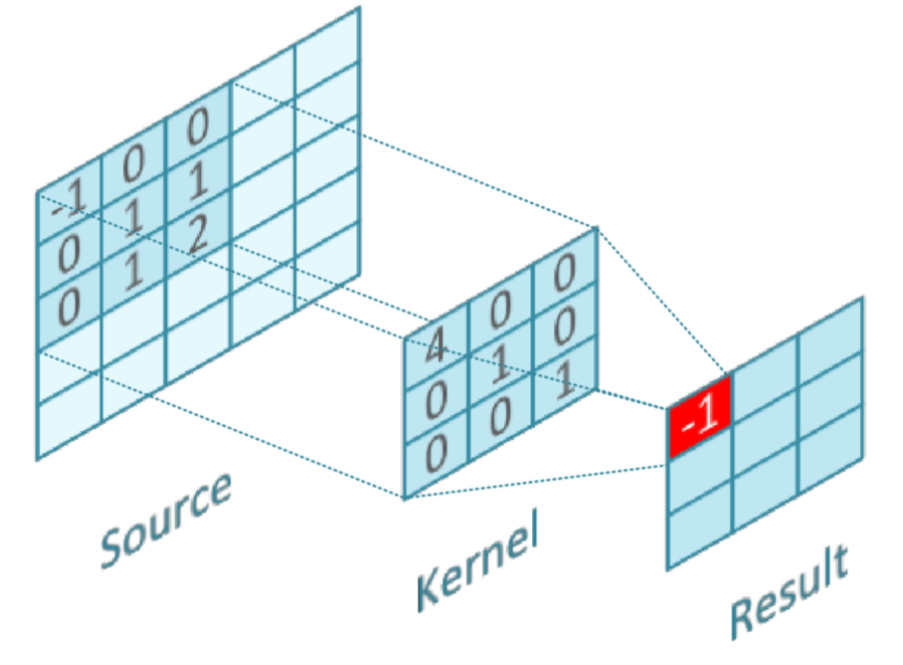
Using canny detection algorithm to detect edge, and it analyzed data based on Noise Reduction and Edge detection is sensitive to image noise, the first step is to eliminate the noise with a 5x5 or 3x3 Gaussian filter. Then, finding the Image’s Intensity Gradient. The smoothed picture is then filtered in both the horizontal and vertical directions with a Sobel kernel to obtain the first derivative in both the horizontal (Gy) and vertical (Gx) directions.
| (1) |

IV-D Piezoelectric and Voltage generator
For Powering the prosthetic leg we used Piezoelectric to generate required voltage with each movement, we stored the overcome voltage in capacitor and battery cells We collected average 1.48 volts with for each .45 Kg. Each Trail average consist of 6 trails. The utilization of piezoelectric devices for battery recharging has garnered significant attention. Extensive testing is imperative to determine their viability as the primary voltage generator. The accompanying graph showcases the relationship between the load and voltage, revealing that as the load increases, the voltage also rises. This observation establishes the adequacy of the device’s output as a main voltage generator. Such findings contribute valuable insights into the potential application of piezoelectric devices in sustainable energy harvesting and battery charging systems.


IV-E Early diagnosis of dystonia
Runner’s dystonia (RD) is a task-specific focal dystonia of the lower limbs that occurs when running to diagnostic of dystonia. In this retrospective case series, we present surface electromyography (EMG) and joint kinematic data from thirteen patients who underwent instrumented gait analysis (IGA) at the Functional and Biomechanics Laboratory at the National Institutes of Health[1]. Four cases of RD are described in greater detail to demonstrate the potential utility of EMG with kinematic studies to identify dystonia muscle groups in RD. Lateral heel whip, a proposed novel presentation of lower-limb dystonia, is also described. Surface EMG is showing continuous activity in the left hamstrings (b) and early activity in the left tibialis anterior during running (a). Showing how left leg delayed in activation of motor neuron with respect to other leg (A) in the time domain and in (B) in the frequency domain. The signal was recorded from the vastus lateralis (VL) muscle during Whole body vibration (WBV) at 30 Hz. While the sEMG signal in the time domain does not highlight any specific characteristics to WBV. The sEMG signal in the frequency domain clearly shows excessive spikes at the vibration frequency and at a few multiple harmonics and that means how dystonia is diagnostic obviously. At the same time, and also for preliminary purposes, the electrophysiological signal was recorded from the patella during WBV. Such a signal obtained during WBV at 30 Hz is shown in Figure 9. In the time domain, the patella signal resembles a sinusoidal wave at 30 Hz. In frequency domain, excessive spikes are observed at the vibration frequency and to a lower extent at its multiple harmonics. No myoelectrical activity is shown for all the other frequencies.

A surface electromyography (sEMG) spectrum of the Vastus Lateralis during whole-body vibration at 30 Hz. sEMG signals were processed using the no-filter method (black solid line), linear interpolation (grey solid line), band-stop filter (grey dotted line) and band-pass filter (black dashed line). As shown in Figure 10, The crucial role of filter especially High Pass filtration (HPF) and Low Pass filtration (LPF).
V Real testing on subjects


Based on the previous discussion, it can be concluded that digital filters overtake analog filters in EMG filtration in terms of flexibility, adaptability, and stability. The utilization of digital filters led to an enhancement of the signal processing stage by approximately 200The RMSE value obtained from the analog filter was 0.0686, while the digital filter was 0.0327, indicating a significant improvement in signal accuracy. These findings suggest that digital filters are highly effective for EMG filtration and can provide substantial improvements in signal processing performance.
V-A Equations
| (2) |
An infinite impulse response (IIR) filter is a type of digital filter where the output of the filter depends not only on current and past input samples but also on past output samples.

In digital signal processing, the filter function is expressed mathematically, while analog filters are designed using schematic.
VI Discussion and conclusion
Digital filters outperformed analog filters for filtering EMG signals, resulting in significant improvements in signal processing performance. Also, VGG16-V1 model was more efficient and accurate than VGG16-V2. These advancements enhanced the use of sEMG signals for controlling prosthetic legs and early diagnosis of dystonia.
VII Future Plans
- •
Increase the dataset size and diversity to enhance accuracy and generalization.
- •
User Experience optimization and usability of the sEMG system based on user feedback.
- •
Implement Real-time Feedback by Integrating sensory feedback mechanisms for enhanced control and user experience.
- •
Utilizing more advancing filtration method of digital filtering like wavelet-based or adaptive filters, for more filtering improvements.
References
- [1] O. F. Ahmad, P. Ghosh, C. Stanley, B. Karp, M. Hallett, C. Lungu, and K. Alter, ”Electromyographic and joint kinematic patterns in runner’s dystonia,” Toxins, vol. 10, no. 4, p. 166, 2018.
- [2] T. J. Bates, J. R. Fergason, and S. N. Pierrie, ”Technological advances in prosthesis design and rehabilitation following upper extremity limb loss,” Current Reviews in Musculoskeletal Medicine, vol. 13, no. 4, pp. 485-493, 2020.
- [3] T. Beyrouthy, S. Al Kork, J. A. Korbane, and M. Abouelela, ”EEG mind controlled smart prosthetic arm-a comprehensive study,” Advances in Science, Technology and Engineering Systems Journal, vol. 2, no. 3, pp. 891-899, 2017.
- [4] J. Cheng, Q. Song, H. Peng, J. Huang, H. Wu, and B. Jia, ”Optimization of VGG16 Algorithm Pattern Recognition for Signals of Michelson–Sagnac Interference Vibration Sensing System,” Photonics, vol. 9, no. 8, p. 535, July 2022.
- [5] K. H. Cheah, H. Nisar, V. V. Yap, C. Y. Lee, and G. R. Sinha, ”Optimizing residual networks and VGG for classification of EEG signals: Identifying ideal channels for emotion recognition,” Journal of Healthcare Engineering, 2021.
- [6] B. D. Farnsworth, R. J. Triolo, and D. J. Young, ”Wireless implantable EMG sensing microsystem,” in IEEE, pp. 1245-1248, 2008.
- [7] L. Gan, W. Liu, Y. Luo, X. Wu, and B. L. Lu, ”A cross-culture study on multimodal emotion recognition using deep learning,” in Neural Information Processing: 26th International Conference, ICONIP 2019, Sydney, NSW, Australia, December 12–15, 2019, Proceedings, Part IV, vol. 26, pp. 670-680, Springer International Publishing.
- [8] W. L. Zheng and B. L. Lu, ”Investigating critical frequency bands and channels for EEG-based emotion recognition with deep neural networks,” IEEE Transactions on Autonomous Mental Development, 2015.
- [9] J. U. Chu and I. Moon, ”A supervised feature-projection-based real-time EMG pattern recognition for multifunction myoelectric hand control,” IEEE/ASME Transactions on Mechatronics, vol. 12, no. 3, pp. 282-290, June 2007.
- [10] K. Ziegler-Graham et al., ”Estimating the Prevalence of Limb Loss in the United States - 2005 to 2050,” Archives of Physical Medicine and Rehabilitation, vol. 89, 2008, pp. 422-429.
- [11] K. T. Kim, S. Park, T. H. Lim, and S. J. Lee, ”Upper-Limb Electromyogram Classification of Reaching-to-Grasping Tasks Based on Convolutional Neural Networks for Control of a Prosthetic Hand,” Frontiers in Neuroscience, vol. 15, 2021.
- [12] M. L. Edwards, D. A. Olson, and F. DeRuyter, ”Chapter 18 - Lower Limb Prosthetics,” in Clinician’s guide to assistive technology, Mosby, 2002.
- [13] C. L. McDonald et al., ”Prosthetics and Orthotics International,” Prosthetics and Orthotics International, vol. 45, no. 2, pp. 105-114, 2020. doi: 10.1177/0309364620972258
- [14] L. Rozaqi, A. Nugroho, K. H. Sanjaya, and A. I. Simbolon, ”Design of analog and digital filter of electromyography,” in 2019 International Conference on Sustainable Energy Engineering and Application (ICSEEA), IEEE, 2019, pp. 186-192.
- [15] E. D. Setioningsih, ”The Impact of Using Digital Filter and Analog Filter on Surface Electromyography Signal,” International Journal of Advanced Health Science and Technology, vol. 1, no. 2, pp. 68-73, 2021.