Trustworthy clinical AI solutions: a unified review
of uncertainty quantification in deep learning models for medical image analysis
Benjamin Lambert
Affiliation: Univ. Grenoble Alpes, Inserm, U1216, Grenoble Institut des Neurosciences, Grenoble, 38000, France
Affiliation: Pixyl Research and Development Laboratory, Grenoble, 38000, France
Florence Forbes
Affiliation: Univ. Grenoble Alpes, Inria, CNRS, Grenoble INP, LJK, Grenoble, 38000, France
Alan Tucholka
Affiliation: Pixyl Research and Development Laboratory, Grenoble, 38000, France
Senan Doyle
Affiliation: Pixyl Research and Development Laboratory, Grenoble, 38000, France
Harmonie Dehaene
Affiliation: Pixyl Research and Development Laboratory, Grenoble, 38000, France
Michel Dojat
Affiliation: Univ. Grenoble Alpes, Inserm, U1216, Grenoble Institut des Neurosciences, Grenoble, 38000, France
Abstract
The full acceptance of Deep Learning (DL) models in the clinical field is rather low with respect to the quantity of high-performing solutions reported in the literature. Particularly, end users are reluctant to rely on the rough
predictions of DL models. Uncertainty quantification methods have been proposed in the literature as a potential response to reduce the rough decision provided by the DL black box and thus increase the interpretability and the acceptability of the result by the final user. In this review, we propose an overview of the existing methods to quantify uncertainty associated to DL predictions. We focus on applications to medical image analysis, which present specific challenges due to the high dimensionality of images and their quality variability, as well as constraints associated to real-life clinical routine. We then discuss the evaluation protocols to validate the relevance of uncertainty estimates. Finally, we highlight the open challenges of uncertainty quantification in the medical field.
1 Introduction
These past years, many Deep Learning (DL) medical applications were proposed for the automatic analysis of various imaging modalities, including Magnetic Resonance Imaging (MRI), Computed Tomography (CT), Ultrasound (US) or histopathological images (see Puttagunta and Ravi 2021 for a review). To be accepted and routinely used by clinicians, however, these algorithms must provide robust and trustable predictions. This is of particular importance in the context of clinical applications, where the automated prediction may have a direct impact on patient care. Yet, DL models are often considered and used as black-boxes, due to the absence of clear decision rules, as well as to the lack of reliable confidence estimates associated with their predictions (Guo et al. 2017). Additionally, DL models proved to be overconfident about their predictions on outliers data (Nguyen et al. 2015), and very sensitive to adversarial attacks (Ma et al. 2021), which suggests a global lack of robustness of this type of models. Due to these limitations, detecting failures or inconsistencies produced by DL models is complex, raising concerns regarding the reliability and safety of using these algorithms in clinical practice (Ford et al. 2016). To tackle this essential aspect, several research directions have emerged in order to mitigate the "black-box issue", including Explainable Artificial Intelligence (XAI) and Uncertainty Quantification (UQ). XAI methods (Arrieta et al. 2020) propose to explain the prediction of the DL model in a way that is understandable to humans. In the context of medical image analysis, an example of XAI approach is the computing of saliency maps showing the image’s relevant features identified by the DL model, or example-based explanations consisting in the presentation of cases similar to the one considered, e.g. medical images of patients with the same condition, (van der Velden et al. 2022). However, concerns have been raised concerning the fidelity and intelligibility of the explanations provided by XAI methods, which may give the misleading impression of a better understanding of the black-box Adebayo et al. 2018; Rudin 2019. On the other side, UQ methods (Abdar et al. 2021a) were developed to quantify the predictive uncertainty of a given DL model. Enhancing an automated prediction with an estimation of its confidence has numerous benefits. First, it allows the identification of uncertain samples that need human reviewing. In a medical setting, this is particularly crucial to prevent silent errors, that may lead to inaccurate diagnosis or treatment. Second, it enables the identification of the model’s pitfalls. For example, unconfident predictions can indicate an incomplete training dataset. It gives insights regarding the knowledge captured by the model, and can be used to extend the training set with supplementary data, if needed. High uncertainty can also reveal anomalies within the input data, which is critical for Quality Control (QC). Overall, UQ increases trust in the algorithm, and facilitates the interaction between the algorithm and the user. Moreover, UQ benefits from strong theoretical foundations and has emerged, from the clinical point of view, as one of the expected property of a deployed AI algorithm (Tonekaboni et al. 2019). As a result, the medical-imaging community is becoming increasingly interested in incorporating UQ to image processing pipelines in order to highlight model failures or weaknesses. In this work, we propose a comprehensive overview of such an UQ integration in medical image processing pipelines.
1.1 Research Outline
Several review articles focusing on uncertainty in DL can be found in the literature. In Abdar et al. 2021a, authors propose a complete review of UQ methods, as well as their various concrete applications. Hüllermeier and Waegeman 2021 focus their article on the definition of the two main categories of uncertainty, namely aleatoric and epistemic uncertainties, in the context of machine learning applications. In Gawlikowski et al. 2021, insights about the various sources of uncertainty are presented. Reviews focusing on Bayesian DL (Jospin et al. 2022; Wang and Yeung 2020) and prediction intervals Kabir et al. 2018 have been also published. More recently, Zhou et al. 2022 present a review of the latest advances considering epistemic uncertainty quantification in DL from the perspective of generalization error. While these various works propose a complete overview of UQ methods in DL from a general point of view, we have noticed the lack of reviews focusing on medical image processing applications, where being able to correctly identify the confidence of the model is crucial. Kurz et al. 2022 presented a first work in this direction, using a corpus of 22 papers. Their study, however, is restricted to medical image classification. With the present review, we propose to extend the latter by presenting a complete review of 130 peer-reviewed papers implementing UQ applications in supervised DL-based pipelines, for both medical image classification and segmentation. We also aim at providing an in-depth discussion of UQ methods’ evaluation procedures, as well as pointing out the challenges of the field and potential future directions. Our review differentiates from other previously published ones by the following contributions:
•
A review of UQ methods dedicated to DL medical image processing classification and segmentation.
•
A focus on the proposed metrics for uncertainty estimates evaluation.
•
Discussion on the current challenges and limitations of UQ for medical image analysis, and suggestion of future work directions.
1.2 Organization of this Review
This report is divided into four sections. Section 2 introduces the key concepts addressed in this study, namely the application of DL models to medical image classification and segmentation (subsection 2.2), as well as the main notions of UQ (subsection 2.3). Section 3 presents the most popular UQ methods applied in the context of medical image analysis. Section 4 then focuses on the evaluation procedures that can be implemented to assess the usefulness of uncertainty estimates. Finally, Section 5 proposes a discussion of the current challenges and gaps in the literature in the field of UQ for DL medical image processing.
2 Framework
2.1 Problem setting
In this work, we focus on supervised learning approaches. With this classical setting, the goal of the DL algorithm is to learn a task based on a training dataset composed of pairs of input images , and their associated ground truth . This target represents a class in the context of classification (e.g., healthy, pathological), whereas it consists in a mask for segmentation tasks (e.g., the manual delineation of tumors). By observing multiple examples of pairs of images and their corresponding labels during training, the learning agent estimates the mapping function from the data.
2.2 Deep Learning for medical image analysis
The common approach for supervised DL medical image processing is the training of a Convolutional Neural Network (CNN) using an annotated dataset (i.e. the ground truth). The building block of CNNs is the convolutional layer, which convolves the input data with learnable weighted kernels. This enables the extraction of features within the image, while being insensitive to the position, scale and shape.
For medical image classification, popular convolutional architectures comprises Residual and Dense CNNs (Huang et al. 2017) or EfficientNets (Tan and Le 2019). These architectures consist of a succession of convolutional layers that extract features from the image at different scales while reducing its size, thus its spatial resolution. For medical image segmentation, popular choices include U-Net (Ronneberger et al. 2015) and its variants, such as Residual U-Net (Kerfoot et al. 2018), V-Net (Milletari et al. 2016), Attention U-Net (Oktay et al. 2018) or Dynamic U-Net (Isensee et al. 2021). These segmentation models are composed of two branches, an encoder and a decoder, forming the U shape. The encoder compresses the dimension of the input image, while the decoder decompresses the signal until it recovers its original size. Between the two modules, skip connections are usually added so that the features learned in the encoder part can be used to generate the segmentation in the decoder part. Similarly to medical images that can be either 2-dimensional (e.g. 2D CT, Optical coherence tomography (OCT), microscopy or colonoscopy) or 3D (e.g MRI, 3D CT, PET…), the CNNs can be implemented in 2D or 3D.
During the supervised training stage, the CNN uses images from the training set to produces predictions, which are compared to the ground truth targets in order to estimate the error of the model. To do so, a loss function is introduced to estimate the discrepancy between predicted and true labels. Standard choices for both image classification and segmentation include the cross-entropy loss or focal loss (Lin et al. 2020). For segmentation tasks, specific loss functions can also be used such as the popular Dice loss (Milletari et al. 2016) and variants (Generalized Dice loss (Fidon et al. 2017) or Tversky loss (Salehi et al. 2017)).
In the context of medical image classification, CNNs provide a categorical probability distribution over the different observable classes, by applying a softmax function on the model’s output. The final assigned class corresponds to the one having the highest probability. The same process is applied for medical image segmentation, except that the CNN predicts one class per pixel or voxel. UQ aims at completing these predictions with uncertainty estimates, allowing a better interpretation of the results with respect to the model’s confidence. In the following section, the main concepts of uncertainty are introduced.
2.3 The specific language of uncertainty
Predictive uncertainty, meaning the uncertainty associated with the prediction of a DL model, is typically divided in two parts: model (or epistemic) and data (or aleatoric) uncertainty.
Epistemic uncertainty describes uncertainty arising from the lack of knowledge about the perfect predictor, considering the current input (Hüllermeier and Waegeman 2021). In complex scenarios, there is often not a single model, but rather a multitude of models that can explain the observed data (Gal et al. 2016). Thus, uncertainty arises regarding the choice of the model parameters. Epistemic uncertainty is considered to be reducible, meaning that it can be reduced by using additional data. In practice, epistemic uncertainty is expected to be high for images far from the training data distribution (referred to as out-of-distribution (OOD) samples). Such discrepancy between test and training datasets is frequent in medical image analysis, where there may be significant variations between images acquired at different hospitals or using different machines. Additionally, unexpected patterns can be encountered in test images, such as diseases not encountered during training, or artifacts. Popular approaches to improve the generalizability of models to unseen domains include data augmentation (Chen et al. 2020; Ouyang et al. 2021; Zhang et al. 2020) or transfer learning (Ghafoorian et al. 2017).
Aleatoric uncertainty describes intrinsic noise and random effects within the data (Hüllermeier and Waegeman 2021). It is not intrinsic to the model, but rather a property of the underlying generative distribution of the data. In the context of classification or segmentation, aleatoric uncertainty increases when the number of classes is high and when these classes are fine-grained (Malinin 2019). Aleatoric uncertainty is considered to be irreducible, meaning that it cannot be reduced with more data. Actually, the only way to diminish aleatoric uncertainty would be to increase the measurement system precision to reduce noise that corrupts the dataset (Gal et al. 2016). Finally, aleatoric uncertainty can be further split into two categories: homoscedastic uncertainty, which is identical for each sample of the dataset, and heteroscedastic uncertainty, which depends on the query input.
Lastly, closely linked to this notion of data uncertainty, the notion of label uncertainty was introduced for segmentation tasks. It has been observed that inter-rater variability in the context of manual delineations of medical images was important (Becker et al. 2019; Joskowicz et al. 2019). This has a direct impact on the model’s overall uncertainty as the same object of interest (e.g. a brain tumor) may have significantly different ground truth delineations depending on the rater.
In the next section, we propose an overview of the most employed UQ methods for medical image classification and segmentation, in light of the selected corpus of papers.
3 Review of Uncertainty-Quantification methods for medical image analysis using Deep Learning
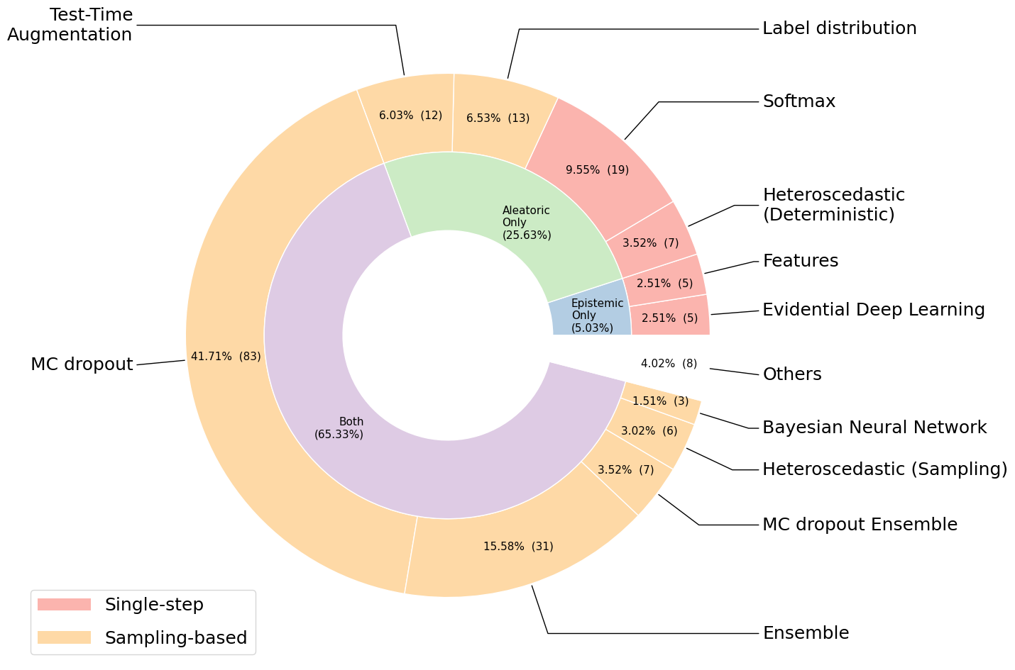
We performed a systematic search on June 2022 using Google Scholar and PubMed to identify DL studies implementing UQ methods for medical image classification and segmentation published from 2015 (included) to June 2022. The following combination of keywords was used for the search: "Deep Learning", "Uncertainty", "MRI", "CT", "PET", "X-RAY", "Medical image". Studies were included if they 1) implemented supervised DL models for medical image classification or segmentation; and 2) proposed a quantification of the uncertainty of their algorithms. The following exclusion criteria were applied 1) non-peer-reviewed studies (exception were made for papers with more than 30 citations); 2) non-English papers; 3) review articles and 4) animal studies. 130 papers were finally selected for analysis. It resulted a total of 199 UQ models, implemented either as principal contributions or as comparison methods (the exhaustive list of methods can be found in A). We first clustered them according to the method used for uncertainty estimation. We further proposed a categorization of these methods according to the type of uncertainty that is modeled, namely epistemic or aleatoric. Moreover, for real-world deployment of a DL model in a clinical setting, speed is crucial for integration into the routine. This means that the implementation of a UQ protocol should not come with prohibitive inference time or computational cost. Thus, we further distinguish between sampling methods, which require multiple inferences per input image (and thus tend to be slow and/or computationally costly), and single-step methods, which produce uncertainty estimate at the cost of a single inference step (and thus tend to be faster). The resulting taxonomy is presented in Figure 1. In the following of the section, we briefly present each UQ framework.
Figure 1: Implemented UQ methods in the 130 selected papers. The percentage (and the number) of the selected papers for each class of methods is indicated in the outer ring and the corresponding percentage below the class name. Orange identifies sampling-based methods, and red single-step methods. The inner ring classifies methods according to the type of uncertainty modeled: aleatoric, epistemic or both.
3.1 Softmax methods
An immediate and simple approach to obtain uncertainty estimates from classification or segmentation by neural network (NN) techniques is to consider the output predicted probabilities . Naively, the higher this probability, the more certain is the prediction. Works have been proposed to improve the calibration of the probabilities predicted by a DL model, and ensure that these scores match the true performance of the model (Guo et al. 2017; Kumar et al. 2019; Nixon et al. 2019). More formally, a model is calibrated if, for each prediction with the associated probability p, the model is correct 100 x p of the time. However, UQ based on softmax probabilities only consider the distribution over the model’s outputs, and not on the model’s weights. Thus, this type of deterministic uncertainty estimates only consider aleatoric uncertainty Hüllermeier and Waegeman 2021; Kendall and Gal 2017.
3.2 Bayesian Neural Network methods
In Bayesian Deep Learning (BDL), each weight of the NN is replaced by a distribution, rather than having a single fixed value (Blundell et al. 2015). To achieve this, a prior distribution (usually Gaussian) is first initialized over the NN weights. It follows that each weight is represented by a mean and a variance (thus doubling the number of parameters of the model). Then, during training, the model learns the posterior distribution given the training dataset and the prior distribution, which account for the less and more likely parameters given the observed data. The trained Bayesian Neural Network (BNN) is akin to a virtually infinite ensemble of NNs, where each instance has its weights drawn from the learned posterior distribution. During inference, the distribution is marginalized by repeatedly sampling weights from the shared distribution and averaging the predictions. Uncertainty estimates such as the entropy of the predictive distribution, its variance or its mutual information can be computed. BNN places a distribution on the model’s outputs as well as on the model’s weights, hence is able to model both aleatoric and epistemic uncertainties. While being theoretically founded, BNN requires extensive changes to both the model architecture and training paradigm, and also significantly increases the computational cost of training and inference.
3.3 Monte Carlo dropout methods
In Gal and Ghahramani 2016, authors demonstrated that a NN trained with dropout is able to efficiently approximate Bayesian inference without the associated prohibitive computational cost. Based on this principle, the Monte Carlo Dropout (MC dropout) technique proposes to train a model with dropout and keeps it activated during inference. For a given query input, multiple forward passes are then performed. Each time, a different dropout mask is randomly sampled, producing different predictions. Following this process, a predictive distribution is obtained, similarly to BNN. MC dropout allowing to approximate a BNN in any network trained with dropout, it thus rapidly gained popularity. As a counterpart, finding the optimal dropout strategy (rate and position within the NN) is not straight-forward (Jungo et al. 2020).
3.4 Ensemble methods
Deep Ensemble (DE) (Lakshminarayanan et al. 2017) proposes to sequentially train a series of NN. As the weights of the neural network are initialized randomly, the models reach different optimums during training. As a result, they produce diverse predictions for the same query input. As for BDL and MC dropout, uncertainty estimates can then be extracted from the ensemble’s predictive distribution. A DE does not require any changes to model architecture or training paradigm. Yet, it requires to repeat the training several times, and the aggregation of each individual prediction at inference, which increases the computational cost of this approach. Finally, it is worth noticing that some works propose to associate ensemble and MC dropout in order to get the best of both methods. This allows the combination of individual models uncertainty (though Monte Carlo dropout) as well as the overall ensemble uncertainty (though Deep Ensemble).
3.5 Heteroscedastic model-based methods
As its denomination indicates, an heteroscedastic model aims at evaluating the heteroscedastic part of aleatoric uncertainty. Within this framework, uncertainty is directly learned during training from the data itself, without the need for ground truth labels for uncertainty. Heteroscedastic models can be categorized into two subtypes: sampling methods, which extend MC dropout models, and deterministic approaches, which are a recent improvement and produce the prediction together with its related uncertainty in a single-step.
3.5.1 Sampling Heteroscedastic models
In Kendall and Gal 2017, authors hypothesize that the network output logits are corrupted by Gaussian noise with mean equal to 0 and variance . The higher the variance, the higher is the aleatoric uncertainty. A model can then be trained to predict the mean logits , as well as the noise variance . To do so, authors duplicate the outputs of a MC dropout network: one for the logits, and one for the variance. At each training step, the loss of the model is evaluated by integrating over multiple samples of noise. Paired with the MC dropout framework, this sampling formulation of heteroscedastic models enables the modeling of both epistemic and aleatoric uncertainties.
3.5.2 Deterministic Heteroscedastic models
Recently, deterministic variants of the heteroscedastic model have been proposed. In this approach, uncertainty is still learned during training from the data itself, but do not require the integration of the loss over multiple samples of noise. As in the sampling approach, the NN is modified by adding a dedicated output for uncertainty. Then, an uncertainty-augmented loss function is used to learn the predictive task (classification or segmentation), while also learning to predict high uncertainty scores for samples that are likely to be incorrect. In this context, aleatoric uncertainty is learned without any additional cost, as it simply exploits the ground truth label (class or segmentation).
3.6 Label-distribution model-based methods
A branch of the UQ literature focuses on modeling label uncertainty in the context of image segmentation. These approaches focus on datasets for which multiple expert manual segmentations are provided for each image, interpreting the inter-rater variability as a form of ground truth uncertainty. In this setting, it becomes possible to approximate the expert label distribution using generative segmentation neural networks (Kohl et al. 2018). At inference, sampling from the learned distribution produces diverse segmentation masks, which reproduce the inter-rater variability. We refer to this family of methods as label-distribution models. Despite being intuitive, it is not clear whether or not inter-rater variability can be used as ground truth for uncertainty. In the context of medical image segmentation, there are many cases where a unique segmentation cannot be obtained, for instance due to partial volume effect observed in MRI at the boundaries between healthy tissue and lesions. In that context, experts segmentations exhibit somewhat random variations around the boundaries of the target object. Moreover, experts can over-segment or alternatively under-segment the same object of interest, based on their annotation style. This inter-rater variability is thus rather linked to contextual biases (e.g, radiologist experience or annotation habits) rather that on the true uncertainty of the label (Mehta et al. 2022).
3.7 Test-Time Augmentation
Test-Time Augmentation (TTA) (Ayhan and Berens 2018) was proposed as an UQ method to evaluate aleatoric uncertainty. At test time, multiple variants of the input image are generated using Data Augmentation. This can include spatial transformations (e.g. flipping, rotation) as well as intensity augmentations (e.g. contrast modification, noise injection, or artifacts). This process aims at exploring the impact of input-image transformations on the prediction. Using TTA, the model generates a set of predictions for the same initial input image. From this distribution of predictions, uncertainty metrics can be extracted such as the mediane or the variance.
3.8 Feature-based methods
From a practical point-of-view, epistemic uncertainty is expected to be high for Out-of-distribution (OOD) images, e.g. images that are far from the training image distribution. Based on this concrete application, efficient epistemic-uncertainty techniques were recently proposed to detect OOD from the feature map signature of a trained NN (Postels et al. 2021). This builds on the hypothesis that feature maps contain information regarding the correctness of a prediction. Despite being efficient for OOD detection, it has been observed that feature-based uncertainty estimates are generally poorly calibrated (Postels et al. 2021). These methods are computationally efficient, however their application to medical images remains rare.
3.9 Evidential Deep Learning
The Dempster–Shafer Theory of Evidence (DST) is a framework for dealing with epistemic uncertainty (Dempster 1968). In a K-class classification (respectively, segmentation) problem, DST proposes to assign belief masses to each possible class, as well as an overall uncertainty mass.
When there is no evidence collected guiding to any of the classes, the beliefs reach their minimal values , while the overall uncertainty reaches its maximal value . In practice, DST can be applied to Deep Learning models by fitting a Dirichlet distribution on the model’s outputs, in place of the standard categorical distribution. Additionally, the Bayes-Risk loss function (Sensoy et al. 2018) is used to train the model in replacement of the standard cross-entropy loss.
3.10 Other UQ methods
Finally, we found a few methods not conforming to any of the frameworks previously introduced. We list these applications in B, with a short description of each UQ approach proposed.
4 How to evaluate uncertainty quantification approaches
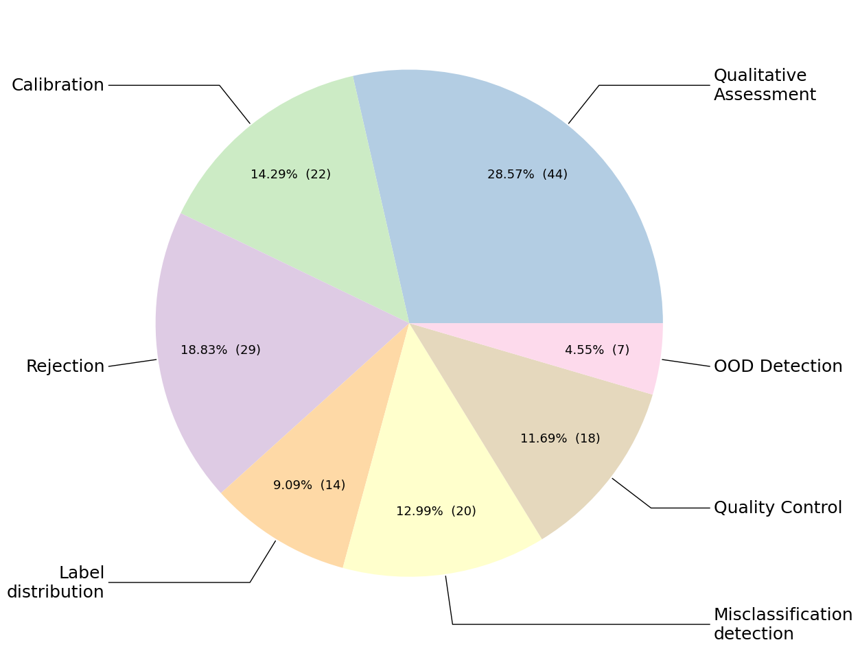
In the previous section, we have presented the main UQ approaches that are applied to DL-based medical image classification and segmentation. In this section, we now propose to introduce the different protocols that are implemented in these papers to evaluate the relevance of the UQ approaches. Evaluating UQ approaches is not straight forward, as we typically do not dispose of ground-truth uncertainty values. Proxy metrics are thus developed to estimate the performances of uncertainty quantification methods. We have identified 7 types of evaluation protocols (see Figure 2). In the following, we present each protocol and identify their use cases. Table C lists use cases of each metrics in the reviewed corpus of papers.
Figure 2: Implemented UQ evaluation protocols in the reviewed papers. The percentage (and the number) of the reviewed papers per class is mentioned in the Pie chart.
4.1 Qualitative assessment protocol
As computing quantitative metrics for uncertainty is not direct, several works focused on a qualitative assessment of the computed uncertainty estimates. In this context, a visual inspection of the cases considered as certain/uncertain is usually performed to verify whether they correspond to cases that a human would consider as uncertain. Alternatively, the pertinence of the incorporation of UQ in a medical image processing pipeline can be assessed via the monitoring of its beneficial impact on a downstream task (e.g. training-image selection in a semi-supervised learning, or improvement of the predictive performance).
4.2 Calibration protocol
As presented in Section 3.1, the output softmax probabilities of a NN can directly be used as a marker of (un)certainty. A popular way of estimating the accuracy of such uncertainty estimates is the use of calibration metrics, that verify the correspondence between predicted probabilities and error rates. Usual choices consist of the Expected Calibration Error (ECE) (Guo et al. 2017), the Brier Score, or the Negative Log-Likelihood (NLL) score.
4.3 Misclassification detection protocol
A direct downstream application of uncertainty in an automated pipeline is the detection of samples for which the prediction is likely to be incorrect. This is crucial to prevent silent errors that could have dramatic impact, especially in real-world medical image applications. In that sense, the uncertainty estimates can be turned into a binary classifier that aims at distinguishing between correct and incorrect predictions (i.e., sample for which the predicted label and the ground truth label differ). As in the binary classification setting, an uncertainty threshold is applied to distinguish between positive (i.e. certain) and negative (i.e. uncertain) samples. The result of this classification is then compared to the true label of each sample, namely correct or incorrect. In that context, a confusion matrix from the uncertainty point of view can be constructed, by distinguishing 4 possibles cases, as shown in Figure 3. Usual classification metrics can then be computed based on the counts of True Positive (TP): the classification is uncertain and the expected label and the prediction differ, False Negative (FP): the classification is certain but the expected label and the prediction differ, True Negative (TN): the classification is certain and the expected label and the prediction are identical, and False Negative (FN): the classification is uncertain but the prediction and the expected label are identical.
Figure 3: Confusion matrix for uncertainty-based misclassification detection. Desired cases are represented in white, while undesired cases are presented in gray.
4.4 Rejection protocol
Another way of exploiting uncertainty estimates in an automated pipeline is the rejection mechanism. In this context, predictions of the model are ordered from the most certain to the most uncertain. A fraction of the most uncertain predictions are then rejected, and the performance of the model is computed on the remaining predictions. If uncertainty estimates efficiently identify uncertain cases that are more likely to be incorrect, then the performance and the remaining prediction should improve. Multiple fractions can be used, producing a curve showing the performance of the model with respect to the fraction of rejected data. The area under the resulting curve is used as a qualitative score. This rejection-based evaluation protocol essentially highlights the same trends as the previous misclassification detection setting.
4.5 OOD detection protocol
A desired property of uncertainty is to be high for abnormal images that are different from the images seen during training. Similarly to the misclassification detection setting, the uncertainty estimates can be translated into a binary classifier that aim at distinguishing between in-distribution (ID) and OOD images. Standard classification metrics can further be computed from the confusion matrix, as presented above in Section 4.3.
4.6 Quality-control protocol
For segmentation tasks, uncertainty estimates are obtained for every pixel (or voxel) in the medical image. These scores can then be aggregated into image-wise uncertainty, for instance by taking their mean. The correlation between this image-wise uncertainty, and image-wise metrics quantifying the quality of the segmentation, such as the Dice score, can be computed. In an automated medical image segmentation pipeline, this process can be used to detect images for which the produced segmentation does not meet quality standards. We refer to this mode of evaluation, specific to segmentation tasks, as QC-based evaluation protocols.
4.7 Label-distribution protocol
Finally, label-distribution protocol consists in comparing the predicted distribution of labels with the ground truth distribution of the experts . A popular choice of metric is the Generalized Energy Distance (Kohl et al. 2018) between both distributions.
5 Discussion
We reviewed the most popular UQ methods for DL-based medical image analysis and the associated evaluation protocols. In this section, we list the keys insights of this review, and identify potential future research directions.
First, the large number of studies incorporating UQ in their medical analysis pipeline proves that the need for UQ is well taken into account by the DL community. This shows that efforts are being made to develop AI tools that are not only powerful, but also useful in a real clinical setting. In this context, the predictive performance of the model only is not enough to reach a good acceptance. UQ is key to facilitate the human-machine collaboration and break the black-box effect.
Bayesian methodology, although providing a strong theoretical background for uncertainty, is scarcely implemented for medical image analysis. This can be explained by the complex implementation that requires (i) the modification of the NN (weights are replaced by distributions, thus doubling the number of parameters to estimate) and (ii) the modification of the training paradigm. Additionally, convergence tends to be slow for complex scenarios (Osawa et al. 2019) and gradient descent more noisy and unstable using a Bayesian NN than with a standard non-deterministic NN (Jospin et al. 2022). Finally, it has also been observed that Bayesian NNs tend to underfit (Dusenberry et al. 2020) and that their predictive performances are lower than standard NNs (Wenzel et al. 2020). Approximations of the Bayesian framework, such as dropout-based methods, are thus generally preferred.
Overall, MC dropout method seems to be the most popular approach for UQ in medical image analysis, representing nearly half of the implemented methods (, considering both the standard MC dropout methods () as well as sampling Heteroscedastic models (), which are an MC-dropout extension). This popularity can be explained by its easy implementation in any NN trained with dropout, indeed a large majority of NNs. Additionally, dropout helps preventing over-fitting during training, which is a common problem in medical domain, where training-dataset size is limited. However, the performance of MC dropout is highly dependent on the applied dropout rate (Osawa et al. 2019), which can makes it impractical to tune. Moreover, it requires multiple inferences for the same input image, considerably extending the inference time, which may not be compatible with AI applications in clinical environments.
Ensembling approaches are also commonly employed for UQ, although less common than MC dropout models. Aggregating the predictions of multiple models is a popular trick to improve the predictive performance, while also providing quality uncertainty estimates. The drawback is an increased computational cost and time as it requires multiple training and their predictions aggregation at testing.
In the literature, a large variety of evaluation protocols are reported, aiming at assessing the quality of uncertainty estimates. In the context of medical image segmentation, if multiple manual expert delineations are available for a given input image, the inter-rater variability can be used as ground truth uncertainty, to be compared with the predicted one. However, most of the time, the corresponding uncertainty values are not provided. Thus, evaluation of UQ usually relies on proxy tasks, such as the detection of misclassification, Out-of-distribution, or Quality Control. These methods are inspired from concrete applications of uncertainty in a real-world scenario. Yet, although commonly used, UQ evaluation based on misclassification detection should not directly be used for ranking methods. Indeed, the set of correct and incorrect predictions is specific to each model that produces its own binary misclassification. It is then inappropriate to compare them directly (Ashukha et al. 2020).
In this review, we have distinguished between sampling and single-step UQ methods. The latter approaches offer to compute uncertainty in a quick and efficient manner, which is generally required for medical applications. Although they currently represent a niche, their easy practical considerations may promote their rapid adoption. These methods, however, only partially model uncertainty, by computing either aleatoric or epistemic uncertainties, but not both.
Finally, it must be acknowledged that the effort of the community is promoted by challenges, such as the 2020 edition of the BraTS challenge (Mehta et al. 2022; Menze et al. 2014) that included an UQ task, the MICCAI QUBIQ challenge111
https://qubiq21.grand-challenge.org/ that focused on label uncertainty, and the SHIFT 2022 challenge222
https://shifts.grand-challenge.org/ that will contain a task of uncertainty quantification for Multiple Sclerosis lesions segmentation (Malinin et al. 2022).
5.1 Future directions
Based on the gaps identified above, we suggest several future research directions for UQ in DL-based medical image analysis.
As shown, the vast majority () of the implemented UQ methods are based on a sampling protocol, aiming at generating multiple predictions for the same query input. Yet, this significantly increases the computational burden of UQ, which may prevent its adoption in an automated pipeline in medical domain. Deterministic UQ methods requiring a single-step to compute uncertainty, are very promising and should be more intensively explored.
Overall, the detection of Out-of-distribution (OOD) predictions using uncertainty concerns few studies, despite of being crucial in a real-world medical scenarios. In an automated medical image pipeline, input samples can exhibit various anomalies that may disturb the functioning of the NN, thus resulting in very poor predictions. Real clinical cases may include artifacts, present a pathology unseen during training, or an unusual contrast setting due to a particular acquisition protocol. In such situations, uncertainty associated to the computed predictions are expected to be high and should represent a warning for the user (e.g., the medical practitioner). In practice, this is usually not the case with standard approaches such as softmax uncertainty, MC dropout or Deep Ensemble, which have limited performance in terms of OOD detection (Snoek et al. 2019; Ulmer and Cinà 2021). This motivates the development of feature-based methods, specially tailored for OOD detection. Note that OOD detection is a very active research topic not specific to the UQ field (Bulusu et al. 2020), but currently rarely exploited for medical image analysis. We hypothesize that the lack of OOD-based evaluation protocol may be due to the difficulty of gathering relevant data. A simple solution may be to use two distinct datasets, one for training the neural network and its evaluation on in-distribution data, and one during testing for OOD detection (Karimi and Gholipour 2020). Another approach would be to use data augmentation to corrupt images with synthetic artifacts, helping to achieve a more realistic setting (Shaw et al. 2021).
Finally, while the need for UQ in medical applications is unquestionable, we argue that being able to understand the prediction process of the DL model is also crucial to promote a trustable usage of AI in medicine. Then, the link between explainability and uncertainty should be studied, which would allow to understand both how the prediction is made, and whether or not it should be trusted. An interesting research direction would be to complement uncertainty estimates with explanations, helping the user to understand the sources of uncertainty in an intelligible way and possibly contribute to its improvement.
6 Conclusion
In this review, we have proposed an overview of the most popular UQ methods implemented in DL-based medical image applications, a specific domain with inherent uncertainty. Numerous phenomenons can cause predictive uncertainty, such as noisy images, imperfect ground truth labels, lack or incomplete data, and inter-site image variability. The literature proposes various methods to quantify this uncertainty which are applied to a very large range of medical image applications. As demonstrated in this review, developing trustable AI solutions integrating uncertainty quantification of the computed predictions is an active research topics.
7 Declaration of competing interest
BL, AT, SD, HD are employees of the Pixyl company. MD and FF serve on Pixyl scientific advisory board.
8 Acknowledgments
Benjamin Lambert is supported by a CIFRE convention (ANRT 2020/1555). The authors declare that they have no known competing financial interests or personal relationships that could have appeared to influence the work reported in this paper.
Appendix A Classification of the reviewed papers with respect to the UQ methods implemented.
plausible classes for a given image with coverage guarantees.
Appendix C Evaluation protocols proposed in the corpus of papers and classified according to their framework.
References
Abdar et al. (2021a)
Abdar, M., Pourpanah, F.,
Hussain, S., Rezazadegan, D.,
Liu, L., Ghavamzadeh, M.,
Fieguth, P., Cao, X.,
Khosravi, A., Acharya, U.R., et al.,
2021a.
A review of uncertainty quantification in deep
learning: Techniques, applications and challenges.
Information Fusion 76,
243–297.
Abdar et al. (2021b)
Abdar, M., Salari, S.,
Qahremani, S., Lam, H.K.,
Karray, F., Hussain, S.,
Khosravi, A., Acharya, U.R.,
Nahavandi, S., 2021b.
Uncertaintyfusenet: Robust uncertainty-aware
hierarchical feature fusion with ensemble monte carlo dropout for covid-19
detection.
arXiv e-prints .
Abdar et al. (2021c)
Abdar, M., Samami, M.,
Mahmoodabad, S.D., Doan, T.,
Mazoure, B., Hashemifesharaki, R.,
Liu, L., Khosravi, A.,
Acharya, U.R., Makarenkov, V., et al.,
2021c.
Uncertainty quantification in skin cancer
classification using three-way decision-based bayesian deep learning.
Computers in biology and medicine
135, 104418.
Abideen et al. (2020)
Abideen, Z.U., Ghafoor, M.,
Munir, K., Saqib, M.,
Ullah, A., Zia, T.,
Tariq, S.A., Ahmed, G.,
Zahra, A., 2020.
Uncertainty assisted robust tuberculosis
identification with bayesian convolutional neural networks.
IEEE Access 8,
22812–22825.
Adebayo et al. (2018)
Adebayo, J., Gilmer, J.,
Muelly, M., Goodfellow, I.J.,
Hardt, M., Kim, B., 2018.
Sanity checks for saliency maps.
Advances in Neural Information Processing Systems
31: Annual Conference on Neural Information Processing Systems 2018 ,
9525–9536.
Ahsan et al. (2022)
Ahsan, M.A., Qayyum, A.,
Razi, A., Qadir, J.,
2022.
An active learning method for diabetic retinopathy
classification with uncertainty quantification.
Medical & Biological Engineering & Computing ,
1–15.
Arrieta et al. (2020)
Arrieta, A.B., Díaz-Rodríguez,
N., Del Ser, J., Bennetot, A.,
Tabik, S., Barbado, A.,
García, S., Gil-López, S.,
Molina, D., Benjamins, R., et al.,
2020.
Explainable artificial intelligence (xai): Concepts,
taxonomies, opportunities and challenges toward responsible ai.
Information fusion 58,
82–115.
Asgharnezhad et al. (2022)
Asgharnezhad, H., Shamsi, A.,
Alizadehsani, R., Khosravi, A.,
Nahavandi, S., Sani, Z.A.,
Srinivasan, D., Islam, S.M.S.,
2022.
Objective evaluation of deep uncertainty predictions
for covid-19 detection.
Scientific Reports 12,
1–11.
Ashukha et al. (2020)
Ashukha, A., Lyzhov, A.,
Molchanov, D., Vetrov, D.P.,
2020.
Pitfalls of in-domain uncertainty estimation and
ensembling in deep learning.
8th International Conference on Learning
Representations, ICLR 2020 .
Ayhan and Berens (2018)
Ayhan, M.S., Berens, P.,
2018.
Test-time data augmentation for estimation of
heteroscedastic aleatoric uncertainty in deep neural networks.
International conference on Medical Imaging with
Deep Learning .
Ayhan et al. (2020)
Ayhan, M.S., Kühlewein, L.,
Aliyeva, G., Inhoffen, W.,
Ziemssen, F., Berens, P.,
2020.
Expert-validated estimation of diagnostic uncertainty
for deep neural networks in diabetic retinopathy detection.
Medical Image Analysis 64,
101724.
Balagopal et al. (2021)
Balagopal, A., Nguyen, D.,
Morgan, H., Weng, Y.,
Dohopolski, M., Lin, M.H.,
Barkousaraie, A.S., Gonzalez, Y.,
Garant, A., Desai, N., et al.,
2021.
A deep learning-based framework for segmenting
invisible clinical target volumes with estimated uncertainties for
post-operative prostate cancer radiotherapy.
Medical image analysis 72,
102101.
Baumgartner et al. (2019)
Baumgartner, C.F., Tezcan, K.C.,
Chaitanya, K., Hötker, A.M.,
Muehlematter, U.J., Schawkat, K.,
Becker, A.S., Donati, O.,
Konukoglu, E., 2019.
Phiseg: Capturing uncertainty in medical image
segmentation.
International Conference on Medical Image Computing
and Computer-Assisted Intervention , 119–127.
Becker et al. (2019)
Becker, A.S., Chaitanya, K.,
Schawkat, K., Muehlematter, U.J.,
Hötker, A.M., Konukoglu, E.,
Donati, O.F., 2019.
Variability of manual segmentation of the prostate in
axial t2-weighted mri: A multi-reader study.
European journal of radiology
121, 108716.
Belharbi et al. (2021)
Belharbi, S., Rony, J.,
Dolz, J., Ayed, I.B.,
McCaffrey, L., Granger, E.,
2021.
Deep interpretable classification and
weakly-supervised segmentation of histology images via max-min uncertainty.
IEEE Transactions on Medical Imaging
41, 702–714.
Berger et al. (2021)
Berger, C., Paschali, M.,
Glocker, B., Kamnitsas, K.,
2021.
Confidence-based out-of-distribution detection: a
comparative study and analysis.
Uncertainty for Safe Utilization of Machine
Learning in Medical Imaging, and Perinatal Imaging, Placental and Preterm
Image Analysis , 122–132.
Bhat and Kuijf (2022)
Bhat, I., Kuijf, H.J.,
2022.
Extending probabilistic u-net using mc-dropout to
quantify data and model uncertainty.
International MICCAI Brainlesion Workshop ,
555–559.
Bhat et al. (2021)
Bhat, I., Kuijf, H.J.,
Cheplygina, V., Pluim, J.P.,
2021.
Using uncertainty estimation to reduce false
positives in liver lesion detection.
2021 IEEE 18th International Symposium on
Biomedical Imaging (ISBI) , 663–667.
Blundell et al. (2015)
Blundell, C., Cornebise, J.,
Kavukcuoglu, K., Wierstra, D.,
2015.
Weight uncertainty in neural network.
International Conference on Machine Learning ,
1613–1622.
Bulusu et al. (2020)
Bulusu, S., Kailkhura, B.,
Li, B., Varshney, P.K.,
Song, D., 2020.
Anomalous example detection in deep learning: A
survey.
IEEE Access 8,
132330–132347.
Calderon-Ramirez et al. (2021a)
Calderon-Ramirez, S., Murillo-Hernandez,
D., Rojas-Salazar, K., Calvo-Valverd,
L.A., Yang, S., Moemeni, A.,
Elizondo, D., Lopez-Rubio, E.,
Molina-Cabello, M.A., 2021a.
Improving uncertainty estimations for mammogram
classification using semi-supervised learning.
2021 International Joint Conference on Neural
Networks (IJCNN) , 1–8.
Calderon-Ramirez et al. (2021b)
Calderon-Ramirez, S., Yang, S.,
Moemeni, A., Colreavy-Donnelly, S.,
Elizondo, D.A., Oala, L.,
Rodríguez-Capitán, J.,
Jiménez-Navarro, M., López-Rubio,
E., Molina-Cabello, M.A., 2021b.
Improving uncertainty estimation with semi-supervised
deep learning for covid-19 detection using chest x-ray images.
IEEE Access 9,
85442–85454.
Cao et al. (2021)
Cao, X., Chen, H., Li,
Y., Peng, Y., Wang, S.,
Cheng, L., 2021.
Dilated densely connected u-net with uncertainty
focus loss for 3d abus mass segmentation.
Computer Methods and Programs in Biomedicine
209, 106313.
Carneiro et al. (2020)
Carneiro, G., Pu, L.Z.C.T.,
Singh, R., Burt, A.,
2020.
Deep learning uncertainty and confidence calibration
for the five-class polyp classification from colonoscopy.
Medical Image Analysis 62,
101653.
Cetindag et al. (2022)
Cetindag, S.C., Yergin, M.,
Alis, D., Oksuz, I.,
2022.
Meta-learning for medical image segmentation
uncertainty quantification.
International MICCAI Brainlesion Workshop ,
578–584.
Chen et al. (2020)
Chen, C., Qin, C., Qiu,
H., Ouyang, C., Wang, S.,
Chen, L., Tarroni, G.,
Bai, W., Rueckert, D.,
2020.
Realistic adversarial data augmentation for mr image
segmentation.
International Conference on Medical Image Computing
and Computer-Assisted Intervention , 667–677.
Combalia et al. (2020)
Combalia, M., Hueto, F.,
Puig, S., Malvehy, J.,
Vilaplana, V., 2020.
Uncertainty estimation in deep neural networks for
dermoscopic image classification.
Proceedings of the IEEE/CVF Conference on Computer
Vision and Pattern Recognition Workshops , 744–745.
Dempster (1968)
Dempster, A.P., 1968.
A generalization of bayesian inference.
Journal of the Royal Statistical Society: Series B
(Methodological) 30, 205–232.
DeVries and Taylor (2018)
DeVries, T., Taylor, G.W.,
2018.
Leveraging uncertainty estimates for predicting
segmentation quality.
arXiv e-prints .
Dhakal and Joshi (2021)
Dhakal, P., Joshi, S.R.,
2021.
Uncertainty estimation in detecting knee
abnormalities on mri using bayesian deep learning.
Proceedings of 10th IOE Graduate Conference
10.
Diao et al. (2022)
Diao, Z., Jiang, H., Shi,
T., 2022.
A unified uncertainty network for tumor segmentation
using uncertainty cross entropy loss and prototype similarity.
Knowledge-Based Systems 246,
108739.
Dusenberry et al. (2020)
Dusenberry, M., Jerfel, G.,
Wen, Y., Ma, Y., Snoek,
J., Heller, K., Lakshminarayanan, B.,
Tran, D., 2020.
Efficient and scalable bayesian neural nets with
rank-1 factors.
International Conference on Machine Learning ,
2782–2792.
Eaton-Rosen et al. (2018)
Eaton-Rosen, Z., Bragman, F.,
Bisdas, S., Ourselin, S.,
Cardoso, M.J., 2018.
Towards safe deep learning: accurately quantifying
biomarker uncertainty in neural network predictions.
International Conference on Medical Image Computing
and Computer-Assisted Intervention , 691–699.
Fidon et al. (2017)
Fidon, L., Li, W.,
Garcia-Peraza-Herrera, L.C., Ekanayake,
J., Kitchen, N., Ourselin, S.,
Vercauteren, T., 2017.
Generalised wasserstein dice score for imbalanced
multi-class segmentation using holistic convolutional networks.
International MICCAI Brainlesion workshop ,
64–76.
Filos et al. (2019)
Filos, A., Farquhar, S.,
Gomez, A.N., Rudner, T.G.,
Kenton, Z., Smith, L.,
Alizadeh, M., De Kroon, A.,
Gal, Y., 2019.
A systematic comparison of bayesian deep learning
robustness in diabetic retinopathy tasks.
arXiv e-prints .
Föllmer et al. (2022)
Föllmer, B., Biavati, F.,
Wald, C., Stober, S.,
Ma, J., Dewey, M.,
Samek, W., 2022.
Active multi-task learning with uncertainty weighted
loss for coronary calcium scoring.
Medical Physics .
Ford et al. (2016)
Ford, R.A., Price, W.,
Nicholson, I., 2016.
Privacy and accountability in black-box medicine.
Mich. Telecomm. & Tech. L. Rev.
23, 1.
Gal and Ghahramani (2016)
Gal, Y., Ghahramani, Z.,
2016.
Dropout as a Bayesian approximation: Representing
model uncertainty in deep learning.
International Conference on Machine Learning ,
1050–1059.
Gal et al. (2016)
Gal, Y., et al., 2016.
Uncertainty in deep learning.
Ph.D. thesis. University of Cambridge.
Gantenbein et al. (2020)
Gantenbein, M., Erdil, E.,
Konukoglu, E., 2020.
Revphiseg: A memory-efficient neural network for
uncertainty quantification in medical image segmentation.
Uncertainty for Safe Utilization of Machine
Learning in Medical Imaging, and Graphs in Biomedical Image Analysis ,
13–22.
Gawlikowski et al. (2021)
Gawlikowski, J., Tassi, C.R.N.,
Ali, M., Lee, J., Humt,
M., Feng, J., Kruspe, A.,
Triebel, R., Jung, P.,
Roscher, R., et al., 2021.
A survey of uncertainty in deep neural networks.
arXiv e-prints .
Ghafoorian et al. (2017)
Ghafoorian, M., Mehrtash, A.,
Kapur, T., Karssemeijer, N.,
Marchiori, E., Pesteie, M.,
Guttmann, C.R., Leeuw, F.E.d.,
Tempany, C.M., Ginneken, B.v., et al.,
2017.
Transfer learning for domain adaptation in mri:
Application in brain lesion segmentation.
International Conference on Medical Image Computing
and Computer-Assisted Intervention , 516–524.
Ghesu et al. (2019)
Ghesu, F.C., Georgescu, B.,
Gibson, E., Guendel, S.,
Kalra, M.K., Singh, R.,
Digumarthy, S.R., Grbic, S.,
Comaniciu, D., 2019.
Quantifying and leveraging classification uncertainty
for chest radiograph assessment.
International Conference on Medical Image Computing
and Computer-Assisted Intervention , 676–684.
Ghesu et al. (2021)
Ghesu, F.C., Georgescu, B.,
Mansoor, A., Yoo, Y.,
Gibson, E., Vishwanath, R.,
Balachandran, A., Balter, J.M.,
Cao, Y., Singh, R., et al.,
2021.
Quantifying and leveraging predictive uncertainty for
medical image assessment.
Medical Image Analysis 68,
101855.
Ghosal et al. (2021)
Ghosal, S., Xie, A., Shah,
P., 2021.
Uncertainty quantified deep learning for predicting
dice coefficient of digital histopathology image segmentation.
arXiv e-prints .
Ghoshal and Tucker (2020)
Ghoshal, B., Tucker, A.,
2020.
Estimating uncertainty and interpretability in deep
learning for coronavirus (covid-19) detection.
arXiv e-prints .
Ghoshal et al. (2021)
Ghoshal, B., Tucker, A.,
Sanghera, B., Lup Wong, W.,
2021.
Estimating uncertainty in deep learning for reporting
confidence to clinicians in medical image segmentation and diseases
detection.
Computational Intelligence 37,
701–734.
Gou and He (2021)
Gou, X., He, X., 2021.
Deep learning-based detection and diagnosis of
subarachnoid hemorrhage.
Journal of Healthcare Engineering
2021.
Guo et al. (2017)
Guo, C., Pleiss, G., Sun,
Y., Weinberger, K.Q., 2017.
On calibration of modern neural networks.
International Conference on Machine Learning ,
1321–1330.
Guo et al. (2022)
Guo, F., Ng, M., Kuling,
G., Wright, G., 2022.
Cardiac mri segmentation with sparse annotations:
Ensembling deep learning uncertainty and shape priors.
Medical Image Analysis , 102532.
Hasan and Linte (2021)
Hasan, S.K., Linte, C.A.,
2021.
A multi-task cross-task learning architecture for ad
hoc uncertainty estimation in 3d cardiac mri image segmentation.
2021 Computing in Cardiology (CinC)
48, 1–4.
Hasan and Linte (2022)
Hasan, S.K., Linte, C.A.,
2022.
Calibration of cine mri segmentation probability for
uncertainty estimation using a multi-task cross-task learning architecture.
Medical Imaging 2022: Image-Guided Procedures,
Robotic Interventions, and Modeling 12034,
174–179.
Herzog et al. (2020)
Herzog, L., Murina, E.,
Dürr, O., Wegener, S.,
Sick, B., 2020.
Integrating uncertainty in deep neural networks for
mri based stroke analysis.
Medical Image Analysis 65,
101790.
Hiasa et al. (2019)
Hiasa, Y., Otake, Y.,
Takao, M., Ogawa, T.,
Sugano, N., Sato, Y.,
2019.
Automated muscle segmentation from clinical ct using
bayesian u-net for personalized musculoskeletal modeling.
IEEE Transactions on Medical Imaging
39, 1030–1040.
Hoebel et al. (2020)
Hoebel, K., Andrearczyk, V.,
Beers, A., Patel, J.,
Chang, K., Depeursinge, A.,
Müller, H., Kalpathy-Cramer, J.,
2020.
An exploration of uncertainty information for
segmentation quality assessment.
Medical Imaging 2020: Image Processing
11313, 381–390.
Hu et al. (2019)
Hu, S., Worrall, D.,
Knegt, S., Veeling, B.,
Huisman, H., Welling, M.,
2019.
Supervised uncertainty quantification for
segmentation with multiple annotations.
International Conference on Medical Image Computing
and Computer-Assisted Intervention , 137–145.
Hu et al. (2020)
Hu, X., Guo, R., Chen,
J., Li, H., Waldmannstetter, D.,
Zhao, Y., Li, B., Shi,
K., Menze, B., 2020.
Coarse-to-fine adversarial networks and zone-based
uncertainty analysis for nk/t-cell lymphoma segmentation in ct/pet images.
IEEE journal of biomedical and health informatics
24, 2599–2608.
Huang et al. (2017)
Huang, G., Liu, Z., Van
Der Maaten, L., Weinberger, K.Q., 2017.
Densely connected convolutional networks.
Proceedings of the IEEE/CVF Conference on Computer
Vision and Pattern Recognition , 4700–4708.
Huang et al. (2021)
Huang, L., Ruan, S.,
Decazes, P., Denoeux, T.,
2021.
Evidential segmentation of 3d pet/ct images.
International Conference on Belief Functions ,
159–167.
Huang et al. (2020)
Huang, Z., Gan, Y., Lye,
T., Zhang, H., Laine, A.,
Angelini, E.D., Hendon, C.,
2020.
Heterogeneity measurement of cardiac tissues
leveraging uncertainty information from image segmentation.
International Conference on Medical Image Computing
and Computer-Assisted Intervention , 782–791.
Hüllermeier and Waegeman (2021)
Hüllermeier, E., Waegeman, W.,
2021.
Aleatoric and epistemic uncertainty in machine
learning: An introduction to concepts and methods.
Machine Learning 110,
457–506.
Isensee et al. (2021)
Isensee, F., Jaeger, P.F.,
Kohl, S.A., Petersen, J.,
Maier-Hein, K.H., 2021.
nnu-net: a self-configuring method for deep
learning-based biomedical image segmentation.
Nature methods 18,
203–211.
Iwamoto et al. (2021)
Iwamoto, S., Raytchev, B.,
Tamaki, T., Kaneda, K.,
2021.
Improving the reliability of semantic segmentation of
medical images by uncertainty modeling with bayesian deep networks and
curriculum learning.
Uncertainty for Safe Utilization of Machine
Learning in Medical Imaging, and Perinatal Imaging, Placental and Preterm
Image Analysis , 34–43.
Javadi et al. (2022)
Javadi, G., Bayat, S.,
Kazemi Esfeh, M.M., Samadi, S.,
Sedghi, A., Sojoudi, S.,
Hurtado, A., Chang, S.,
Black, P., Mousavi, P., et al.,
2022.
Towards targeted ultrasound-guided prostate biopsy by
incorporating model and label uncertainty in cancer detection.
International Journal of Computer Assisted
Radiology and Surgery 17, 121–128.
Jensen et al. (2019)
Jensen, M.H., Jørgensen, D.R.,
Jalaboi, R., Hansen, M.E.,
Olsen, M.A., 2019.
Improving uncertainty estimation in convolutional
neural networks using inter-rater agreement.
International Conference on Medical Image Computing
and Computer-Assisted Intervention , 540–548.
Ji et al. (2020)
Ji, W., Chen, W., Yu, S.,
Ma, K., Cheng, L., Shen,
L., Zheng, Y., 2020.
Uncertainty quantification for medical image
segmentation using dynamic label factor allocation among multiple raters.
MICCAI on QUBIQ Workshop .
Jiménez-Sánchez et al. (2022)
Jiménez-Sánchez, A., Mateus, D.,
Kirchhoff, S., Kirchhoff, C.,
Biberthaler, P., Navab, N.,
Ballester, M.A.G., Piella, G.,
2022.
Curriculum learning for improved femur fracture
classification: Scheduling data with prior knowledge and uncertainty.
Medical Image Analysis 75,
102273.
Joskowicz et al. (2019)
Joskowicz, L., Cohen, D.,
Caplan, N., Sosna, J.,
2019.
Inter-observer variability of manual contour
delineation of structures in ct.
European radiology 29,
1391–1399.
Jospin et al. (2022)
Jospin, L.V., Laga, H.,
Boussaid, F., Buntine, W.,
Bennamoun, M., 2022.
Hands-on bayesian neural networks—a tutorial for
deep learning users.
IEEE Computational Intelligence Magazine
17, 29–48.
Ju et al. (2022)
Ju, L., Wang, X., Wang,
L., Mahapatra, D., Zhao, X.,
Zhou, Q., Liu, T., Ge,
Z., 2022.
Improving medical images classification with label
noise using dual-uncertainty estimation.
IEEE Trans. Medical Imaging
41, 1533–1546.
Judge et al. (2022)
Judge, T., Bernard, O.,
Porumb, M., Chartsias, A.,
Beqiri, A., Jodoin, P.M.,
2022.
Crisp - reliable uncertainty estimation for medical
image segmentation , 492–502.
Jungo et al. (2020)
Jungo, A., Balsiger, F.,
Reyes, M., 2020.
Analyzing the quality and challenges of uncertainty
estimations for brain tumor segmentation.
Frontiers in neuroscience , 282.
Jungo et al. (2017)
Jungo, A., McKinley, R.,
Meier, R., Knecht, U.,
Vera, L., Pérez-Beteta, J.,
Molina-García, D.,
Pérez-García, V.M., Wiest, R.,
Reyes, M., 2017.
Towards uncertainty-assisted brain tumor segmentation
and survival prediction.
International MICCAI Brainlesion Workshop ,
474–485.
Jungo et al. (2018a)
Jungo, A., Meier, R.,
Ermis, E., Blatti-Moreno, M.,
Herrmann, E., Wiest, R.,
Reyes, M., 2018a.
On the effect of inter-observer variability for a
reliable estimation of uncertainty of medical image segmentation.
International Conference on Medical Image Computing
and Computer-Assisted Intervention , 682–690.
Jungo et al. (2018b)
Jungo, A., Meier, R.,
Ermis, E., Herrmann, E.,
Reyes, M., 2018b.
Uncertainty-driven sanity check: Application to
postoperative brain tumor cavity segmentation.
arXiv e-prints .
Kabir et al. (2018)
Kabir, H.D., Khosravi, A.,
Hosen, M.A., Nahavandi, S.,
2018.
Neural network-based uncertainty quantification: A
survey of methodologies and applications.
IEEE Access 6,
36218–36234.
Karimi and Gholipour (2020)
Karimi, D., Gholipour, A.,
2020.
Improving calibration and out-of-distribution
detection in medical image segmentation with convolutional neural networks.
arXiv e-prints .
Kendall and Gal (2017)
Kendall, A., Gal, Y., 2017.
What uncertainties do we need in bayesian deep
learning for computer vision?
Advances in Neural Information Processing Systems
30: Annual Conference on Neural Information Processing Systems 2017 ,
5574–5584.
Kerfoot et al. (2018)
Kerfoot, E., Clough, J.,
Oksuz, I., Lee, J.,
King, A.P., Schnabel, J.A.,
2018.
Left-ventricle quantification using residual u-net.
International Workshop on Statistical Atlases and
Computational Models of the Heart , 371–380.
Kohl et al. (2018)
Kohl, S., Romera-Paredes, B.,
Meyer, C., Fauw, J.D.,
Ledsam, J.R., Maier-Hein, K.H.,
Eslami, S.M.A., Rezende, D.J.,
Ronneberger, O., 2018.
A probabilistic u-net for segmentation of ambiguous
images , 6965–6975.
Kohl et al. (2019)
Kohl, S.A., Romera-Paredes, B.,
Maier-Hein, K.H., Rezende, D.J.,
Eslami, S., Kohli, P.,
Zisserman, A., Ronneberger, O.,
2019.
A hierarchical probabilistic u-net for modeling
multi-scale ambiguities.
arXiv e-prints .
Kumar et al. (2019)
Kumar, A., Liang, P., Ma,
T., 2019.
Verified uncertainty calibration.
Advances in Neural Information Processing Systems
32: Annual Conference on Neural Information Processing Systems 2019 ,
3787–3798.
Kurz et al. (2022)
Kurz, A., Hauser, K.,
Mehrtens, H.A., Krieghoff-Henning, E.,
Hekler, A., Kather, J.N.,
Fröhling, S., von Kalle, C.,
Brinker, T.J., et al., 2022.
Uncertainty estimation in medical image
classification: Systematic review.
JMIR Medical Informatics 10,
e36427.
Kushibar et al. (2022)
Kushibar, K., Campello, V.,
Garrucho, L., Linardos, A.,
Radeva, P., Lekadir, K.,
2022.
Layer ensembles: A single-pass uncertainty estimation
in deep learning for segmentation , 514–524.
Kwon et al. (2020)
Kwon, Y., Won, J.H., Kim,
B.J., Paik, M.C., 2020.
Uncertainty quantification using bayesian neural
networks in classification: Application to biomedical image segmentation.
Computational Statistics & Data Analysis
142, 106816.
Lakshminarayanan et al. (2017)
Lakshminarayanan, B., Pritzel, A.,
Blundell, C., 2017.
Simple and scalable predictive uncertainty estimation
using deep ensembles.
Advances in Neural Information Processing Systems
30: Annual Conference on Neural Information Processing Systems 2017 ,
6402–6413.
Lambert et al. (2022)
Lambert, B., Forbes, F.,
Doyle, S., Tucholka, A.,
Dojat, M., 2022.
Beyond voxel prediction uncertainty: Identifying
brain lesions you can trust 13611.
Laves et al. (2019)
Laves, M.H., Ihler, S.,
Ortmaier, T., 2019.
Uncertainty quantification in computer-aided
diagnosis: Make your model say" i don’t know" for ambiguous cases.
arXiv e-prints .
Lee et al. (2022)
Lee, J., Shin, D., Oh,
S.H., Kim, H., 2022.
Method to minimize the errors of ai: Quantifying and
exploiting uncertainty of deep learning in brain tumor segmentation.
Sensors 22,
2406.
Leibig et al. (2017)
Leibig, C., Allken, V.,
Ayhan, M.S., Berens, P.,
Wahl, S., 2017.
Leveraging uncertainty information from deep neural
networks for disease detection.
Scientific reports 7,
1–14.
Li and Luo (2020)
Li, H., Luo, H., 2020.
Uncertainty quantification in medical image
segmentation.
2020 IEEE 6th International Conference on Computer
and Communications (ICCC) , 1936–1940.
Li et al. (2021)
Li, Y., Chen, X., Quan,
L., Zhang, N., 2021.
Uncertainty-guided robust training for medical image
segmentation.
2021 IEEE 18th International Symposium on
Biomedical Imaging (ISBI) , 1471–1475.
Liang et al. (2020)
Liang, G., Zhang, Y.,
Jacobs, N., 2020.
Neural network calibration for medical imaging
classification using dca regularization.
International Conference on Machine Learning,
Workshop on Uncertainty & Robustness in Deep Learning .
Lin et al. (2021)
Lin, H., Li, Z., Yang,
Z., Wang, Y., 2021.
Variance-aware attention u-net for multi-organ
segmentation.
Medical Physics 48,
7864–7876.
Lin et al. (2020)
Lin, T.Y., Goyal, P.,
Girshick, R., He, K.,
Dollár, P., 2020.
Focal loss for dense object detection.
IEEE Transactions on Pattern Analysis and Machine
Intelligence 42, 318–327.
Linmans et al. (2020)
Linmans, J., van der Laak, J.,
Litjens, G., 2020.
Efficient out-of-distribution detection in digital
pathology using multi-head convolutional neural networks.
Medical Imaging with Deep Learning ,
465–478.
Liu et al. (2020)
Liu, Y., Yang, G.,
Hosseiny, M., Azadikhah, A.,
Mirak, S.A., Miao, Q.,
Raman, S.S., Sung, K.,
2020.
Exploring uncertainty measures in bayesian deep
attentive neural networks for prostate zonal segmentation.
IEEE Access 8,
151817–151828.
Lourenço-Silva and Oliveira (2022)
Lourenço-Silva, J., Oliveira, A.L.,
2022.
Using soft labels to model uncertainty in medical
image segmentation.
International MICCAI Brainlesion Workshop ,
585–596.
Lu et al. (2022)
Lu, C., Angelopoulos, A.N.,
Pomerantz, S., 2022.
Improving trustworthiness of ai disease severity
rating in medical imaging with ordinal conformal prediction sets ,
545–554.
Ma et al. (2021)
Ma, X., Niu, Y., Gu, L.,
Wang, Y., Zhao, Y.,
Bailey, J., Lu, F., 2021.
Understanding adversarial attacks on deep learning
based medical image analysis systems.
Pattern Recognition 110,
107332.
Mahapatra et al. (2021)
Mahapatra, D., Poellinger, A.,
Shao, L., Reyes, M.,
2021.
Interpretability-driven sample selection using self
supervised learning for disease classification and segmentation.
IEEE Transactions on Medical Imaging
40, 2548–2562.
Malinin (2019)
Malinin, A., 2019.
Uncertainty estimation in deep learning with
application to spoken language assessment.
Ph.D. thesis. University of Cambridge.
Malinin et al. (2022)
Malinin, A., Athanasopoulos, A.,
Barakovic, M., Cuadra, M.B.,
Gales, M.J., Granziera, C.,
Graziani, M., Kartashev, N.,
Kyriakopoulos, K., Lu, P.J., et al.,
2022.
Shifts 2.0: Extending the dataset of real
distributional shifts.
arXiv preprint arXiv:2206.15407 .
McClure et al. (2019)
McClure, P., Rho, N., Lee,
J.A., Kaczmarzyk, J.R., Zheng, C.Y.,
Ghosh, S.S., Nielson, D.M.,
Thomas, A.G., Bandettini, P.,
Pereira, F., 2019.
Knowing what you know in brain segmentation using
bayesian deep neural networks.
Frontiers in neuroinformatics
13, 67.
McKinley et al. (2018)
McKinley, R., Meier, R.,
Wiest, R., 2018.
Ensembles of densely-connected cnns with
label-uncertainty for brain tumor segmentation.
International MICCAI Brainlesion Workshop ,
456–465.
McKinley et al. (2020a)
McKinley, R., Rebsamen, M.,
Daetwyler, K., Meier, R.,
Radojewski, P., Wiest, R.,
2020a.
Uncertainty-driven refinement of tumor-core
segmentation using 3d-to-2d networks with label uncertainty.
International MICCAI Brainlesion Workshop ,
401–411.
McKinley et al. (2019)
McKinley, R., Rebsamen, M.,
Meier, R., Wiest, R.,
2019.
Triplanar ensemble of 3d-to-2d cnns with
label-uncertainty for brain tumor segmentation.
International MICCAI Brainlesion workshop ,
379–387.
McKinley et al. (2020b)
McKinley, R., Wepfer, R.,
Grunder, L., Aschwanden, F.,
Fischer, T., Friedli, C.,
Muri, R., Rummel, C.,
Verma, R., Weisstanner, C., et al.,
2020b.
Automatic detection of lesion load change in multiple
sclerosis using convolutional neural networks with segmentation confidence.
NeuroImage: Clinical 25,
102104.
Mehrtash et al. (2021)
Mehrtash, A., Kapur, T.,
Tempany, C.M., Abolmaesumi, P.,
Wells, W.M., 2021.
Prostate cancer diagnosis with sparse biopsy data and
in presence of location uncertainty.
2021 IEEE 18th International Symposium on
Biomedical Imaging (ISBI) , 443–447.
Mehrtash et al. (2020)
Mehrtash, A., Wells, W.M.,
Tempany, C.M., Abolmaesumi, P.,
Kapur, T., 2020.
Confidence calibration and predictive uncertainty
estimation for deep medical image segmentation.
IEEE Transactions on Medical Imaging
39, 3868–3878.
Mehta et al. (2019)
Mehta, R., Christinck, T.,
Nair, T., Lemaitre, P.,
Arnold, D., Arbel, T.,
2019.
Propagating uncertainty across cascaded medical
imaging tasks for improved deep learning inference.
Uncertainty for Safe Utilization of Machine
Learning in Medical Imaging and Clinical Image-Based Procedures ,
23–32.
Mehta et al. (2022)
Mehta, R., Filos, A.,
Baid, U., Sako, C.,
McKinley, R., Rebsamen, M.,
Dätwyler, K., Meier, R.,
Radojewski, P., Murugesan, G.K., et al.,
2022.
Qu-brats: Miccai brats 2020 challenge on
quantifyinguncertainty in brain tumor segmentation-analysis of ranking scores
and benchmarking results.
Journal of Machine Learning for Biomedical Imaging
1.
Mehta et al. (2020)
Mehta, R., Filos, A., Gal,
Y., Arbel, T., 2020.
Uncertainty evaluation metric for brain tumour
segmentation.
arXiv e-prints .
Menze et al. (2014)
Menze, B.H., Jakab, A.,
Bauer, S., Kalpathy-Cramer, J.,
Farahani, K., Kirby, J.,
Burren, Y., Porz, N.,
Slotboom, J., Wiest, R., et al.,
2014.
The multimodal brain tumor image segmentation
benchmark (brats).
IEEE Transactions on Medical Imaging
34, 1993–2024.
Milletari et al. (2016)
Milletari, F., Navab, N.,
Ahmadi, S.A., 2016.
V-net: Fully convolutional neural networks for
volumetric medical image segmentation.
2016 fourth international conference on 3D vision
(3DV) , 565–571.
Mishra et al. (2021)
Mishra, S., Chen, D.Z.,
Hu, X.S., 2021.
Objective-dependent uncertainty driven retinal vessel
segmentation.
2021 IEEE 18th International Symposium on
Biomedical Imaging (ISBI) , 453–457.
Mobiny et al. (2019)
Mobiny, A., Singh, A.,
Van Nguyen, H., 2019.
Risk-aware machine learning classifier for skin
lesion diagnosis.
Journal of clinical medicine 8,
1241.
Mojabi et al. (2020)
Mojabi, P., Khoshdel, V.,
Lovetri, J., 2020.
Tissue-type classification with uncertainty
quantification of microwave and ultrasound breast imaging: A deep learning
approach.
IEEE Access 8,
182092–182104.
Mojiri Forooshani et al. (2022)
Mojiri Forooshani, P., Biparva, M.,
Ntiri, E.E., Ramirez, J.,
Boone, L., Holmes, M.F.,
Adamo, S., Gao, F.,
Ozzoude, M., Scott, C.J., et al.,
2022.
Deep Bayesian networks for uncertainty estimation and
adversarial resistance of white matter hyperintensity segmentation.
Technical Report. Wiley Online Library.
Molle et al. (2019)
Molle, P.V., Verbelen, T.,
Boom, C.D., Vankeirsbilck, B.,
Vylder, J.D., Diricx, B.,
Kimpe, T., Simoens, P.,
Dhoedt, B., 2019.
Quantifying uncertainty of deep neural networks in
skin lesion classification.
Uncertainty for Safe Utilization of Machine
Learning in Medical Imaging and Clinical Image-Based Procedures ,
52–61.
Monteiro et al. (2020)
Monteiro, M., Le Folgoc, L.,
Coelho de Castro, D., Pawlowski, N.,
Marques, B., Kamnitsas, K.,
van der Wilk, M., Glocker, B.,
2020.
Stochastic segmentation networks: Modelling spatially
correlated aleatoric uncertainty.
Advances in Neural Information Processing Systems
33, 12756–12767.
Nair et al. (2020)
Nair, T., Precup, D.,
Arnold, D.L., Arbel, T.,
2020.
Exploring uncertainty measures in deep networks for
multiple sclerosis lesion detection and segmentation.
Medical image analysis 59,
101557.
Natekar et al. (2020)
Natekar, P., Kori, A.,
Krishnamurthi, G., 2020.
Demystifying brain tumor segmentation networks:
interpretability and uncertainty analysis.
Frontiers in computational neuroscience
14, 6.
Nguyen et al. (2015)
Nguyen, A., Yosinski, J.,
Clune, J., 2015.
Deep neural networks are easily fooled: High
confidence predictions for unrecognizable images.
Proceedings of the IEEE/CVF Conference on Computer
Vision and Pattern Recognition , 427–436.
Nixon et al. (2019)
Nixon, J., Dusenberry, M.W.,
Zhang, L., Jerfel, G.,
Tran, D., 2019.
Measuring calibration in deep learning.
Proceedings of the IEEE/CVF Conference on Computer
Vision and Pattern Recognition Workshops , 38–41.
Norouzi et al. (2019)
Norouzi, A., Emami, A.,
Najarian, K., Karimi, N.,
Soroushmehr, S.R., et al., 2019.
Exploiting uncertainty of deep neural networks for
improving segmentation accuracy in mri images.
ICASSP 2019-2019 IEEE International Conference on
Acoustics, Speech and Signal Processing (ICASSP) ,
2322–2326.
Oktay et al. (2018)
Oktay, O., Schlemper, J.,
Folgoc, L.L., Lee, M.,
Heinrich, M., Misawa, K.,
Mori, K., McDonagh, S.,
Hammerla, N.Y., Kainz, B., et al.,
2018.
Attention u-net: Learning where to look for the
pancreas.
Medical Imaging with Deep Learning .
Orlando et al. (2019)
Orlando, J.I., Seeböck, P.,
Bogunović, H., Klimscha, S.,
Grechenig, C., Waldstein, S.,
Gerendas, B.S., Schmidt-Erfurth, U.,
2019.
U2-net: A bayesian u-net model with epistemic
uncertainty feedback for photoreceptor layer segmentation in pathological oct
scans.
2019 IEEE 16th International Symposium on
Biomedical Imaging (ISBI 2019) , 1441–1445.
Osawa et al. (2019)
Osawa, K., Swaroop, S.,
Khan, M.E., Jain, A.,
Eschenhagen, R., Turner, R.E.,
Yokota, R., 2019.
Practical deep learning with bayesian principles.
Advances in Neural Information Processing Systems
32: Annual Conference on Neural Information Processing Systems 2019 ,
4289–4301.
Ouyang et al. (2021)
Ouyang, C., Chen, C., Li,
S., Li, Z., Qin, C.,
Bai, W., Rueckert, D.,
2021.
Causality-inspired single-source domain
generalization for medical image segmentation.
arXiv e-prints .
Ozdemir et al. (2019)
Ozdemir, O., Russell, R.L.,
Berlin, A.A., 2019.
A 3d probabilistic deep learning system for detection
and diagnosis of lung cancer using low-dose ct scans.
IEEE Transactions on Medical Imaging
39, 1419–1429.
Ozdemir et al. (2017)
Ozdemir, O., Woodward, B.,
Berlin, A.A., 2017.
Propagating uncertainty in multi-stage bayesian
convolutional neural networks with application to pulmonary nodule
detection.
arXiv e-prints .
Pal (2022)
Pal, J.B., 2022.
Holistic network for quantifying uncertainties in
medical images.
International MICCAI Brainlesion Workshop ,
560–569.
Pan et al. (2019)
Pan, H., Feng, Y., Chen,
Q., Meyer, C., Feng, X.,
2019.
Prostate segmentation from 3d mri using a two-stage
model and variable-input based uncertainty measure.
2019 IEEE 16th International Symposium on
Biomedical Imaging (ISBI 2019) , 468–471.
Pocevičiūtė et al. (2022)
Pocevičiūtė, M., Eilertsen,
G., Jarkman, S., Lundström, C.,
2022.
Generalisation effects of predictive uncertainty
estimation in deep learning for digital pathology.
Scientific Reports 12,
1–15.
Postels et al. (2021)
Postels, J., Segu, M.,
Sun, T., Van Gool, L.,
Yu, F., Tombari, F.,
2021.
On the practicality of deterministic epistemic
uncertainty.
International Conference on Machine Learning .
Puttagunta and Ravi (2021)
Puttagunta, M., Ravi, S.,
2021.
Medical image analysis based on deep learning
approach.
Multimedia Tools and Applications
80, 24365–24398.
Rączkowski et al. (2019)
Rączkowski, Ł., Możejko, M.,
Zambonelli, J., Szczurek, E.,
2019.
Ara: accurate, reliable and active histopathological
image classification framework with bayesian deep learning.
Scientific reports 9,
1–12.
Rajaraman et al. (2022)
Rajaraman, S., Zamzmi, G.,
Yang, F., Xue, Z.,
Jaeger, S., Antani, S.K.,
2022.
Uncertainty quantification in segmenting
tuberculosis-consistent findings in frontal chest x-rays.
Biomedicines 10,
1323.
Redekop and Chernyavskiy (2021)
Redekop, E., Chernyavskiy, A.,
2021.
Uncertainty-based method for improving poorly labeled
segmentation datasets.
2021 IEEE 18th International Symposium on
Biomedical Imaging (ISBI) , 1831–1835.
Ronneberger et al. (2015)
Ronneberger, O., Fischer, P.,
Brox, T., 2015.
U-net: Convolutional networks for biomedical image
segmentation.
International Conference on Medical image computing
and computer-assisted intervention , 234–241.
Rosas-Gonzalez et al. (2021)
Rosas-Gonzalez, S., Birgui-Sekou, T.,
Hidane, M., Zemmoura, I.,
Tauber, C., 2021.
Asymmetric ensemble of asymmetric u-net models for
brain tumor segmentation with uncertainty estimation.
Frontiers in Neurology , 1421.
Rousseau et al. (2021)
Rousseau, A.J., Becker, T.,
Bertels, J., Blaschko, M.B.,
Valkenborg, D., 2021.
Post training uncertainty calibration of deep
networks for medical image segmentation.
2021 IEEE 18th International Symposium on
Biomedical Imaging (ISBI) , 1052–1056.
Roy et al. (2019)
Roy, A.G., Conjeti, S.,
Navab, N., Wachinger, C.,
Initiative, A.D.N., et al., 2019.
Bayesian quicknat: Model uncertainty in deep
whole-brain segmentation for structure-wise quality control.
NeuroImage 195,
11–22.
Rudin (2019)
Rudin, C., 2019.
Stop explaining black box machine learning models for
high stakes decisions and use interpretable models instead.
Nature Machine Intelligence 1,
206–215.
Salehi et al. (2017)
Salehi, S.S.M., Erdogmus, D.,
Gholipour, A., 2017.
Tversky loss function for image segmentation using 3d
fully convolutional deep networks.
International Workshop on Machine Learning in
Medical Imaging , 379–387.
Sander et al. (2019)
Sander, J., de Vos, B.D.,
Wolterink, J.M., Isgum, I.,
2019.
Towards increased trustworthiness of deep learning
segmentation methods on cardiac MRI.
Medical Imaging 2019: Image Processing
10949, 1094919.
Sedai et al. (2018)
Sedai, S., Antony, B.,
Mahapatra, D., Garnavi, R.,
2018.
Joint segmentation and uncertainty visualization of
retinal layers in optical coherence tomography images using bayesian deep
learning.
Computational Pathology and Ophthalmic Medical
Image Analysis , 219–227.
Sedai et al. (2019)
Sedai, S., Antony, B.,
Rai, R., Jones, K.,
Ishikawa, H., Schuman, J.,
Gadi, W., Garnavi, R.,
2019.
Uncertainty guided semi-supervised segmentation of
retinal layers in oct images.
International Conference on Medical Image Computing
and Computer-Assisted Intervention , 282–290.
Selvan et al. (2020)
Selvan, R., Faye, F.,
Middleton, J., Pai, A.,
2020.
Uncertainty quantification in medical image
segmentation with normalizing flows.
International Workshop on Machine Learning in
Medical Imaging , 80–90.
Senousy et al. (2021)
Senousy, Z., Abdelsamea, M.M.,
Gaber, M.M., Abdar, M.,
Acharya, U.R., Khosravi, A.,
Nahavandi, S., 2021.
Mcua: Multi-level context and uncertainty aware
dynamic deep ensemble for breast cancer histology image classification.
IEEE Transactions on Biomedical Engineering
69, 818–829.
Sensoy et al. (2018)
Sensoy, M., Kaplan, L.M.,
Kandemir, M., 2018.
Evidential deep learning to quantify classification
uncertainty.
Advances in Neural Information Processing Systems
31: Annual Conference on Neural Information Processing Systems 2018 ,
3183–3193.
Shamsi et al. (2021)
Shamsi, A., Asgharnezhad, H.,
Jokandan, S.S., Khosravi, A.,
Kebria, P.M., Nahavandi, D.,
Nahavandi, S., Srinivasan, D.,
2021.
An uncertainty-aware transfer learning-based
framework for covid-19 diagnosis.
IEEE Transactions on neural networks and learning
systems 32, 1408–1417.
Shaw et al. (2021)
Shaw, R., Sudre, C.H.,
Ourselin, S., Cardoso, M.J.,
Pemberton, H.G., 2021.
A decoupled uncertainty model for mri segmentation
quality estimation.
arXiv e-prints .
Snoek et al. (2019)
Snoek, J., Ovadia, Y.,
Fertig, E., Lakshminarayanan, B.,
Nowozin, S., Sculley, D.,
Dillon, J.V., Ren, J.,
Nado, Z., 2019.
Can you trust your model’s uncertainty? evaluating
predictive uncertainty under dataset shift.
Advances in Neural Information Processing Systems
32: Annual Conference on Neural Information Processing Systems 2019 ,
13969–13980.
Soberanis-Mukul et al. (2020)
Soberanis-Mukul, R.D., Navab, N.,
Albarqouni, S., 2020.
Uncertainty-based graph convolutional networks for
organ segmentation refinement.
Medical Imaging with Deep Learning ,
755–769.
Song et al. (2021)
Song, B., Sunny, S., Li,
S., Gurushanth, K., Mendonca, P.,
Mukhia, N., Patrick, S.,
Gurudath, S., Raghavan, S.,
Tsusennaro, I., et al., 2021.
Bayesian deep learning for reliable oral cancer image
classification.
Biomedical Optics Express 12,
6422–6430.
Tan and Le (2019)
Tan, M., Le, Q., 2019.
Efficientnet: Rethinking model scaling for
convolutional neural networks.
International Conference on Machine Learning ,
6105–6114.
Tardy et al. (2019)
Tardy, M., Scheffer, B.,
Mateus, D., 2019.
Uncertainty measurements for the reliable
classification of mammograms.
International Conference on Medical Image Computing
and Computer-Assisted Intervention , 495–503.
Thagaard et al. (2020)
Thagaard, J., Hauberg, S.,
Vegt, B.v.d., Ebstrup, T.,
Hansen, J.D., Dahl, A.B.,
2020.
Can you trust predictive uncertainty under real
dataset shifts in digital pathology?
International Conference on Medical Image Computing
and Computer-Assisted Intervention , 824–833.
Toledo-Cortés et al. (2020)
Toledo-Cortés, S., De La Pava, M.,
Perdómo, O., González, F.A.,
2020.
Hybrid deep learning gaussian process for diabetic
retinopathy diagnosis and uncertainty quantification.
International Workshop on Ophthalmic Medical Image
Analysis , 206–215.
Tonekaboni et al. (2019)
Tonekaboni, S., Joshi, S.,
McCradden, M.D., Goldenberg, A.,
2019.
What clinicians want: contextualizing explainable
machine learning for clinical end use.
Machine learning for healthcare conference ,
359–380.
Tousignant et al. (2019)
Tousignant, A., Lemaître, P.,
Precup, D., Arnold, D.L.,
Arbel, T., 2019.
Prediction of disease progression in multiple
sclerosis patients using deep learning analysis of mri data.
International conference on medical imaging with
deep learning , 483–492.
Ulmer and Cinà (2021)
Ulmer, D., Cinà, G.,
2021.
Know your limits: Uncertainty estimation with relu
classifiers fails at reliable ood detection.
Uncertainty in Artificial Intelligence ,
1766–1776.
van der Velden et al. (2022)
van der Velden, B.H., Kuijf, H.J.,
Gilhuijs, K.G., Viergever, M.A.,
2022.
Explainable artificial intelligence (xai) in deep
learning-based medical image analysis.
Medical Image Analysis , 102470.
Vu et al. (2020)
Vu, M.H., Nyholm, T.,
Löfstedt, T., 2020.
Multi-decoder networks with multi-denoising inputs
for tumor segmentation.
International MICCAI Brainlesion Workshop ,
412–423.
Wang et al. (2019a)
Wang, G., Li, W., Aertsen,
M., Deprest, J., Ourselin, S.,
Vercauteren, T., 2019a.
Aleatoric uncertainty estimation with test-time
augmentation for medical image segmentation with convolutional neural
networks.
Neurocomputing 338,
34–45.
Wang et al. (2019b)
Wang, G., Li, W.,
Ourselin, S., Vercauteren, T.,
2019b.
Automatic brain tumor segmentation based on cascaded
convolutional neural networks with uncertainty estimation.
Frontiers in computational neuroscience
13, 56.
Wang et al. (2018)
Wang, G., Li, W., Zuluaga,
M.A., Pratt, R., Patel, P.A.,
Aertsen, M., Doel, T.,
David, A.L., Deprest, J.,
Ourselin, S., et al., 2018.
Interactive medical image segmentation using deep
learning with image-specific fine tuning.
IEEE Transactions on Medical Imaging
37, 1562–1573.
Wang and Yeung (2020)
Wang, H., Yeung, D.Y.,
2020.
A survey on bayesian deep learning.
ACM Computing Surveys (CSUR) 53,
1–37.
Wang et al. (2020)
Wang, X., Tang, F., Chen,
H., Luo, L., Tang, Z.,
Ran, A.R., Cheung, C.Y.,
Heng, P.A., 2020.
Ud-mil: uncertainty-driven deep multiple instance
learning for oct image classification.
IEEE journal of biomedical and health informatics
24, 3431–3442.
Wenzel et al. (2020)
Wenzel, F., Roth, K.,
Veeling, B.S., Swiatkowski, J.,
Tran, L., Mandt, S.,
Snoek, J., Salimans, T.,
Jenatton, R., Nowozin, S.,
2020.
How good is the bayes posterior in deep neural
networks really?
International Conference on Machine Learning
119, 10248–10259.
Wickstrøm et al. (2020)
Wickstrøm, K., Kampffmeyer, M.,
Jenssen, R., 2020.
Uncertainty and interpretability in convolutional
neural networks for semantic segmentation of colorectal polyps.
Medical image analysis 60,
101619.
Xia et al. (2020)
Xia, Y., Yang, D., Yu,
Z., Liu, F., Cai, J.,
Yu, L., Zhu, Z., Xu,
D., Yuille, A., Roth, H.,
2020.
Uncertainty-aware multi-view co-training for
semi-supervised medical image segmentation and domain adaptation.
Medical Image Analysis 65,
101766.
Xiang et al. (2022)
Xiang, J., Qiu, P., Yang,
Y., 2022.
Fussnet: Fusing two sources of uncertainty for
semi-supervised medical image segmentation , 481–491.
Yang et al. (2021)
Yang, J., Liang, Y.,
Zhang, Y., Song, W.,
Wang, K., He, L., 2021.
Exploring instance-level uncertainty for medical
detection.
2021 IEEE 18th International Symposium on
Biomedical Imaging (ISBI) , 448–452.
Yang et al. (2017)
Yang, L., Zhang, Y., Chen,
J., Zhang, S., Chen, D.Z.,
2017.
Suggestive annotation: A deep active learning
framework for biomedical image segmentation.
International Conference on Medical Image Computing
and Computer-Assisted Intervention , 399–407.
Yang and Fevens (2021)
Yang, S., Fevens, T., 2021.
Uncertainty quantification and estimation in medical
image classification.
International Conference on Artificial Neural
Networks , 671–683.
Yang et al. (2022)
Yang, Y., Guo, X., Pan,
Y., Shi, P., Lv, H.,
Ma, T., 2022.
Uncertainty quantification in medical image
segmentation with multi-decoder u-net.
International MICCAI Brainlesion Workshop ,
570–577.
Yu et al. (2019)
Yu, L., Wang, S., Li, X.,
Fu, C.W., Heng, P.A.,
2019.
Uncertainty-aware self-ensembling model for
semi-supervised 3d left atrium segmentation.
International Conference on Medical Image Computing
and Computer-Assisted Intervention , 605–613.
Zhang et al. (2022)
Zhang, G., Dang, H., Xu,
Y., 2022.
Epistemic and aleatoric uncertainties reduction with
rotation variation for medical image segmentation with convnets.
SN Applied Sciences 4,
1–11.
Zhang et al. (2020)
Zhang, L., Wang, X., Yang,
D., Sanford, T., Harmon, S.,
Turkbey, B., Wood, B.J.,
Roth, H., Myronenko, A.,
Xu, D., et al., 2020.
Generalizing deep learning for medical image
segmentation to unseen domains via deep stacked transformation.
IEEE Transactions on Medical Imaging
39, 2531–2540.
Zhao et al. (2022)
Zhao, Y., Yang, C.,
Schweidtmann, A., Tao, Q.,
2022.
Efficient bayesian uncertainty estimation for
nnu-net , 535–544.
Zhou et al. (2022)
Zhou, X., Liu, H.,
Pourpanah, F., Zeng, T.,
Wang, X., 2022.
A survey on epistemic (model) uncertainty in
supervised learning: Recent advances and applications.
Neurocomputing 489,
449–465.
Zou et al. (2022)
Zou, K., Yuan, X., Shen,
X., Wang, M., Fu, H.,
2022.
Tbrats: Trusted brain tumor segmentation.
International Conference on Medical Image Computing
and Computer-Assisted Intervention 13438,
503–513.