Ultra-High Q Nanomechanical Resonators for Force SensingPreprint: APS/123-QED
Abstract
Nanomechanical resonators with ultra-high quality factors have become a central element in fundamental research, enabling measurements below the standard quantum limit and the preparation of long-lived quantum states. Here, I propose that such resonators will allow the detection of electron and nuclear spins with high spatial resolution, paving the way to future nanoscale magnetic resonance imaging instruments. The article lists the challenges that must be overcome before this vision can become reality, and indicates potential solutions.
I Introduction
The detection of electron and nuclear spins is an important open frontier in scanning force microscopy. The demonstration of single electron spin detection in 2004 already hinted at the capabilities of nanomechanical resonators as force sensors Rugar et al. 2004. The measurement apparatus used in that experiment, however, required an integration time of 13 hours per point, which is unpractical for applications.
One goal of the community is to improve scanning force microscopy to a point where electron spin states can be detected within their coherence time or lifetime, establishing strong spin-mechanics coupling Rabl et al. 2009 and enabling scanning instruments to address spin quantum memories Rabl et al. 2010. A second long-term vision is force-detected nanoscale magnetic resonance imaging (NanoMRI). In this application, nuclear spins are detected over distances of tens of nanometers to form a three-dimensional image of complex molecules and other nanoscale objects Degen et al. 2009; Nichol et al. 2013; Grob et al. 2019. Once the spatial resolution approaches the atomic scale, NanoMRI can become an important tool for structural biology.
Strong spin-mechanics coupling and NanoMRI both require nanomechanical sensors with the capability to detect zeptonewton forces within seconds, translating into a force sensitivity below . Such a sensitivity is difficult to achieve. The main obstacle to overcome is usually the thermomechanical force noise, which has a single-sided power spectral density (PSD) of , where is Boltzmann’s constant, is the sensor temperature, and is the damping coefficient defined by the effective resonator mass , the resonance frequency , and the quality factor . The spin signal to be detected is in direct competition with the displacement fluctuations generated by this force noise, which has a frequency-dependent PSD of with the resonator susceptibility
| (1) |
The most widely used strategy to reduce the force noise (at a given temperature) is to reduce the resonator mass and resonance frequency, resulting in long and thin geometries. Following this approach, researchers explored top-down fabricated pendulum-style cantilevers Mamin and Rugar 2001; Tao et al. 2014; Héritier et al. 2018 as well as bottom-up-grown devices such as nanowires Nichol et al. 2012; Rossi et al. 2017; de Lepinay et al. 2017; Sahafi et al. 2019, carbon nanotubes Moser et al. 2013; de Bonis et al. 2018, and graphene sheets Weber et al. 2016. All of these resonators have spring constants on the order of or below, causing them to vibrate with sizeable amplitudes in response to tiny forces. At the same time, their low masses and frequencies render them susceptible to interactions that can degrade their performances. Most notably, non-contact friction sets in when the sensor tip approaches a surface, increasing the force noise PSD via a reduction of Stipe et al. 2001; Volokitin and Persson 2003; Zurita-Sánchez et al. 2004; Kuehn et al. 2006; Volokitin and Persson 2007; Yazdanian et al. 2008; Kisiel et al. 2011; She and Balatsky 2012; den Haan et al. 2015; de Voogd et al. 2017; Héritier et al. 2021. In addition, conservative tip-surface interactions can cause bending and mechanical instability of cantilevers and impede high-resolution scans Krass et al. 2022. In general, the impact of non-contact friction is thought to scale roughly with Yazdanian et al. 2008, while static bending decreases with increasing spring constant as .
II Dissipation Dilution
Between 1994 and 2011, various studies investigated the impact of tensile stress on the quality factor in doubly-clamped resonators González and Saulson 1994; Southworth et al. 2009; Unterreithmeier et al. 2010; Schmid et al. 2011. It was found that tensile stress can increase the angular frequency without affecting the overall damping coefficient (or the mass ), implying that . This effect became known under the term ‘dissipation dilution’, illustrating the fact that the dissipation remains the same while the ratio is reduced Fedorov et al. 2019. For a force sensor, this provides an opportunity for engineering higher spring constants without any loss in sensitivity.
Dissipation dilution has become a key feature of optomechanical resonators. Quality factors in excess of were implemented in silicon nitride membranes Zwickl et al. 2008; Thompson et al. 2008 and strings Verbridge et al. 2008 in 2008. Paired with optomechanical detection schemes for precise displacement measurements Jayich et al. 2008; Anetsberger et al. 2009; Unterreithmeier et al. 2009; Gavartin et al. 2012, these devices enabled high-impact results in fundamental Faust et al. 2013; Purdy et al. 2013; Yuan et al. 2015; Wilson et al. 2010 and applied science Andrews et al. 2014; Bagci et al. 2014. In an additional step, phononic crystal engineering allowed researchers to reach the material dissipation limits of nanomechanical resonators by elimination of radiation to the substrate Wilson et al. 2009; Schmid et al. 2011; Suhel et al. 2012; Yu et al. 2012; Villanueva and Schmid 2014; Chakram et al. 2014; Tsaturyan et al. 2014; Yu et al. 2014; Ghadimi et al. 2017 and clamping losses Tsaturyan et al. 2017; Ghadimi et al. 2018. Further improvement was made by the realization that the dissipation dilution of a resonator can be increased by appropriate mode-shape engineering, resulting in various geometrical solutions Norte et al. 2016; Reinhardt et al. 2016; Reetz et al. 2019; Catalini et al. 2020; Fedorov et al. 2020; Bereyhi et al. 2022a; Bereyhi et al. 2022b; Shin et al. 2022. In parallel, experimental demonstrations of the properties of silicon nitride resonators in extreme parameter regimes laid the basis for future applications Rossi et al. 2018; Mason et al. 2019; Rossi et al. 2019; Guo et al. 2019; Fischer et al. 2019; Hälg et al. 2021; Seis et al. 2022; Hälg et al. 2022; Gieseler et al. 2012.
| Reference | type | (K) | (kg) | (MHz) | () | (kg/s) |
| Thompson et al. 2008 Thompson et al. | membrane | |||||
| Zwickl et al. 2008 Zwickl et al. | membrane | |||||
| Verbridge et al. 2008 Verbridge et al. | string | - | - | |||
| Wilson et al. 2009 Wilson et al. | membrane | - | - | |||
| Schmid et al. 2011 Schmid et al. | string | |||||
| Yu et al. 2012 Purdy et al. | membrane | - | - | |||
| Gavartin et al. 2012 Gavartin et al. | string | |||||
| Faust et al. 2013 Faust et al. | string | - | - | |||
| Purdy et al. 2013 Purdy et al. | membrane | |||||
| Chakram et al. 2014 Chakram et al. | membrane | - | - | |||
| Tsaturyan et al. 2014 Tsaturyan et al. | membrane | - | - | |||
| Yuan et al. 2015 Yuan et al. | membrane | |||||
| Reinhardt et al. 2016 Reinhardt et al. | trampoline | |||||
| Norte et al. 2016 Norte et al. | trampoline | |||||
| Tsaturyan et al. 2017 Tsaturyan et al. | membrane | |||||
| Ghadimi et al. 2018 Ghadimi et al. | string | |||||
| Rossi et al. 2018 Rossi et al. | membrane | |||||
| Guo et al. 2019 Guo et al. | string | |||||
| Fischer et al. 2019 Fischer et al. | trampoline | |||||
| Gisler et al. 2022 Gisler et al. | string | |||||
| Seis et al. 2022 Seis et al. | membrane | |||||
| Bereyhi et al. 2022a Bereyhi et al. | string (perimeter) | |||||
| Bereyhi et al. 2022b Bereyhi et al. | string (hierarchical) | |||||
| Shin et al. 2022 Shin et al. | string (spiderweb) | |||||
| Beccari et al. 2022 Beccari et al.∗ | sSi string |
Table 1 contains a selection of silicon nitride device parameters sorted by the publication year. It is worth noting that increased from ‘simple’ dissipation-diluted devices to the latest generation of resonators by more than three orders of magnitudes, while the typical resonance frequency and the mass remained in a similar range (for similar resonator types). This signifies a reduction of and , as desired for force sensing applications. Silicon nitride resonators therefore offer the prospect of unprecedented force experiments: the best device by Bereyhi et al. Bereyhi et al. 2022a has a room-temperature force noise PSD of . In principle, this would be enough to detect a single proton spin with a magnetic moment of inside a nanomagnetic field gradient of (achievable with a sharp magnetic tip Košata et al. 2020) within an averaging time of . The device by Gisler et al. Gisler et al. 2022, characterized in a dilution refrigerator, offers a nominal PSD of , reducing the required averaging time for single-proton spin detection to .
III Steps to Applications
The bare sensitivity values shown in Table 1 are impressive, but translating these numbers into real scanning force applications requires a number of steps that we address in the following.
Additional noise sources:
while thermal fluctuations (due to ) usually dominate the noise budget of a nanomechanical sensor, there can be other important noise sources in a force sensing experiment. Most importantly, detector noise with a PSD denoted by adds uncertainty to a measurement of the resonator displacement. Typically, is white to a good approximation and appears in the same way as an additional force noise of the form .
In order to reduce , an efficient detection mechanism is required. In cavity optomechanics Aspelmeyer et al. 2014, efficient readout is achieved by coupling the resonator displacement dispersively to an electromagnetic cavity. Both optical and microwave cavities have been applied to silicon nitride membranes Jayich et al. 2008; Wilson et al. 2009; Yuan et al. 2015; Andrews et al. 2014; Bagci et al. 2014 and strings Anetsberger et al. 2009; Gavartin et al. 2012; Faust et al. 2012, and it is currently not clear which cavity variant will be most useful for scanning force microscopy: while optical cavities are versatile and generally offer the lowest detection uncertainty, the absorption of photons by the resonator may lead to heating at cryogenic temperatures Gisler et al. 2022. Superconducting microwave cavities, on the other hand, have been tested to very low temperatures Yuan et al. 2015; Seis et al. 2022, but have the drawback that they require metallization of the resonator and cannot operate at ambient temperatures or in high magnetic fields.
As the detection uncertainty becomes low, quantum backaction sets in. The force noise PSD added by quantum backaction is given by Aspelmeyer et al. 2014, where is the reduced Planck constant and quantifies the measurement efficiency, which can approach unity Rossi et al. 2018. Note that corresponds to an (approximately) white force noise, while the contribution of increases with the detuning from resonance roughly as . For this reason, the optimal balance between the detector noise and quantum backaction depends on the bandwidth (i.e., the temporal resolution) desired in a measurement.
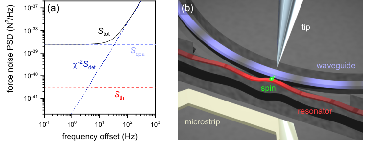
A calculated example for different force noise contributions is shown in Fig. 1(a). We note that with the parameters chosen for this calculation, the total force noise is dominated by up to a frequency offset of about . The optimal compromise between high detection precision and low quantum backaction is termed the ‘standard quantum limit’ (SQL). While representing a general property of interferometers, the SQL is not a fundamental limitation to quantum measurements. Various methods have been proposed and tested to enable displacement detection with an imprecision below the SQL Braginsky et al. 1980; Braginsky and Khalili 1996; Kimble et al. 2001; Giovannetti et al. 2004; Clerk et al. 2008; Lecocq et al. 2015; Kampel et al. 2017; Shomroni et al. 2019; Mason et al. 2019. In the context of scanning force microscopy, these methods offer a strategy for improving the sensitivity beyond backaction-dominated situations such as shown in Fig. 1(a).
For very low temperatures, the thermal noise becomes negligible and the resonator approaches its ground-state motion set by quantum fluctuations. For the purpose of this article, quantum fluctuations are no important limit, as we expect to remain in the regime , where is the number of average phonons occupying the bath at the resonance frequency. Quantum fluctuations pose also no fundamental limit for feedback damping Courty et al. 2001, which is often used to match the sensor bandwidth to that of the signal Poggio et al. 2007a; Degen et al. 2009.
One additional aspect we need to keep in mind is non-contact friction. Even at MHz frequencies, this effect can eventually become dominant when all other noise sources are sufficiently small. Previous experiments found an added non-contact dissipation coefficient of at room temperature and at tip-surface distances around Hälg et al. 2021; Hälg et al. 2022. It is to be expected that this value can be significantly reduced at cryogenic temperatures, larger tip-surface distances, and with suitable surface treatment techniques that minimize the presence of adsorbant layers Héritier et al. 2021.
Instrumentation:
to become a scanning force sensor, the resonator must be mounted in a dedicated measurement apparatus. In contrast to singly-clamped cantilevers, doubly-clamped beams or membranes are more difficult to utilize as scanning probes. This problem can be solved by inverting the typical geometry found in most atomic force microscopes: using the resonator as a sample plate and as the vibrating sensor at the same time, a sharp (but rigid) scanning tip is employed to probe interactions with the samples Hälg et al. 2021; Hälg et al. 2022. The same design can potentially be used with trampolines and strings, as long as the resonator surface is large enough to allow sample deposition, see Fig. 1(b). In the case of NanoMRI, the relevant sample scale is on the order of Degen et al. 2009; Krass et al. 2022.
Sample deposition:
suitable methods to load samples onto fragile resonators must be developed. One possibility is the direct deposition from liquid droplets through micropipettes Hälg et al. 2021 or hollow cantilevers Meister et al. 2009. The main drawback of this method is the creation of residues from the liquid that can potentially reduce the resonator’s sensitivity. Alternative methods that avoid contamination include laser-induced desorption of particles from a substrate in vacuum Bykov et al. 2019 or spray injection through a finely tuned valve Chaste et al. 2012; Tavernarakis et al. 2014. For biological samples, freeze-drying is advantageous to avoid structural damage during the chamber evacuation Giddings and Wray 1986. However, it appears unlikely that nanomechanical resonators can survive direct dipping into liquid nitrogen. This issue has previously been overcome by a modular sample preparation procedure for cantilevers Overweg et al. 2015; Krass et al. 2022.
Sensing protocols:
in this last section, we take a look at some spin detection protocols. In general, moving from topography scanning force microscopy to the special case of spin detection requires two additional components. The first of these is a magnetic field gradient that translates a magnetic moment into a measurable force . The best nanomagnets used in previous NanoMRI demonstrations yielded values between Grob et al. 2019 and Mamin et al. 2012 at distances of several tens of nanometers. Nanoscale magnets used in harddisk write heads even reach Tao et al. 2016, indicating the potential for further improvements in NanoMRI. From simulations, the field generated by a magnetic coating layer on a sharp tip, as shown in the illustration in Fig. 1(b), is expected to reach or more Košata et al. 2020.
The second component that is indispensable in most NanoMRI protocols is a source for oscillating magnetic field pulses. These pulses are used to periodically manipulate the spins at their Larmor frequencies (in the MHz to GHz range) to induce spin-mechanics coupling. In order to minimize the amount of electric current needed to create the magnetic field pulses, and hence the heating imposed on the instrument, it is advantageous to place the field source as close as possible to the sample. Metallic microstrips have become a popular choice because they can easily be fabricated on chips and cover a wide range of frequencies Poggio et al. 2007b.
With cantilevers in the kHz range, various protocols have been developed and tested Sidles 1991; Mamin et al. 2003; Rugar et al. 2004; Mamin et al. 2007; Degen et al. 2009; Poggio and Degen 2010; Wagenaar et al. 2017. They typically rely on periodic spin inversions (induced by ac magnetic field pulses) to create a force acting on the cantilever. When contemplating resonators in the range of to as force sensors, most of these protocols are impractical because adiabatic inversion of nuclear spins at such high frequencies requires too much power to be implemented in a cryogenic environment.
A technique developed especially for nanowires at a frequency close to makes use of switchable magnetic field gradients to create a force resonant with the sensor Nichol et al. 2012; Nichol et al. 2013. This protocol could be translated to membrane and string resonators if switchable magnetic tips can be fabricated. Alternatively, it was proposed to invert spins at the frequency difference between two modes, giving rise to parametric coupling Dougherty et al. 1996; Moore et al. 2010. Membrane resonators can be engineered to have two normal modes split by a few kHz in frequency Catalini et al. 2020; Hälg et al. 2022, which offers a convenient tool for nuclear spin detection Košata et al. 2020. Finally, nanoscale resonant coupling between nuclear spins and a mechanical resonator, as originally envisioned Sidles 1992, could at long last be realized with this new class of sensors. Resonant coupling allows not only to detect nuclear spins, but also to manipulate them via the mechanics. Such manipulations have already been demonstrated with the electron spin ensembles of large atomic clouds Karg et al. 2020; Thomas et al. 2021.
In general, ensembles of electron spins are more accessible to mechanical detection than nuclear spins, both due to their larger magnetic moment and because adiabatic inversion can be achieved much faster. For this reason, pioneering experimental demonstrations of spin detection with membrane Scozzaro et al. 2016 and trampoline Fischer et al. 2019 resonators targeted electron spins in diphenyl picrahydrazyl samples with a size of a few tens of micrometers. These experiments, albeit not on the nanoscale, fuel our optimism that silicon nitride resonators will make excellent NanoMRI sensors.
IV Outlook
Looking forward, we can only speculate what the next step in the evolution of nanomechanical sensors will be. Pushing the limits to lower temperatures or cleaner surfaces will certainly allow for yet another increase in sensitivity. A major breakthrough could be achieved if the microscopic origin of intrinsic nanomechanical dissipation is precisely understood and can be addressed. With regards to concrete spin detection protocols, the possibilities afforded by geometrical design and pulsed spin control are far from exhausted. Here, we will hopefully see a host of interesting new ideas over the next few years.
Much remains to be done before silicon nitride membranes or strings can be used for scanning force microscopy, and in particular for nuclear spin detection and NanoMRI. Nevertheless, the prospects offered by this new class of mechanical sensors are truly encouraging. With all of the exciting developments taking place in the optomechanics community, it may well be that this new material paves the way to realizing an old dream.
Acknowledgements.
The author would like to thank Nils Engelsen and Letizia Catalini for critical reading and feedback on the manuscript.References
- Rugar et al. (2004) D. Rugar, R. Budakian, H. J. Mamin, and B. W. Chui, Nature 430, 329 (2004), https://www.nature.com/articles/nature02658.pdf .
- Rabl et al. (2009) P. Rabl, P. Cappellaro, M. V. G. Dutt, L. Jiang, J. R. Maze, and M. D. Lukin, Phys. Rev. B 79, 041302 (2009).
- Rabl et al. (2010) P. Rabl, S. J. Kolkowitz, F. Koppens, J. Harris, P. Zoller, and M. D. Lukin, Nature Physics 6, 602 (2010).
- Degen et al. (2009) C. L. Degen, M. Poggio, H. J. Mamin, C. T. Rettner, and D. Rugar, Proc. Nat. Acad. Sci. U.S.A. 106, 1313 (2009).
- Nichol et al. (2013) J. M. Nichol, T. R. Naibert, E. R. Hemesath, L. J. Lauhon, and R. Budakian, Phys. Rev. X 3, 031016 (2013).
- Grob et al. (2019) U. Grob, M.-D. Krass, M. Héritier, R. Pachlatko, J. Rhensius, J. Kosata, B. Moores, H. Takahashi, A. Eichler, and C. Degen, Nano letters 19, 7935 (2019).
- Mamin and Rugar (2001) H. J. Mamin and D. Rugar, Applied Physics Letters 79, 3358 (2001), https://doi.org/10.1063/1.1418256 .
- Tao et al. (2014) Y. Tao, J. M. Boss, B. A. Moores, and C. L. Degen, Nature Comm. 5, 3638 (2014).
- Héritier et al. (2018) M. Héritier, A. Eichler, Y. Pan, U. Grob, I. Shorubalko, M. D. Krass, Y. Tao, and C. L. Degen, Nano letters 18, 1814 (2018).
- Nichol et al. (2012) J. M. Nichol, E. R. Hemesath, L. J. Lauhon, and R. Budakian, Phys. Rev. B 85, 054414 (2012).
- Rossi et al. (2017) N. Rossi, F. R. Braakman, D. Cadeddu, D. Vasyukov, G. Tutuncuoglu, A. F. i Morral, and M. Poggio, Nat. Nano. 12, 150 (2017).
- de Lepinay et al. (2017) L. M. de Lepinay, B. Pigeau, B. Besga, P. Vincent, P. Poncharal, and O. Arcizet, Nature Nanotechnology 12, 156 (2017).
- Sahafi et al. (2019) P. Sahafi, W. Rose, A. Jordan, B. Yager, M. Piscitelli, and R. Budakian, Nano Letters 20, 218 (2019).
- Moser et al. (2013) J. Moser, J. Güttinger, A. Eichler, M. J. Esplandiu, D. E. Liu, M. I. Dykman, and A. Bachtold, Nature Nanotechnology 8, 493 (2013).
- de Bonis et al. (2018) S. L. de Bonis, C. Urgell, W. Yang, C. Samanta, A. Noury, J. Vergara-Cruz, Q. Dong, Y. Jin, and A. Bachtold, Nano Letters 18, 5324 (2018).
- Weber et al. (2016) P. Weber, J. Güttinger, A. Noury, J. Vergara-Cruz, and A. Bachtold, Nature Communications 7, 1 (2016).
- Stipe et al. (2001) B. C. Stipe, H. J. Mamin, T. D. Stowe, T. W. Kenny, and D. Rugar, Physical Review Letters 87, 096801 (2001).
- Volokitin and Persson (2003) A. I. Volokitin and B. N. J. Persson, Physical Review B 68, 155420 (2003).
- Zurita-Sánchez et al. (2004) J. R. Zurita-Sánchez, J.-J. Greffet, and L. Novotny, Physical Review A 69, 022902 (2004).
- Kuehn et al. (2006) S. Kuehn, R. F. Loring, and J. A. Marohn, Physical Review Letters 96, 156103 (2006).
- Volokitin and Persson (2007) A. I. Volokitin and B. N. J. Persson, Reviews of Modern Physics 79, 1291 (2007).
- Yazdanian et al. (2008) S. M. Yazdanian, J. A. Marohn, and R. F. Loring, The Journal of Chemical Physics 128, 224706 (2008).
- Kisiel et al. (2011) M. Kisiel, E. Gnecco, U. Gysin, L. Marot, S. Rast, and E. Meyer, Nature Materials 10, 119 (2011).
- She and Balatsky (2012) J.-H. She and A. V. Balatsky, Physical Review Letters 108, 136101 (2012).
- den Haan et al. (2015) A. M. J. den Haan, J. J. T. Wagenaar, J. M. de Voogd, G. Koning, and T. H. Oosterkamp, Physical Review B 92, 235441 (2015).
- de Voogd et al. (2017) J. M. de Voogd, J. J. T. Wagenaar, and T. H. Oosterkamp, Scientific Reports 7, 42239 (2017).
- Héritier et al. (2021) M. Héritier, R. Pachlatko, Y. Tao, J. M. Abendroth, C. L. Degen, and A. Eichler, Physical Review Letters 127, 216101 (2021).
- Krass et al. (2022) M.-D. Krass, N. Prumbaum, R. Pachlatko, U. Grob, H. Takahashi, Y. Yamauchi, C. L. Degen, and A. Eichler, arXiv preprint arXiv:2206.03168 (2022).
- González and Saulson (1994) G. I. González and P. R. Saulson, The Journal of the Acoustical Society of America 96, 207 (1994).
- Southworth et al. (2009) D. R. Southworth, R. A. Barton, S. S. Verbridge, B. Ilic, A. D. Fefferman, H. G. Craighead, and J. M. Parpia, Phys. Rev. Lett. 102, 225503 (2009).
- Unterreithmeier et al. (2010) Q. P. Unterreithmeier, T. Faust, and J. P. Kotthaus, Phys. Rev. Lett. 105, 027205 (2010).
- Schmid et al. (2011) S. Schmid, K. D. Jensen, K. H. Nielsen, and A. Boisen, Phys. Rev. B 84, 165307 (2011).
- Fedorov et al. (2019) S. A. Fedorov, N. J. Engelsen, A. H. Ghadimi, M. J. Bereyhi, R. Schilling, D. J. Wilson, and T. J. Kippenberg, Phys. Rev. B 99, 054107 (2019).
- Zwickl et al. (2008) B. Zwickl, W. Shanks, A. Jayich, C. Yang, A. Bleszynski Jayich, J. Thompson, and J. Harris, Applied Physics Letters 92, 103125 (2008).
- Thompson et al. (2008) J. Thompson, B. Zwickl, A. Jayich, F. Marquardt, S. Girvin, and J. Harris, Nature 452, 72 (2008).
- Verbridge et al. (2008) S. S. Verbridge, H. G. Craighead, and J. M. Parpia, Applied Physics Letters 92, 013112 (2008).
- Jayich et al. (2008) A. Jayich, J. Sankey, B. Zwickl, C. Yang, J. Thompson, S. Girvin, A. Clerk, F. Marquardt, and J. Harris, New Journal of Physics 10, 095008 (2008).
- Anetsberger et al. (2009) G. Anetsberger, O. Arcizet, Q. P. Unterreithmeier, R. Rivière, A. Schliesser, E. M. Weig, J. P. Kotthaus, and T. J. Kippenberg, Nature Physics 5, 909 (2009).
- Unterreithmeier et al. (2009) Q. P. Unterreithmeier, E. M. Weig, and J. P. Kotthaus, Nature 458, 1001 (2009).
- Gavartin et al. (2012) E. Gavartin, P. Verlot, and T. J. Kippenberg, Nature nanotechnology 7, 509 (2012).
- Faust et al. (2013) T. Faust, J. Rieger, M. J. Seitner, J. P. Kotthaus, and E. M. Weig, Nature Physics 9, 485 (2013).
- Purdy et al. (2013) T. P. Purdy, R. W. Peterson, and C. Regal, Science 339, 801 (2013).
- Yuan et al. (2015) M. Yuan, M. A. Cohen, and G. A. Steele, Applied Physics Letters 107, 263501 (2015).
- Wilson et al. (2010) C. M. Wilson, T. Duty, M. Sandberg, F. Persson, V. Shumeiko, and P. Delsing, Phys. Rev. Lett. 105, 233907 (2010).
- Andrews et al. (2014) R. W. Andrews, R. W. Peterson, T. P. Purdy, K. Cicak, R. W. Simmonds, C. A. Regal, and K. W. Lehnert, Nature Physics 10, 321 (2014).
- Bagci et al. (2014) T. Bagci, A. Simonsen, S. Schmid, L. G. Villanueva, E. Zeuthen, J. Appel, J. M. Taylor, A. Sørensen, K. Usami, A. Schliesser, and E. S. Polzik, Nature 507, 81 (2014).
- Wilson et al. (2009) D. J. Wilson, C. A. Regal, S. B. Papp, and H. J. Kimble, Phys. Rev. Lett. 103, 207204 (2009).
- Suhel et al. (2012) A. Suhel, B. Hauer, T. S. Biswas, K. S. Beach, and J. P. Davis, Applied Physics Letters 100, 173111 (2012).
- Yu et al. (2012) P.-L. Yu, T. P. Purdy, and C. A. Regal, Phys. Rev. Lett. 108, 083603 (2012).
- Villanueva and Schmid (2014) L. G. Villanueva and S. Schmid, Phys. Rev. Lett. 113, 227201 (2014).
- Chakram et al. (2014) S. Chakram, Y. Patil, L. Chang, and M. Vengalattore, Physical review letters 112, 127201 (2014).
- Tsaturyan et al. (2014) Y. Tsaturyan, A. Barg, A. Simonsen, L. G. Villanueva, S. Schmid, A. Schliesser, and E. S. Polzik, Optics express 22, 6810 (2014).
- Yu et al. (2014) P.-L. Yu, K. Cicak, N. Kampel, Y. Tsaturyan, T. Purdy, R. Simmonds, and C. Regal, Applied Physics Letters 104, 023510 (2014).
- Ghadimi et al. (2017) A. H. Ghadimi, D. J. Wilson, and T. J. Kippenberg, Nano letters 17, 3501 (2017).
- Tsaturyan et al. (2017) Y. Tsaturyan, A. Barg, E. S. Polzik, and A. Schliesser, Nature Nanotechnology 12, 776 (2017).
- Ghadimi et al. (2018) A. H. Ghadimi, S. A. Fedorov, N. J. Engelsen, M. J. Bereyhi, R. Schilling, D. J. Wilson, and T. J. Kippenberg, Science 360, 764 (2018).
- Norte et al. (2016) R. A. Norte, J. P. Moura, and S. Gröblacher, Phys. Rev. Lett. 116, 147202 (2016).
- Reinhardt et al. (2016) C. Reinhardt, T. Müller, A. Bourassa, and J. C. Sankey, Phys. Rev. X 6, 021001 (2016).
- Reetz et al. (2019) C. Reetz, R. Fischer, G. Assumpção, D. McNally, P. Burns, J. Sankey, and C. Regal, Phys. Rev. Applied 12, 044027 (2019).
- Catalini et al. (2020) L. Catalini, Y. Tsaturyan, and A. Schliesser, Phys. Rev. Applied 14, 014041 (2020).
- Fedorov et al. (2020) S. A. Fedorov, A. Beccari, N. J. Engelsen, and T. J. Kippenberg, Phys. Rev. Lett. 124, 025502 (2020).
- Bereyhi et al. (2022a) M. J. Bereyhi, A. Arabmoheghi, A. Beccari, S. A. Fedorov, G. Huang, T. J. Kippenberg, and N. J. Engelsen, Phys. Rev. X 12, 021036 (2022a).
- Bereyhi et al. (2022b) M. J. Bereyhi, A. Beccari, R. Groth, S. A. Fedorov, A. Arabmoheghi, T. J. Kippenberg, and N. J. Engelsen, Nature Communications 13, 1 (2022b).
- Shin et al. (2022) D. Shin, A. Cupertino, M. H. de Jong, P. G. Steeneken, M. A. Bessa, and R. A. Norte, Advanced Materials 34, 2106248 (2022).
- Rossi et al. (2018) M. Rossi, D. Mason, J. Chen, Y. Tsaturyan, and A. Schliesser, Nature 563, 53 (2018).
- Mason et al. (2019) D. Mason, J. Chen, M. Rossi, Y. Tsaturyan, and A. Schliesser, Nature Physics 15, 745 (2019).
- Rossi et al. (2019) M. Rossi, D. Mason, J. Chen, and A. Schliesser, Phys. Rev. Lett. 123, 163601 (2019).
- Guo et al. (2019) J. Guo, R. Norte, and S. Gröblacher, Phys. Rev. Lett. 123, 223602 (2019).
- Fischer et al. (2019) R. Fischer, D. P. McNally, C. Reetz, G. G. T. Assumpção, T. Knief, Y. Lin, and C. A. Regal, New Journal of Physics 21, 043049 (2019).
- Hälg et al. (2021) D. Hälg, T. Gisler, Y. Tsaturyan, L. Catalini, U. Grob, M.-D. Krass, M. Héritier, H. Mattiat, A.-K. Thamm, R. Schirhagl, E. C. Langman, A. Schliesser, C. L. Degen, and A. Eichler, Phys. Rev. Applied 15, L021001 (2021).
- Seis et al. (2022) Y. Seis, T. Capelle, E. Langman, S. Saarinen, E. Planz, and A. Schliesser, Nature communications 13, 1 (2022).
- Hälg et al. (2022) D. Hälg, T. Gisler, E. C. Langman, S. Misra, O. Zilberberg, A. Schliesser, C. L. Degen, and A. Eichler, Phys. Rev. Lett. 128, 094301 (2022).
- Gieseler et al. (2012) J. Gieseler, B. Deutsch, R. Quidant, and L. Novotny, Phys. Rev. Lett. 109, 103603 (2012).
- Gisler et al. (2022) T. Gisler, M. Helal, D. Sabonis, U. Grob, M. Héritier, C. L. Degen, A. H. Ghadimi, and A. Eichler, Phys. Rev. Lett. 129, 104301 (2022).
- Beccari et al. (2022) A. Beccari, D. A. Visani, S. A. Fedorov, M. J. Bereyhi, V. Boureau, N. J. Engelsen, and T. J. Kippenberg, Nature Physics 18, 436 (2022).
- Košata et al. (2020) J. Košata, O. Zilberberg, C. L. Degen, R. Chitra, and A. Eichler, Phys. Rev. Applied 14, 014042 (2020).
- Aspelmeyer et al. (2014) M. Aspelmeyer, T. J. Kippenberg, and F. Marquardt, Rev. Mod. Phys. 86, 1391 (2014).
- Faust et al. (2012) T. Faust, P. Krenn, S. Manus, J. P. Kotthaus, and E. M. Weig, Nature communications 3, 1 (2012).
- Braginsky et al. (1980) V. B. Braginsky, Y. I. Vorontsov, and K. S. Thorne, Science 209, 547 (1980).
- Braginsky and Khalili (1996) V. B. Braginsky and F. Y. Khalili, Rev. Mod. Phys. 68, 1 (1996).
- Kimble et al. (2001) H. J. Kimble, Y. Levin, A. B. Matsko, K. S. Thorne, and S. P. Vyatchanin, Phys. Rev. D 65, 022002 (2001).
- Giovannetti et al. (2004) V. Giovannetti, S. Lloyd, and L. Maccone, Science 306, 1330 (2004).
- Clerk et al. (2008) A. A. Clerk, F. Marquardt, and K. Jacobs, New Journal of Physics 10, 095010 (2008).
- Lecocq et al. (2015) F. Lecocq, J. B. Clark, R. W. Simmonds, J. Aumentado, and J. D. Teufel, Phys. Rev. X 5, 041037 (2015).
- Kampel et al. (2017) N. S. Kampel, R. W. Peterson, R. Fischer, P.-L. Yu, K. Cicak, R. W. Simmonds, K. W. Lehnert, and C. A. Regal, Phys. Rev. X 7, 021008 (2017).
- Shomroni et al. (2019) I. Shomroni, L. Qiu, D. Malz, A. Nunnenkamp, and T. J. Kippenberg, Nature communications 10, 1 (2019).
- Courty et al. (2001) J.-M. Courty, A. Heidmann, and M. Pinard, The European Physical Journal D-Atomic, Molecular, Optical and Plasma Physics 17, 399 (2001).
- Poggio et al. (2007a) M. Poggio, C. L. Degen, H. J. Mamin, and D. Rugar, Phys. Rev. Lett. 99, 017201 (2007a).
- Meister et al. (2009) A. Meister, M. Gabi, P. Behr, P. Studer, J. Vörös, P. Niedermann, J. Bitterli, J. Polesel-Maris, M. Liley, H. Heinzelmann, and T. Zambelli, Nano letters 9, 2501 (2009).
- Bykov et al. (2019) D. S. Bykov, P. Mestres, L. Dania, L. Schmöger, and T. E. Northup, Applied Physics Letters 115, 034101 (2019).
- Chaste et al. (2012) J. Chaste, A. Eichler, J. Moser, G. Ceballos, R. Rurali, and A. Bachtold, Nat. Nanotechnol. 7, 300 (2012).
- Tavernarakis et al. (2014) A. Tavernarakis, J. Chaste, A. Eichler, G. Ceballos, M. C. Gordillo, J. Boronat, and A. Bachtold, Phys. Rev. Lett. 112, 196103 (2014).
- Giddings and Wray (1986) T. H. Giddings and G. P. Wray, in Ultrastructure Techniques for Microorganisms (Springer, 1986) pp. 241–265.
- Overweg et al. (2015) H. C. Overweg, A. M. J. den Haan, H. J. Eerkens, P. F. A. Alkemade, A. L. La Rooij, R. J. C. Spreeuw, L. Bossoni, and T. H. Oosterkamp, Applied Physics Letters 107, 072402 (2015).
- Mamin et al. (2012) H. Mamin, C. Rettner, M. Sherwood, L. Gao, and D. Rugar, Applied Physics Letters 100, 013102 (2012).
- Tao et al. (2016) Y. Tao, A. Eichler, T. Holzherr, and C. L. Degen, Nature Communications 7, 12714 (2016).
- Poggio et al. (2007b) M. Poggio, C. Degen, C. Rettner, H. Mamin, and D. Rugar, Applied physics letters 90, 263111 (2007b).
- Sidles (1991) J. A. Sidles, Applied Physics Letters 58, 2854 (1991), https://doi.org/10.1063/1.104757 .
- Mamin et al. (2003) H. J. Mamin, R. Budakian, B. W. Chui, and D. Rugar, Phys. Rev. Lett. 91, 207604 (2003).
- Mamin et al. (2007) H. Mamin, M. Poggio, C. Degen, and D. Rugar, Nature nanotechnology 2, 301 (2007).
- Poggio and Degen (2010) M. Poggio and C. L. Degen, Nanotechnology 21, 342001 (2010).
- Wagenaar et al. (2017) J. J. T. Wagenaar, A. M. J. den Haan, R. J. Donkersloot, F. Marsman, M. de Wit, L. Bossoni, and T. H. Oosterkamp, Phys. Rev. Applied 7, 024019 (2017).
- Dougherty et al. (1996) W. M. Dougherty, K. J. Bruland, J. L. Garbini, and J. A. Sidles, Meas. Sci. Technol. 7, 1733–1739 (1996).
- Moore et al. (2010) E. W. Moore, S. Lee, S. A. Hickman, L. E. Harrell, and J. A. Marohn, Appl. Phys. Lett. 97, 044105 (2010).
- Sidles (1992) J. A. Sidles, Phys. Rev. Lett. 68, 1124 (1992).
- Karg et al. (2020) T. M. Karg, B. Gouraud, C. T. Ngai, G.-L. Schmid, K. Hammerer, and P. Treutlein, Science 369, 174 (2020).
- Thomas et al. (2021) R. A. Thomas, M. Parniak, C. Østfeldt, C. B. Møller, C. Bærentsen, Y. Tsaturyan, A. Schliesser, J. Appel, E. Zeuthen, and E. S. Polzik, Nature Physics 17, 228 (2021).
- Scozzaro et al. (2016) N. Scozzaro, W. Ruchotzke, A. Belding, J. Cardellino, E. C. Blomberg, B. A. McCullian, V. P. Bhallamudi, D. V. Pelekhov, and P. C. Hammel, Journal of Magnetic Resonance 271, 15 (2016).