Knowledge Graph Construction and Its Application in Automatic Radiology Report Generation from Radiologist’s Dictation
Abstract
Conventionally, the radiologist prepares the diagnosis notes and shares them with the transcriptionist. Then the transcriptionist prepares a preliminary formatted report referring to the notes, and finally, the radiologist reviews the report, corrects the errors, and signs off. This workflow causes significant delays and errors in the report. In current research work, we focus on applications of NLP techniques like Information Extraction (IE) and domain-specific Knowledge Graph (KG) to automatically generate radiology reports from radiologists’ dictation. This paper focuses on KG construction for each organ by extracting information from an existing large corpus of free-text radiology reports. We develop an information extraction pipeline that combines rule-based, pattern-based, and dictionary-based techniques with lexical-semantic features to extract entities and relations. Missing information in short dictation can be accessed from the KGs to generate pathological descriptions and hence the radiology report. Generated pathological descriptions evaluated using semantic similarity metrics, which shows 97% similarity with gold standard pathological descriptions. Also, our analysis shows that our IE module is performing better than the OpenIE tool for the radiology domain. Furthermore, we include a manual qualitative analysis from radiologists, which shows that 80-85% of the generated reports are correctly written, and the remaining are partially correct.
1 Introduction
Radiology is an integral part of medical care. Radiological imaging-based evidence (X-ray, MRI, CT, USG, etc.) is crucial in determining the nature of the treatment. The usual radiology process is as follows: A patient gets scanned. Then the radiologist prepares the diagnosis notes (referred to as radiologist’s dictation) by either dictating to a voice recording device or writing it on paper and hands over the notes to a transcriptionist. The transcriptionist opens a scan-specific standardized template corresponding to all normal findings (referred to as normal report template) and edits it based on the measurements and findings reported by the radiologist in more descriptive form (referred to as pathological description).
1.1 Motivation
Radiologists are in big demand since the ratio of radiologists per number of patients is very low. Ratio in India is, 1:100,000, the corresponding ratio in the US is 1:10,000, and for China, it is 1:14,772 (Arora 2014). It results in very high patient inflows, making radiologists incredibly busy and stressed out. Currently adopted workflow causes (i) significant delays in report turnaround time, (ii) errors in the reports, and (iii) burnout. These challenges amplify further, because in densely populated countries, radiologists handle several patients every day. Our interactions with radiologists, diagnostic centers, and hospitals highlight that many radiologists want to eliminate the tedious report typing process and focus on the diagnosis.
To overcome tedious typing, radiologists resort to dictating observations in short or abstract form. Domain specific knowledge is required to generate the correct pathological description from short dictations. Once we generate patient-specific pathological description from radiologist’s dictation then we can add it in normal report template by replacing corresponding normal sentences.
Domain knowledge can be acquired from already existing radiology free-text reports. We need a structured format of all essential medical information to reuse it. There are different technologies used to store the structured data like, relational databases, xml files, KGs, etc. KGs are used solely for deriving insights. Maintaining a KG is worry-free because we don’t have to think about how the additional data stored in the graph will affect the existing data. Using a KG to uncover insights is a better choice when the system needs domain insight.
1.2 Problem Statement
Design a system that generates a structured patient-specific report from radiologist’s dictation and domain knowledge.
- •
Input: (i) Radiologist’s dictation, and (ii) Normal report template.
- •
Output: Radiology report with patient-specific findings.
Domain knowledge will come from KG.
Sub-Problem: Develop a system that automatically constructs a KG of essential medical information in radiology free-text reports.
- •
Input: Radiology free-text report corpus.
- •
Output: Structured representation of the essential medical information contained within the free-text reports in a hierarchical KG.
1.3 Challenges
Radiologists help us to construct a preliminary KG of the skeleton (higher-level hierarchy) for the anatomy and findings of an organ. Manually identifying all the entities in the radiology domain is also tricky for radiologists. Hence to complete the preliminary KG provided by the radiologists, we extract information from the radiology report corpus.
IE is one of the foremost steps in KG construction (Zhao et al. 2018). Information extraction from free-text clinical notes/narratives, such as radiology reports, is difficult due to the nuances of natural language like misspellings, abbreviations, and non-standard terminologies. There are three main approaches for IE: (i) Machine Learning (ML) based technique, (ii) Rule-based technique, (iii) Pattern-based technique, and iv) Lexicon dictionary-based technique. The ML-based technique needs annotated corpus. We do not have annotated data, and annotating data in-house is time-consuming and costly. Hence we are not considering ML-based techniques for IE. Pattern-based IE uses a lexico-syntactic and semantic pattern dictionary to extract information from free-text, as detailed in the paper (Tang et al. 2008). The construction of a pattern dictionary is required in a pattern-based approach. To extract information from text, the rule-based technique employs a set of general rules. The rule-based and pattern-based techniques need domain understanding and language understanding. The lexicon dictionary-based approach extracts entities present in the dictionary, but it fails to extract entities not present in the dictionary. Hence, the only pattern-based, rule-based, or lexicon dictionary-based approach does not fit to the radiology report domain.
We propose an approach for information extraction that combines rule-based, pattern-based, and lexicon dictionary-based techniques with lexical semantic features. We test the efficacy of constructed KGs by calculating evaluation metrics BLEU (Papineni et al. 2002) score and ROUGE (Lin 2004) score of pathological description generated using constructed KG vs. gold standard pathological descriptions generated by radiologists manually. We also evaluate our IE system by calculating precision, recall, and F-score of system extracted triples vs. gold standard triples and compare it with the OpenIE system.
1.4 Our Contributions
The key contributions are as follows:
- 1.
Preliminary KGs construction by radiologist manually.
- 2.
Information extraction pipeline to extract medical entities and relations from radiology reports. It extracts all necessary entities from the radiology text reports (findings, observations, anatomy, modifiers, and properties) and their relations.
- 3.
KG augmentation using extracted triples from reports and store constructed KG in standard Resource Description Framework (RDF) triple format.11 1 There are no KGs constructed before for all body organs, including all necessary information like findings, observations, anatomy, properties, and modifiers related to the organ. In our work, we construct KGs for the Liver, Kidney, Gallbladder, Uterus, Urinary bladder, Ovary, Pancreas, Prostate, Biliary Tree, and Bowel, etc. for Ultrasound scan procedure. We have developed generic pipeline for radiology domain to construct KGs that can be used for CT, MRI, X-ray, etc. scan procedures.
- 4.
Pathological description generation from radiologist’s dictation using constructed KGs.
- 5.
Map generated pathological description in normal report template at the appropriate location to generate patient-specific report.
2 Related Work
Research is done in automatic radiology report generation based on scanned images. Yuan et al. 2019 proposes an automated structured-radiology report generation system using extracted features from images. Loveymi et al. 2021 proposed a system that generate descriptions for natural images by image captioning.
There is a wealth of research done on building medical KG from Electronic Medical Records (EMR). Finlayson et al. 2014 builds a graph from medical text, clinical notes etc. Graph nodes represents diseases, drugs, procedures, and devices. Rotmensch et al. 2017 uses the EMR to construct the graph of diseases and symptoms. Researchers worked on creating medical KG from EMR, but no one has built a KG for the radiology domain except Zhang et al. 2020. Graph embedding module is proposed by Zhang et al. 2020 that helps to generate radiology reports from image reports. Each node in their KG represents disease. Taira et al. 2001 developed an NLP pipeline to structure the critical medical information. Extracted information includes the existence, location, properties, and diagnostic interpretation of findings from radiology free-text documents. Information is not integrated since they store the structured information for each report separately. Also, this system does not accept reports with different reporting styles. However, this is not always the case. Every radiologist has their dictation style and reporting style.
IE systems that are based on IE patterns are surveyed by Muslea et al. 1999. Ghoulam et al. 2015 extracts signs of lung cancer, anatomical location, and relation between the signs and the locations expressed in the radiology reports. Embarek and Ferret 2008 used a morpho-syntactic patterns in their rule-based method to find medical entities like symptoms, disease, exams, medicament, and treatment. Xu et al. 2009 explains that pattern is a sub dependency tree that indicates a relation instance. Pons et al. 2016 gives the overview of NLP techniques that can be used in radiology.
3 Our Approach

Figure 1shows the system architecture. Our system converts the radiologist’s dictation to a patient-specific report. The main tasks of the system are:
- 1.
construct organ wise KGs using preliminary KG provided by radiologists and radiology report corpus
- 2.
generate pathological description using KGs
- 3.
map generated pathological descriptions to the corresponding location in the normal report template
- 4.
eliminate normal sentences from templates where an abnormal finding has been reported
- 5.
output the patient-specific report
3.1 Knowledge Graph Construction
Paulheim and Heiko (Paulheim 2017) defines Knowledge Graph (KG) as "A knowledge graph (i) mainly describes real-world entities and their interrelations, organized in a graph, (ii) defines possible classes and relations of entities in a schema, (iii) allows for potentially interrelating arbitrary entities with each other and (iv) covers various topical domains." KGs are designed with suitable ontology to store domain knowledge. Domain ontology and individual information together form a Knowledge Base (KB).
KGs are constructed based on anonymised radiology reports22
2
Anonymised radiology reports were used as provided by a company collaborating with us, with due consent of the physicians.. The de-identified corpus consists of approximately a hundred thousand reports. These include CT, MRI, Ultrasound, and X-Ray reports. Since reports are collected from different hospitals, these report’s reporting styles and dictation styles are different.
3.1.1 Knowledge Base Creation
Ontology Creation
Ontologies are semantic data models that define the types of things in a specific domain and the properties used to describe those types. Three main components of ontology are Classes, Relationships, and Attributes.
We have created our ontology by integrating several classes of Radlex33
3
http://radlex.org/
RadLex is licensed freely for commercial and non-commercial users. entities. For example, Radlex descriptor subclasses are combined in Modifier class, several subclasses of Radlex observation into Observation class, several subclasses of Radlex anatomical entity into Anatomy class, etc. We believe that leaving the granularity at a coarse level is a practical choice to make ontology more generic. We have defined eight logical relations PartOf, TypeOf, ModifierOf, ObservationOf, DefaultObservationOf, PropertyOf, DefaultPropertyOf and FoundIn. We kept our ontology very simple. However, it covers all entities in the radiology report domain. We define attributes like preferedName, synonyms and wordForms for all classes in our ontology. Figure 2 indicates higher level class hierarchy of the ontology that we have constructed with the help of domain experts. Protégé44
4
https://protege.stanford.edu/ (Musen 2015) suite is used for ontology development.

The purpose of constructing KGs is that we should get all the necessary information which is missing in abstract text from radiology. For example, we should get the anatomical location of finding from KG if it is not mentioned in the text. The lesion is found in segment VI of the liver. Here anatomical location is segment VI of the right lobe of the liver. The right lobe is missing in the text, and the KG gives the missing location. Another example is that if some finding is mentioned without details, we should get all default observations and properties from KG. For example, Acute pancreatitis, its default observations and properties are the presence of peripancreatic fluid, increased size of the pancreas, inhomogeneous echotexture, etc.
We have constructed KGs in hierarchical structures. Root represents the organ, and their children express their anatomy (i.e., parts). The edge between them is labeled by PartOf relation. Similarly, if any particular radiological observation is found for an organ or its parts, that observation can be added as a child of that anatomical entity. Furthermore, we label the edge between them by FoundIn relation.
Preliminary Knowledge Graph

Domain experts provide preliminary KGs for each organ with higher-level anatomy and pathology corresponding to that organ. We convert preliminary KGs into standard RDF (Miller 1998) triples format. Using transformation rules55 5 https://github.com/protegeproject/cellfie-plugin we have loaded preliminary KG triplets as individuals/instances in Protege’ tool. Figure 3 shows the KG of the Liver, which is provided by radiologists.
3.1.2 Data Preprocessor
To construct KGs, we use a radiology report corpus. The data preprocessor module takes radiology reports as input. Radiology reports contain Header, Pathology Description, History, and Conclusion/Impression sections. We use simple heuristics like regular expressions to fetch only the Pathological Description and Impression section. We use spell corrector and word-tokenization algorithms since the extracted sentences contain spelling mistakes, unwanted punctuation marks, etc. Spelling correction and word-segmentation algorithm implemented using Symspell66
6
https://github.com/wolfgarbe/symspell APIs. We use the symmetric delete spelling correction algorithm, which provides much higher speed and lower memory consumption. We apply the domain dictionary of correct words and the frequency of their occurrences in the corpus to correct the spellings.
3.1.3 Information Extraction
The information extraction process extracts the entities from unstructured text and couples those entities with their relations. We use the same logical relations that we have defined in our ontology. For example, the corpus sentence is Right kidney is normal in size, shape, location and cortical echogenicity. Here, size, shape, location and echogenicity have a relation with the right kidney. Cortical and echogenicity are related to each other by relation ModifierOf. Also normal is a modifier of size, shape, location, and echogenicity. However, sate-of-the-art OIE tools like Stanford OpenIE77 7 https://nlp.stanford.edu/software/openie.html are not capable of extracting these concrete relations from free-text (Etzioni et al. 2008). As shown in Table 1, for the sentence 1, OpenIE could not find the relation between calculus and middle calyx, middle calyx and right kidney and for the sentence 2, OpenIE does not consider the shape, location, and cortical echogenicity.
| Input Sentence | Triples Extracted Using OpenIE | ||||||
|---|---|---|---|---|---|---|---|
| A 5 mm calculus is noted in an upper calyx and a 4 x 3 mm calculus is noted in a middle calyx of right kidney. |
| ||||||
| Right kidney is normal in size 9.6 x 4.0 cm, shape, location and cortical echogenicity. |
|
Our approach combines rule-based, lexicon dictionary-based, and pattern-based techniques. The three modules of the IE task are (i) Lexical Analyser, (ii) Dependency Parser, and (iii) Semantic Analyser.
Lexical Analyser
There are three main tasks in the lexical analyser (i) Lexical semantic tagging, (ii) Noun phrase chunking and (iii) Medical lexicon dictionary creation. The input to the lexical analyser is a sentences from the corpus, and it outputs the sentence-wise syntactic and semantic features of each word or phrase in the sentence. Features include POS tag, lemma, supersense, and the root of a noun chunk.
Lexical Semantic Supersense Tagger:
Prepositions play a role in forming relations between two entities. To extract the relation between two entities connected by preposition, the machine should understand the meaning of that preposition. As shown in Figure 4, intuition of in preposition in first sentence is characteristic and in second sentence is locus. Supersenses (Schneider et al. 2015) helps to get the intuition of these prepositions. Supersenses disambiguate the lexical units by semantically classifying them. This (Liu et al. 2020) paper deals with three sets of supersense labels: nominal, verbal, and prepositional/possessive. Lexical Semantic Recognition(LSR)88 8 https://github.com/nelson-liu/lexical-semantic-recognition (Liu et al. 2020) task effectively tags the supersense for each word in sentence. Figure 4 shows the sentences that contain prepositions and their corresponding tags.

We have mapped preposition supersenses with corresponding logical relations. Table 2 shows the examples of supersense classes and their corresponding logical relations.
| Supersense | Relation |
|---|---|
| Locus | FoundIn |
| Gestalt | PartOf |
| PartPortion | PartOf |
| Supersense | Relation |
|---|---|
| Whole | PartOf |
| Manner | PropertyOf |
| Purpose | PropertyOf |
Noun Phrase Chunker:
Noun phrase chunking is applied to get the candidate entity phrases. We use spacy APIs to get the noun phrases and corresponding root elements. For example, in sentence An area of increased echogenicity in the right lobe of liver, suggestive of focal fatty infiltration. noun chunks are: an area, increased echogenicity, the right lobe, liver, and focal fatty infiltration. And their corresponding root words are area, echogenicity, lobe, liver and infiltration respectively.
Table 3shows the combined output of supersense tagger and noun phrase chunker.
| Noun Phrases/Words | Token List | Root Token | POS Tags | Lemmas | Supersences |
| Non-enhancing hypodense lesion | [Non, -, enhancing, hypodense, lesion] | lesion | [ADJ, ADJ, VERB, ADJ, NOUN] | [non, , enhance, hypodense, lesion] | [B-ADV, I_, I ADJ, OADJ, COGNITION] |
| noted | [noted] | noted | [VERB] | [note] | [cognition] |
| in | [in] | in | [ADP] | [in] | [Locus] |
| right lobe | [right, lobe] | lobe | [ADJ, NOUN] | [right, lobe] | [OADJ, LOCATION] |
| of | [of] | of | [ADP] | [of] | [Whole] |
| liver. | [liver, .] | liver | [NOUN, PUNCT] | [liver] | [BODY, OPUNCT ] |
Medical Lexicon Dictionary Creator:
Radiology reports contain large number of medical terms including hundreds of descriptive or modifier terms that are complex in nature, it includes abbreviations, synonyms and proper names. These all medical terms are not found in single medical glossaries. RadLex (Langlotz 2006) lexicon is used to create radiology dictionary. RadLex is a comprehensive glossary of radiology terms. Radlex lexicon contains long phrases e.g. fat homogeneous background echotexture. Here fat, homogeneous and background are the modifiers of echotexture. In radiology reports individual entities such as homegeneous echotexture, fat echotexture, background echotexture or only echotexture appears frequently. Hence, using NLP techniques we have extracted radiological terms from RadLex lexicon at granular level. We have divided these long phrases into individual entities with meaning in the radiology context. Also there are some radiological terms which frequently appear in radiology reports but not present in RadLex lexicon e.g. echopattern, corticomedullary differentiation, etc. Entities that are missing in the Radlex lexicon, we have added from the corpus. We use the same categories that we have defined as ontology classes. Table 4 shows the examples of entities and categories in our dictionary.
| Entity | Category |
|---|---|
| lesion | observation |
| cirrhosis | pathologic-finding |
| hepatitis | inflammation |
| size | property |
| Entity | Category |
|---|---|
| small | size-modifier |
| left lobe | anatomy |
| ankle fracture | injury |
| chronic liver disease | disease |
Dependency Parser
Dependency parser gives us direct or indirect linking between entity phrases. We have written rules over dependency tags and POS-tags by analyzing dependency parser to extract the relations between entities. Spacy99 9 https://spacy.io/api/dependencyparser APIs are used for dependency parsing. Dependencies are established between phrases instead of words. Before applying dependency parser we merge noun chunks into a single token. The dependency tree shown in the Figure 5, Non-enhancing hypodense lesion is linked with right lobe and right lobe is linked with liver.

Semantic Analyser
The previous modules’ outputs help us write lexico-semantic rules and patterns to extract entities and relations from input sentences. In our case, entity extraction and relation extraction are very much dependent tasks. As dependencies are established between phrases, the dependency tree gives us the POS and dependency relation between noun phrases.
Dictionary-based Entity Extractor:
Noun phrase chunker gives us noun phrases that are the candidate entity phrases. However, the entity may not always be a whole noun phrase. Hence to extract proper entities from noun phrases, we search the dictionary for matching entities. If a word or phrase matches with multiple dictionary entry through more than one text spans, we consider the longest text span as the matched entry for entity extraction. For example, in phrase right lobe although individual terms right and lobe exist in our dictionaries, only the longest match, right lobe is used for entity extraction.
Pattern-based Relation Extractor:
Single noun phrase contains multiple entities.
| Pattern | Triple Format | Example | Triples |
|---|---|---|---|
| ADJ* NOUN/root | (ADJ, ModifierOf, NOUN/root) | simple clear cyst | (simple, ModifierOf, cyst) |
| (clear, ModifierOf, cyst) | |||
| Anatomy Anatomy/root | (Anatomy/root, PartOf, Anatomy) | liver right lobe | (right lobe, PartOf, liver) |
| Anatomy Finding/root | (Finding/root, FoundIn, Anatomy) | kidney calculus | (calculus, FoundIn, liver) |
| Anatomy Observation/root | (Observation/root, FoundIn, Anatomy) | urinary bladder cyst | (cyst, FoundIn, urinary bladder) |
| Modifier Observation/root | (Modifier, ModifierOf, Observation/root) | non-enhancing lesion | (non-enhancing, ModifierOf, lesion) |
Patterns are used to extract these entities. Table 5 shows the dictionary of patterns. To extract entities we use look up based approach, that check the matching pattern in pattern dictionary for input noun phrase. For example, non-enhancing and hypodense are the modifiers of lesion in the noun phrase non-enhancing hypodense lesion. As non-enhancing present in the dictionary, it applies pattern Modifier Observation and extracts the triple (non-enhancing, ModifierOf, lesion). Hypodense does not exist in our dictionary; hence, it applies the ADJ NOUN pattern and extracts triple (hypodense, ModifierOf, lesion). Table 3 shows the list of some patterns and examples of triples extracted from noun phrases when patterns are applied to extract relations.
Relation Extraction Using Preposition Supersenses:
If two entities are connected with preposition then we consider its supersense to find the relation. For example, lesion in right lobe here in represents locus supersense and as shown in Table 2, locus is mapped to FoundIn relation. We add new triple ( lesion, FoundIn, right lobe).
Rule-based Relation Extractor:
We discussed how to extract entities and the relation between the entities present in the single noun phrase. However, a relation exists between the entities present in two different noun phrases. The relation exists between root entities in candidate pair of noun phrases. The example shown in Figure 5, there exist relation between lesion and right lobe but not between hypodense and right lobe.
We have written rules using the dependency tree to get the candidate pair of noun phrases that are related to one another. We write an algorithm to traverse the dependency tree from each leaf up to the root node to find the candidate pair of noun phrases. For example, the sentence given in Figure 5 there are two leaf nodes, Non-enhancing hypodense lesion and liver. Hence first it traverse from leaf node Non-enhancing hypodense lesion to root node noted and then it traverse from liver to root node noted. We use different stacks to store important linguistic information like subjects, objects present in the current path, prepositions present in current path, etc.
We first find all the subjects present in path while traversing from leaf node to root node. We save that subjects globally with corresponding verb so that when we traverse different path from leaf to root then we can have access to all subjects that we have already traversed associated with verb. For example, we store lesion (root entity of noun phrase Non-enhancing hypodense lesion) as subject associated with verb noted. Similarly we store all objects globally that occurs in the sentence associated with verb. For example, dependency tree shown in Figure 5 there exist path from leaf node liver to root node noted. In that path two objects are present liver and right lobe. When we process liver it stores liver in object stack and when it comes to right lobe at that instance there is one preposition in preposition stack and one object in object stack. It pops of from preposition stack and liver from object stack. Supersense associated with preposition of is Whole and this supersense is mapped with logical relation PartOf. Hence we define relation between right lobe (root entity of right lobe is right lobe) and liver is PartOf. Add new triple (right lobe, PartOf, liver) in triple set and push new object right lobe in stack. Once it reaches to root node it adds new triples (subject, relation, object). If subjects and objects are not connected by any
preposition, then we use lexical-semantic patterns to extract the relation between them. Patterns such as if subject is Observation (e.g. lesion) and object is Anatomy (e.g. right lobe) then relation exist between them is ObservedIn. Hence we add new triple (lesion, ObservedIn, right lobe). List of patterns used to extract relations between two entities are listed in Table 6.
| Pattern (entity1-category, entity2-category) | Triple Format | Example (entity1, entity2) | Triples |
|---|---|---|---|
| (Anatomy, Anatomy) | (entity1, PartOf, entity2) | (right lobe, liver) | (right lobe, PartOf, liver) |
| (Property, Anatomy) | (entity1, PropertyOf, entity2) | (echotexture, pancreas) | (echotexture, PropertyOf, pancreas) |
| (Finding, Anatomy) | (entity1, FoundIn, entity2) | (medical renal disease, kidney) | (medical renal disease, FoundIn, kidney) |
| (Observation, Anatomy) | (entity1, ObservedIn, entity2) | (pseudo cyst, body) | (pseudo cyst, ObservedIn, body) |
3.1.4 Knowledge Graph Augmentation
Preliminary KGs enhanced using triples extracted by IE module. Steps involved in KG augmentation are explained below:
- •
Step 1: Triples are stored in the file against its input sentence. For example, sentence from corpus is, A lesion of increased echotexture in the right lobe of liver. Triplets extracted corresponding to above sentence are, (increased, ModifierOf, echotexture), (echotexture, PropertyOf, lesion), (right lobe, PartOf, liver), and (lesion, FoundIn, right lobe).
- •
Step 2: Construct dynamic KG for sentence triples. Figure 6 shows the dynamic KG constructed for sentence triples.

Figure 6: Dynamic KG constructed for the triples of sentence A lesion of increased echotexture in the right lobe of liver. - •
Step 3: Find its appropriate matched path in our already built preliminary (static) KG. Figure 7 shows the entities from dynamic KG path matched with static KG path.

Figure 7: The static KG at that instance. Yellow highlighted nodes show the entities from the dynamic KG path matched with the static KG path. - •
Step 4: If a triple is missing in the static KG, then we add a new triple in the static KG. Here in above example triple (increased, ModifierOf, echotexture) is missing in static KG. Hence, we will add this triple in static KG. Figure 8 shows the updated static KG.

Figure 8: Updated static KG after adding new triple. Newly added node highlighted in green.
This is how we update the static KG according to our dynamic KG triples. We repeat above steps for all sentences in our corpus.
In a static KG, we have multiple instances of the same observations, same properties, and same modifiers. For example, acute hepatitis reveals decreased echogenicity of the liver and chronic liver disease reveals increased echogenicity of the liver. Here for both the findings echogenicity is the related observation but their related descriptors/modifiers are not same. Decreased is the echogenicity modifier associated with acute hepatitis and increased is the echogenicity modifier associated with chronic liver disease. Therefore we have created different instances of observation with name echogenicity for both the findings. Hence, we use a path from the dynamic KG to find the appropriate entity with identical names from the static KG. In static KGs, we have arranged findings in such a way that its parents represent the anatomical location and its children represent the properties or states of organs related to that finding. Figure 9 shows the augmented KG of the Liver.

3.2 Generating Pathological Description
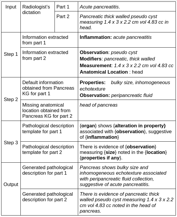
To generate pathological descriptions, we first extract the essential information from dictation using the same IE module we built for the KG construction pipeline. We obtain the missing and default information from KGs and patient-specific information from dictation. Then fill this information in pathological description templates. We have defined pathological description templates based on the type of findings—for example, template for inflammation, lesion, disease, etc. Table in the Figure 10 shows the examples of generated pathological description from radiologist’s dictation.
3.3 Generating Patient Specific Report
One of the crucial tasks of the project is the replacement of normal descriptions with the generated pathological descriptions into the normal report template.
Default normal report templates are provided by the radiologists. We provide an appropriate normal report template to the system, and the system replace generated pathological descriptions with the corresponding location in the normal report template.
3.3.1 Parallel Corpus
At first, we create a parallel corpus for the radiologist’s dictation and the corresponding normal sentences in a normal report template (referred to as normal description).
| Radiologist’s dictation | Normal description | |
|---|---|---|
| i. | Grade 1 fatty liver | Liver is normal in size and echotexture. |
| ii. | Acute pancreatitis | Pancreas is normal in size and echotexture. |
| iii. | Chronic pancreatitis | Pancreas is normal in size and echotexture. |
| iv. | Pancreatic thick walled pseudo cyst in head. | No evidence of focal or diffuse lesion is seen. |
| v. | Pancreatic thick walled pseudo cyst in tail. | No evidence of focal or diffuse lesion is seen. |
Example shown in the Figure 10, generated pathological description for dictation acute pancreatitis, size and echotexture is altered in pathological description, we replace the normal description about the size and echotexture of the Pancreas with generated pathological description in the normal report template.
3.3.2 Workflow
We have implemented a look-up-based approach to achieve this task. The basic approach says that for a radiologist’s dictation, search for the corresponding normal description in a parallel corpus. Then find the location of that normal description in the normal report template and replace the whole sentence with the generated pathological description.
- •
Input:
- 1.
Radiologist’s dictation
- 2.
Generated pathological description
- 3.
Parallel corpus of radiologist’s dictation vs. normal descriptions
- 4.
Normal report template
- 1.
- •
Output: Radiology report with patient-specific findings.
- •
Step 1: Perform a look up into a parallel corpus to find similar radiologist’s dictation with input radiologist’s dictation. We use NLTK BLEU score for matching the dictations. Get the corresponding normal description from corpus. For example, for input dictation Acute pancreatitis we found match Acute pancreatitis in dictionary. Similarly for input dictation Pancreatic thick walled pseudo cyst measuring 1.4 x 3 x 2.2 cm vol 4.83 cc in head. we found corresponding matched dictation Pancreatic thick walled pseudo cyst in head.
As shown in Table 7, pathological description corresponding to matched dictations are Pancreas normal in size and echotexture and No evidence of focal or diffuse lesion is seen.
- •
Step 2: In given normal report template find the appropriate normal sentence to replace with generated pathological description. We find sentences similar to normal description found in step 1 to replace in template (left side of the Figure 11 shows normal report template).
- •
Step 3: Replace matched normal sentences in the template with the corresponding generated pathological description. Table 8 shows the matched sentences in template to replace with generated pathological descriptions.
Sentence to replace Replace by i. Pancreas is normal in size and echotexture. Pancreas shows bulky size and inhomogeneous echotexture associated with peripancreatic fluid collection, suggestive of acute pancreatitis. ii. No evidence of focal or diffuse lesion is seen. There is evidence of pancreatic thick walled pseudo cyst measuring 1.4 x 3 x 2.2 cm vol 4.83 cc noted in the head of pancreas. Table 8: Column 1 shows the sentences in normal report template to replace by corresponding generated pathological descriptions shown in column 2.

Left hand side of the Figure 11 shows normal report template and right hand side of the Figure 11 shows the patient-specific report after replacing normal sentences with generated pathological descriptions. One of the risks of our system is if the information does not exist in KG, then it only considers patient-specific information but not a static one.
4 Evaluation
4.1 Evaluation of Information Extraction Task
To evaluate our IE system, we chose method that calculates the precision, recall, and F-measure by manually comparing the machine annotations against the gold standard. The evaluation method of precision and recall is explained by Fader et al. 2011 and Etzioni et al. 2011 for information retrieval tasks. Etzioni et al. 2011 and Fader et al. 2011 defined precision as the fraction of returned extracted correct triples and recall as the fraction of correct triples in the total corpus. For each extracted triple in a sentence, we manually check whether it is correct or not against the gold standard triples of the corresponding sentence. We calculate precision and recall for each sentence, then calculate average precision and recall. We calculate F1-Score on average precision and average recall. Table 9 shows the results of our IE system and OpenIE system.
| Precision | Recall | F1-Score | |
|---|---|---|---|
| Our System | 0.93 | 0.92 | 0.92 |
| OpenIE | 0.57 | 0.60 | 0.58 |
For OpenIE annotations, we considered only entity pairs from triple but no relation. Relations in our gold standard annotations are the logical relations that we have already defined in our IE pipeline. Relations given by OpenIE are verb-based relations from the sentence itself. Hence, we do not consider relations for calculating precision, recall, and F1-Score of OpenIE annotations vs. gold standard triples.
4.2 Evaluation of Knowledge Graph
The efficacy of KGs is tested after generating pathological descriptions using KG and their corresponding gold standard pathological descriptions generated by radiologists. We calculate BLEU1010 10 https://www.nltk.org/_modules/nltk/translate/bleu_score.html, and ROUGE1111 11 https://pypi.org/project/rouge-score/ score metrics. To calculate the evaluation metrics, we have used 170 samples of Pancreas pathological descriptions. Table Table 10 shows the BLEU score and Table 11 shows the ROUGE scores.
| BLEU score | |
|---|---|
| 1-gram | 0.5839 |
| 2-gram | 0.4011 |
| 3-gram | 0.3206 |
| 4-gram | 0.2843 |
| cumulative 4-gram | 0.3613 |
| Precision | Recall | F-Measure | |
|---|---|---|---|
| ROUGE-1 | 0.7450 | 0.6667 | 0.6937 |
| ROUGE-2 | 0.4848 | 0.4395 | 0.4572 |
| ROUGE-3 | 0.3705 | 0.3374 | 0.3508 |
| ROUGE-4 | 0.2938 | 0.2673 | 0.2779 |
| ROUGE-L | 0.7034 | 0.6311 | 0.6573 |
Two drawbacks of n-gram-based metrics are: i) such methods often fail to match paraphrases robustly, and ii) n-gram models penalize semantically-critical ordering changes since it fail to capture distant dependencies (Zhang et al. 2019). BERT-based embeddings for sentence similarity addressed the above two pitfalls. Hence, we have evaluated the cosine similarity of system-generated pathological descriptions vs. gold standard pathological descriptions using BioBERT (Lee et al. 2020) sentence embeddings. The similarity score is 0.97.
Also, system-generated pathological descriptions are evaluated by radiologists manually. In 80-85 percent of cases, generated pathological descriptions are correct.
5 Conclusion and Future Work
Our approach has established a systematic process to construct organ-wise KGs of radiology concepts from free-text radiology reports for different organs. Also, we have introduced a combined approach based on linguistic rules, dictionaries, patterns, and preposition supersenses to extract radiological entities and their relations. We have constructed generic IE pipeline that can be used for other radiology scan reports like CT, MRI, X-Ray, etc. Domain experts evaluated constructed KGs of static information that is high-quality KGs. The KGs are stored in standard RDF format; hence that can be applied to various medical domain applications. Currently, we are using these constructed KGs to generate structured patient-specific radiology reports. Using domain-specific KG in downstream NLP applications will eliminate the annotated data requirement.
The limitation of our work is that to generate patient-specific reports, we use standard predefined pathological description templates and standard normal report templates. These are not customized according to the radiologists’ reporting style.
We plan to customize generated reports according to the radiologists’ reporting style. Also, we plan to combine all KGs of different organs in a single KG in the future. That would be a single knowledge base for all organs. A single KG will help if a single narrated sentence includes a pathological description of multiple organs. We have constructed KGs for Ultrasound scan procedure. We plan to construct KGs for other scan procedures like CT, MRI, X-Ray, etc., using above mentioned KG construction module.
References
- Arora (2014) Richa Arora. 2014. The training and practice of radiology in india: current trends. Quantitative imaging in medicine and surgery, 4(6):449.
- Embarek and Ferret (2008) Mehdi Embarek and Olivier Ferret. 2008. Learning patterns for building resources about semantic relations in the medical domain. In Proceedings of the Sixth International Conference on Language Resources and Evaluation (LREC’08).
- Etzioni et al. (2008) Oren Etzioni, Michele Banko, Stephen Soderland, and Daniel S Weld. 2008. Open information extraction from the web. Communications of the ACM, 51(12):68–74.
- Etzioni et al. (2011) Oren Etzioni, Anthony Fader, Janara Christensen, Stephen Soderland, et al. 2011. Open information extraction: The second generation. In Twenty-Second International Joint Conference on Artificial Intelligence.
- Fader et al. (2011) Anthony Fader, Stephen Soderland, and Oren Etzioni. 2011. Identifying relations for open information extraction. In Proceedings of the 2011 conference on empirical methods in natural language processing, pages 1535–1545.
- Finlayson et al. (2014) Samuel G Finlayson, Paea LePendu, and Nigam H Shah. 2014. Building the graph of medicine from millions of clinical narratives. Scientific data, 1(1):1–9.
- Ghoulam et al. (2015) Aicha Ghoulam, Fatiha Barigou, and Ghalem Belalem. 2015. Information extraction in the medical domain. Journal of Information Technology Research (JITR), 8(2):1–15.
- Langlotz (2006) Curtis P Langlotz. 2006. Radlex: a new method for indexing online educational materials.
- Lee et al. (2020) Jinhyuk Lee, Wonjin Yoon, Sungdong Kim, Donghyeon Kim, Sunkyu Kim, Chan Ho So, and Jaewoo Kang. 2020. Biobert: a pre-trained biomedical language representation model for biomedical text mining. Bioinformatics, 36(4):1234–1240.
- Lin (2004) Chin-Yew Lin. 2004. Rouge: A package for automatic evaluation of summaries. In Text summarization branches out, pages 74–81.
- Liu et al. (2020) Nelson F Liu, Daniel Hershcovich, Michael Kranzlein, and Nathan Schneider. 2020. Lexical semantic recognition. arXiv preprint arXiv:2004.15008.
- Loveymi et al. (2021) Samira Loveymi, Mir Hossein Dezfoulian, and Muharram Mansoorizadeh. 2021. Automatic generation of structured radiology reports for volumetric computed tomography images using question-specific deep feature extraction and learning. Journal of Medical Signals and Sensors, 11(3):194.
- Miller (1998) Eric Miller. 1998. An introduction to the resource description framework. D-lib Magazine.
- Musen (2015) Mark A Musen. 2015. The protégé project: a look back and a look forward. AI matters, 1(4):4–12.
- Muslea et al. (1999) Ion Muslea et al. 1999. Extraction patterns for information extraction tasks: A survey. In The AAAI-99 workshop on machine learning for information extraction, volume 2. Orlando Florida.
- Papineni et al. (2002) Kishore Papineni, Salim Roukos, Todd Ward, and Wei-Jing Zhu. 2002. Bleu: a method for automatic evaluation of machine translation. In Proceedings of the 40th annual meeting of the Association for Computational Linguistics, pages 311–318.
- Paulheim (2017) Heiko Paulheim. 2017. Knowledge graph refinement: A survey of approaches and evaluation methods. Semantic web, 8(3):489–508.
- Pons et al. (2016) Ewoud Pons, Loes MM Braun, MG Myriam Hunink, and Jan A Kors. 2016. Natural language processing in radiology: a systematic review. Radiology, 279(2):329–343.
- Rotmensch et al. (2017) Maya Rotmensch, Yoni Halpern, Abdulhakim Tlimat, Steven Horng, and David Sontag. 2017. Learning a health knowledge graph from electronic medical records. Scientific reports, 7(1):1–11.
- Schneider et al. (2015) Nathan Schneider, Vivek Srikumar, Jena D Hwang, and Martha Palmer. 2015. A hierarchy with, of, and for preposition supersenses. In Proceedings of The 9th Linguistic Annotation Workshop, pages 112–123.
- Taira et al. (2001) Ricky K Taira, Stephen G Soderland, and Rex M Jakobovits. 2001. Automatic structuring of radiology free-text reports. Radiographics, 21(1):237–245.
- Tang et al. (2008) Jie Tang, Mingcai Hong, Duo Liang Zhang, and Juanzi Li. 2008. Information extraction: Methodologies and applications. In Emerging Technologies of Text Mining: Techniques and Applications, pages 1–33. IGI Global.
- Xu et al. (2009) Hongzhi Xu, Changjian Hu, and Guoyang Shen. 2009. Discovery of dependency tree patterns for relation extraction. In Proceedings of the 23rd Pacific Asia Conference on Language, Information and Computation, Volume 2, pages 851–858.
- Yuan et al. (2019) Jianbo Yuan, Haofu Liao, Rui Luo, and Jiebo Luo. 2019. Automatic radiology report generation based on multi-view image fusion and medical concept enrichment. In International Conference on Medical Image Computing and Computer-Assisted Intervention, pages 721–729. Springer.
- Zhang et al. (2019) Tianyi Zhang, Varsha Kishore, Felix Wu, Kilian Q Weinberger, and Yoav Artzi. 2019. Bertscore: Evaluating text generation with bert. arXiv preprint arXiv:1904.09675.
- Zhang et al. (2020) Yixiao Zhang, Xiaosong Wang, Ziyue Xu, Qihang Yu, Alan Yuille, and Daguang Xu. 2020. When radiology report generation meets knowledge graph. In Proceedings of the AAAI Conference on Artificial Intelligence, volume 34, pages 12910–12917.
- Zhao et al. (2018) Zhanfang Zhao, Sung-Kook Han, and In-Mi So. 2018. Architecture of knowledge graph construction techniques. International Journal of Pure and Applied Mathematics, 118(19):1869–1883.
Appendix A Constructed Knowledge Graphs Examples

