Zero-Shot and Few-Shot Learning for Lung Cancer Multi-Label Classification using Vision Transformer
Abstract
Lung cancer is the leading cause of cancer-related death worldwide. Lung adenocarcinoma (LUAD) and lung squamous cell carcinoma (LUSC) are the most common histologic subtypes of non-small-cell lung cancer (NSCLC). Histology is an essential tool for lung cancer diagnosis. Pathologists make classifications according to the dominant subtypes. Although morphology remains the standard for diagnosis, significant tool needs to be developed to elucidate the diagnosis. In our study, we utilize the pre-trained Vision Transformer (ViT) model to classify multiple label lung cancer on histologic slices (from dataset LC25000), in both Zero-Shot and Few-Shot settings. Then we compare the performance of Zero-Shot and Few-Shot ViT on accuracy, precision, recall, sensitivity and specificity. Our study show that the pre-trained ViT model has a good performance in Zero-Shot setting, a competitive accuracy () in Few-Shot setting (epoch = 1) and an optimal result ( on both validation set and test set) in Few-Shot seeting (epoch = 5).
1 Introduction
Lung cancer is the leading cause of cancer-related death worldwide. It is not only because of smoking, but also exposure to toxic chemicals. Non-small-cell lung cancer (NSCLC) is any malignant epithelial lung tumor that lacks a small-cell component (Mengoli et al. 2018). NSCLC represents approximately of all new lung cancer diagnoses (Gridelli et al. 2015). Lung adenocarcinoma (LUAD) and lung squamous cell carcinoma (LUSC) are the most common histologic subtypes (Herbst et al. 2018) of NSCLC. These subtypes are further subclassified into multiple subtypes according to WHO criteria (Travis et al. 2015). Histology is an essential factor for individualizing treatment based on either safety or efficacy outcomes (Langer et al. 2010). Adenocarcinomas are malignant epithelial tumors with glandular differentiation. It has clear morphologic patterns such as acinar, papillary, lepidic, micropapillary (Travis et al. 2015), although mix pattern adenocarcinomas are most common (Nasim et al. 2019). Squamous cell carcinomas are often centrally located and derived from bronchial epithelial cells. Unequivocal keratinization and well-formed classical bridges can be diagnosed as squamous cell carcinomas (Travis et al. 2015).
Histologic distinctions may be unclear due to poorly differentiated tumors and requires confirmatory immunohistochemical stains. The heterogeneous histology within the same lesion occurs in many NSCLC tumors. Pathologists make classifications according to the dominant subtypes. Although morphology remains the standard for diagnosis, significant tool needs to be developed to elucidate the diagnosis.
Using AI to analyze tissue sections is typically called computational pathology (Fuchs and Buhmann 2011). Research in this area can trace back to the middle of the last century, with the seminal application of image analysis algorithms to medical images. Image analysis algorithms can classify cell images based on quantitative cell characteristics, e.g., size, shape, and chromatin distribution, and support the diagnosis of diseases (Mendelsohn et al. 1965).The early applications implemented computational features matched to a biological process, later replaced by radionics using generic features of texture descriptors (Zwanenburg et al. 2020).
Automated classification of abnormal lesions using images is a challenging task owing to the fine-grained variability in the appearance of abnormal lesions. Deep convolutional neural networks (CNN) (LeCun et al. 1999) show potential for general and highly variable tasks across many fine-grained object categories. In the recent booming (Dean 2022) of Deep Learning(LeCun et al. 2015), a series of CNN-based models unceasingly refreshed the state-of-the-art performance on various computer vision benchmarks (Deng et al. 2009; Krizhevsky et al. 2009). Nowadays, the Transformer (Vaswani et al. 2017) shows an advantage over computer vision tasks (Dosovitskiy et al. 2020) after becoming the de-facto module for natural language processing tasks (Devlin et al. 2019; Brown et al. 2020).

The main advantage of the Deep Learning approach is automatically learning features from the data, instead of crafting meaningful features in feature engineering and conventional image analysis. The automated feature learning from the Deep Learning approach reduced the required domain knowledge and the implementation time. More importantly, the automated Deep Learning approach yields robust, hierarchical feature representations, which outperform traditional image analysis methods in most cases.
Despite the powerful learning ability of the Deep Learning approach, there is still the central issue in the application in the medical domain, the domain shift. How do these state-of-the-art computer vision models perform on medical image analysis tasks? Specifically, the histological analysis of lung cancer in our case. The Zero-Shot Learning (Larochelle et al. 2008;Socher et al. 2013;Wei et al. 2021) and Few-Shot Learning (Brown et al. 2020) are the emerging paradigms to address this issue. Few-shot transfer learning, where a model is first pre-trained on a data-rich task before being fine-tuned on a wide range of downstream tasks, has emerged as a powerful technique in natural languages and computer vision tasks. Pre-trained representations have shown substantial performance improvements using self-supervised learning and transfer learning. There are also emerging zero-shot learning techniques showing outstanding performance, like prompt-tuning (Li and Liang 2021) and instruction tuning (Wei et al. 2021). In our study, we utilize the pre-trained Vision Transformer (ViT from Dosovitskiy et al. 2020) model to classify multiple label lung cancer on histologic slices (from dataset LC25000 in Borkowski et al. 2019), in both Zero-Shot and Few-Shot manners. Then we compare the performance of Zero-Shot and Few-Shot ViT on accuracy, precision, recall, sensitivity and specificity. Our study show that the pre-trained ViT model has a good performance in Zero-Shot setting, a competitive accuracy in Few-Shot setting (epoch = 1) and an optimal result in Few-Shot seeting (epoch = 5).
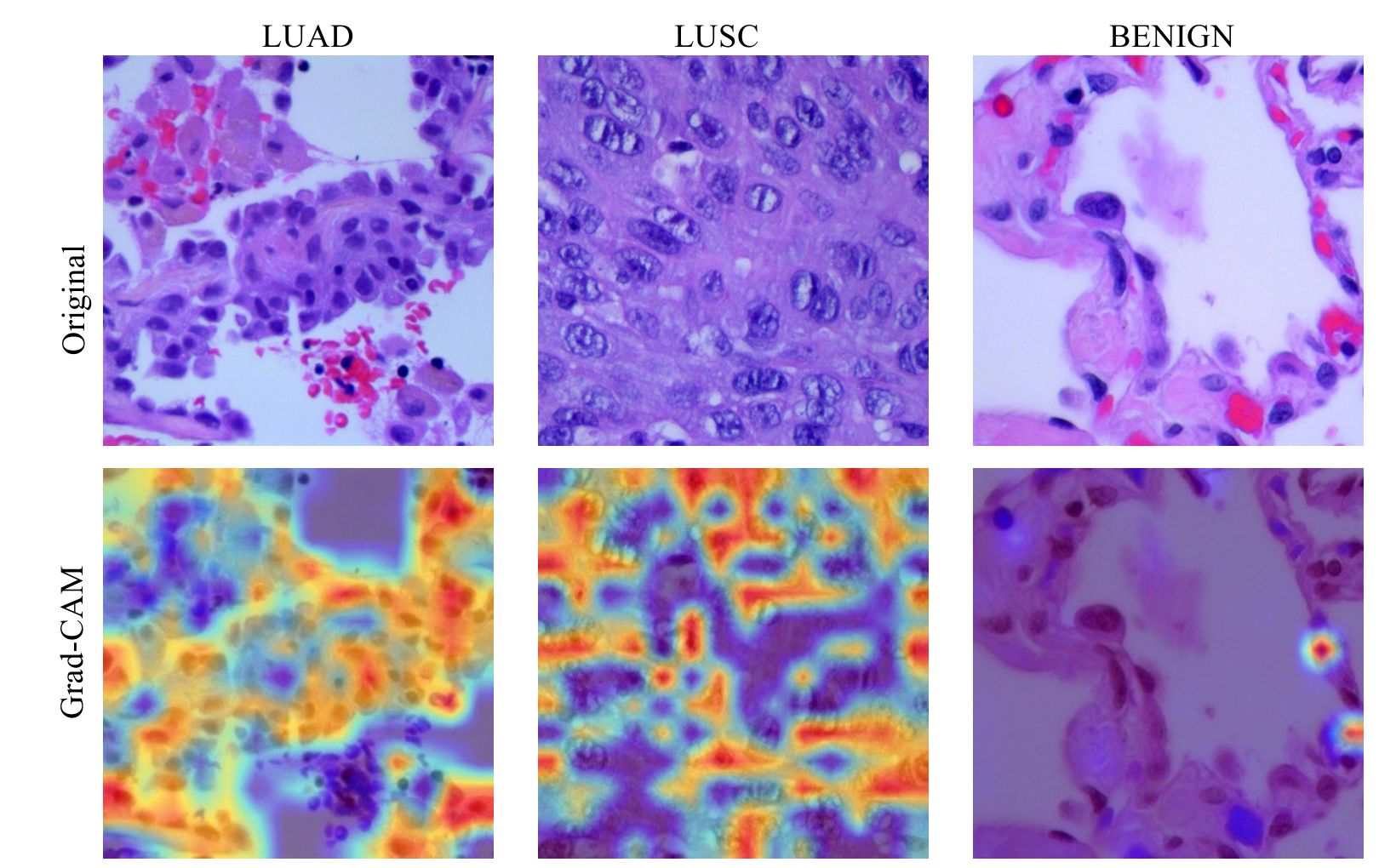
To illustrate the working mechanism behind these models, we utilized the cutting-edge model interpretation work Transformer models (Chefer et al. 2021) compared with the analysis from the human expert. Our pathology expert provides the information about which regions help her decide whether the input image is a lung cancer lesion or not from the professional perspective. The comparison between the pathology cognition and Grad-CAM visualization presents a high degree of consistency. This comparison helps us understand our deep learning model, and rationalizes our proposed method further.
2 Methods
The pre-train-finetune paradigm is the robust defacto pipeline in visual and language learning. In the finetune stage, a Multi-Layer Perceptron (MLP) (Rosenblatt 1958; Aizerman 1964) often follows a thoroughly pre-trained backbone network to function as a projector and aid transfer learning (Wang et al. 2021) on new tasks (medical image classification in our case). Our customized models utilized the paradigm above: ViT as the pre-trained backbone, followed by an MLP projector, and finally, a cross-entropy loss to classify and diagnose the cancer lesion (Figure 1).
2.1 Model Architectures
We utilize the ViT (Dosovitskiy et al. 2020) as our Transformer based backbone. The ViT backbone is pretrained on ImageNet (Deng et al. 2009), ImageNet-21k (Ridnik et al. 2021), and JFT-300M (Sun et al. 2017). ViT backbones holds accuracy of 88.05% on ImageNet (Deng et al. 2009), 94.55% on CIFAR-100 (Krizhevsky et al. 2009) and 77.63% on the VTAB (Zhai et al. 2019) suite of 19 tasks. In Figure 1, we illustrate how the ViT backbone function in our customize d model. The multiple scale input image samples (768 * 768 pixels) are first normalized to 224 * 224 pixels. This follows the input image format in ImageNet, due to ViT is pre-trained on ImageNet. For the 224 x 224 pixels input, the ViT backbone first split the input image into 196 image patches, and each patch is 16 x 16 pixels. Image patches are treated the same way as tokens (words) in natural language processing applications. The image patches are linearly embedded, with positional embeddings. ViT backbone (Dosovitskiy et al. 2020) then feeds the resulting sequence vectors into a standard Transformer encoder (Vaswani et al. 2017). The process is visually illustrated in Figure 1.
3 Experiments
In this section, we describe the dataset we used and our experiments in the Zero-Shot and Few-Shot manners.
3.1 Dataset
We utilized the Lung Cancer part of the dataset LC25000 (Borkowski et al. 2019). LC25000 has 25,000 color images in 5 classes. Each class contains 5,000 images of the histologic categories: colon adenocarcinoma, benign colonic tissue, lung adenocarcinoma, lung squamous cell carcinoma and benign lung tissue. All images are de-identified, HIPPA11 1 Health Insurance Portability and Accountability Act of 1996 (HIPAA), https://www.cdc.gov/phlp/publications/topic/hipaa.html compliant and validated. We utilize the lung cancer categories: lung adenocarcinoma, lung squamous cell carcinoma and benign lung tissue. We split the 15,000 images in lung cancer categories into training set (), validation set () and test set (), by the ratios , after random sampling.
3.2 Zero-Shot transfer learning
First, we conducted the experiments in a Zero-Shot manner. The well pre-trained ViT model functions as frozen wights in this setting. We directly use the frozen pre-trained ViT model to make the prediction on test set (). We report the accuracy on validation set and test set in Table 1.
3.3 Few-Shot transfer learning
Then we conducted the experiments in Few-Shot manners. We fine-tuned the pre-trained ViT model on training set () for 5 epochs, and validation the best model on validation set () at each epoch. We find that the pre-trained ViT has prompt and strong learning ability, so that it quick achieves the optimal accuracy after fine-tuning of only 5 epochs ( on both validation set () and test set ()). We report the accuracies on validation set and test set in Table 1.
| Part | ||
|---|---|---|
| Name | Validation set (acc) | Test set (acc) |
| Zero-Shot | ||
| Few-Shot () | ||
| Few-Shot () | ||
| Few-Shot () | ||
| Few-Shot () | ||
| Few-Shot () | ||
3.4 Receiver operating characteristic (ROC) curve
The Receiver operating characteristic (ROC) curve is used to evaluate the quality of a classifier (Green et al. 1966; Fawcett 2006). The x axis of ROC curve is the ‘false positive rate’ (FPR), and the y axis means the ‘true positive rate’ (TPR). The point or line of a classifier locates more top-left on the ROC curve, the better this classifier is. Area under the Curve of ROC (AUC ROC), tests whether positives are ranked higher than negatives. We reported the AUC values of each model (of all the three classes, LUAD, BENIGH and LUSC) on Figure 2. The AUC values of ViT model (Few-Shot ) is 1.00000000 for all the three classes (LUAD, BENIGN abd LUSC), showing that the ViT model (Few-Shot ) is an optimal classifier in our case.

4 Discussion
A lot of work endeavors to make deep learning more sensible and explainable. In various deep learning applications especially into medical imaging, it is crucial to make the deep learning model more interpretative. Selvaraju et al. 2017 have introduced a Gradient Weighted Class Activation Mapping (Grad-CAM) technique which provides the interpretative view of deep learning models. (Chefer et al. 2021) extends the research of Grad-CAM to Transformer Vaswani et al. 2017 models. Grad-CAM uses the gradients of any target concept, flowing into the final convolutional layer to produce the coarse localization map highlighting important regions in the image for predicting the concept. To illustrate the working mechanism behind these models, we utilized the cutting-edge model interpretation work Transformer models (Chefer et al. 2021) compared with the analysis from the human expert. In our application to classify the histology images, our visualizations (Figure 3) lend insights into failure modes of these models, showing that seemingly unreasonable predictions have reasonable explanations. Our visualization (Figure 3) is robust to adversarial perturbations, are more faithful to the underlying model and help achieve model generalizations by identifying dataset bias. The interpretation work of deep learning models is essential to the understanding of the mechanism behind the success of our model, and making the model more transparent. In our case, we utilize Grad-CAM to generate the visualized explanations via gradient-based localization. The localizations in Grad-CAM help explain which regions (confirmed salient features) in the image input contribute more to the model’s final decision and which regions are less (Figure 3). Furthermore, our pathology expert provides the information about which regions help him decide whether the input image is an infection or not from the professional perspective. The comparison between the pathology cognition and Grad-CAM visualization presents a high degree of consistency. This comparison is novel, helps us understand our deep learning model, and rationalizes our proposed method further.
Acknowledgments and Disclosure of Funding
We thank the computing resource support from the TPU Research Cloud. Both Guo FM and Fan Y contributed to manuscript revision, read, and approved the submitted version.
References
- Aizerman [1964] Mark A Aizerman. Theoretical foundations of the potential function method in pattern recognition learning. Automation and remote control, 25:821–837, 1964.
- Borkowski et al. [2019] Andrew A Borkowski, Marilyn M Bui, L Brannon Thomas, Catherine P Wilson, Lauren A DeLand, and Stephen M Mastorides. Lung and colon cancer histopathological image dataset (lc25000). arXiv preprint arXiv:1912.12142, 2019.
- Brown et al. [2020] Tom B Brown, Benjamin Mann, Nick Ryder, Melanie Subbiah, Jared Kaplan, Prafulla Dhariwal, Arvind Neelakantan, Pranav Shyam, Girish Sastry, Amanda Askell, et al. Language models are few-shot learners. arXiv preprint arXiv:2005.14165, 2020.
- Chefer et al. [2021] Hila Chefer, Shir Gur, and Lior Wolf. Transformer interpretability beyond attention visualization. In Proceedings of the IEEE/CVF Conference on Computer Vision and Pattern Recognition, pages 782–791, 2021.
- Dean [2022] Jeffrey Dean. A golden decade of deep learning: Computing systems & applications. Daedalus, 151(2):58–74, 2022.
- Deng et al. [2009] Jia Deng, Wei Dong, Richard Socher, Li-Jia Li, Kai Li, and Li Fei-Fei. Imagenet: A large-scale hierarchical image database. In 2009 IEEE conference on computer vision and pattern recognition, pages 248–255. Ieee, 2009.
- Devlin et al. [2019] Jacob Devlin, Ming-Wei Chang, Kenton Lee, and Kristina Toutanova. Bert: Pre-training of deep bidirectional transformers for language understanding. In Proceedings of the 2019 Conference of the North American Chapter of the Association for Computational Linguistics: Human Language Technologies, Volume 1 (Long and Short Papers), pages 4171–4186, 2019.
- Dosovitskiy et al. [2020] Alexey Dosovitskiy, Lucas Beyer, Alexander Kolesnikov, Dirk Weissenborn, Xiaohua Zhai, Thomas Unterthiner, Mostafa Dehghani, Matthias Minderer, Georg Heigold, Sylvain Gelly, et al. An image is worth 16x16 words: Transformers for image recognition at scale. arXiv preprint arXiv:2010.11929, 2020.
- Fawcett [2006] Tom Fawcett. An introduction to roc analysis. Pattern recognition letters, 27(8):861–874, 2006.
- Fuchs and Buhmann [2011] Thomas J Fuchs and Joachim M Buhmann. Computational pathology: challenges and promises for tissue analysis. Computerized Medical Imaging and Graphics, 35(7-8):515–530, 2011.
- Green et al. [1966] David Marvin Green, John A Swets, et al. Signal detection theory and psychophysics, volume 1. Wiley New York, 1966.
- Gridelli et al. [2015] Cesare Gridelli, Antonio Rossi, David P Carbone, Juliana Guarize, Niki Karachaliou, Tony Mok, Francesco Petrella, Lorenzo Spaggiari, and Rafael Rosell. Non-small-cell lung cancer. Nature reviews Disease primers, 1(1):1–16, 2015.
- Herbst et al. [2018] Roy S Herbst, Daniel Morgensztern, and Chris Boshoff. The biology and management of non-small cell lung cancer. Nature, 553(7689):446–454, 2018.
- Krizhevsky et al. [2009] Alex Krizhevsky, Geoffrey Hinton, et al. Learning multiple layers of features from tiny images. 2009.
- Langer et al. [2010] Corey J Langer, Benjamin Besse, Antonio Gualberto, Elizabeth Brambilla, and Jean-Charles Soria. The evolving role of histology in the management of advanced non–small-cell lung cancer. Journal of clinical oncology, 28(36):5311–5320, 2010.
- Larochelle et al. [2008] Hugo Larochelle, Dumitru Erhan, and Yoshua Bengio. Zero-data learning of new tasks. In AAAI, volume 1, page 3, 2008.
- LeCun et al. [1999] Yann LeCun, Patrick Haffner, Léon Bottou, and Yoshua Bengio. Object recognition with gradient-based learning. In Shape, contour and grouping in computer vision, pages 319–345. Springer, 1999.
- LeCun et al. [2015] Yann LeCun, Yoshua Bengio, and Geoffrey Hinton. Deep learning. nature, 521(7553):436–444, 2015.
- Li and Liang [2021] Xiang Lisa Li and Percy Liang. Prefix-tuning: Optimizing continuous prompts for generation. In Proceedings of the 59th Annual Meeting of the Association for Computational Linguistics and the 11th International Joint Conference on Natural Language Processing (Volume 1: Long Papers), pages 4582–4597, Online, August 2021. Association for Computational Linguistics. doi: 10.18653/v1/2021.acl-long.353.
- Mendelsohn et al. [1965] Mortimer L Mendelsohn, Wilfred A Kolman, Benson Perry, and Judith MS Prewitt. Computer analysis of cell images. Postgraduate Medicine, 38(5):567–573, 1965.
- Mengoli et al. [2018] MC Mengoli, FR Longo, F Fraggetta, A Cavazza, A Dubini, G Ali, F Guddo, E Gilioli, G Bogina, N Nannini, et al. The 2015 world health organization classification of lung tumors: new entities since the 2004 classification. Pathologica-Journal of the Italian Society of Anatomic Pathology and Diagnostic Cytopathology, 110(1):39–67, 2018.
- Nasim et al. [2019] F. Nasim, B. F. Sabath, and G. A. Eapen. Lung Cancer. Med Clin North Am, 103(3):463–473, May 2019.
- Ridnik et al. [2021] Tal Ridnik, Emanuel Ben-Baruch, Asaf Noy, and Lihi Zelnik-Manor. Imagenet-21k pretraining for the masses. In Thirty-fifth Conference on Neural Information Processing Systems Datasets and Benchmarks Track (Round 1), 2021. URL https://openreview.net/forum?id=Zkj_VcZ6ol.
- Rosenblatt [1958] Frank Rosenblatt. The perceptron: a probabilistic model for information storage and organization in the brain. Psychological review, 65(6):386, 1958.
- Selvaraju et al. [2017] Ramprasaath R Selvaraju, Michael Cogswell, Abhishek Das, Ramakrishna Vedantam, Devi Parikh, and Dhruv Batra. Grad-cam: Visual explanations from deep networks via gradient-based localization. In Proceedings of the IEEE international conference on computer vision, pages 618–626, 2017.
- Socher et al. [2013] Richard Socher, Milind Ganjoo, Christopher D Manning, and Andrew Ng. Zero-shot learning through cross-modal transfer. Advances in neural information processing systems, 26, 2013.
- Sun et al. [2017] Chen Sun, Abhinav Shrivastava, Saurabh Singh, and Abhinav Gupta. Revisiting unreasonable effectiveness of data in deep learning era. In Proceedings of the IEEE international conference on computer vision, pages 843–852, 2017.
- Travis et al. [2015] William D Travis, Elisabeth Brambilla, Andrew G Nicholson, Yasushi Yatabe, John HM Austin, Mary Beth Beasley, Lucian R Chirieac, Sanja Dacic, Edwina Duhig, Douglas B Flieder, et al. The 2015 world health organization classification of lung tumors: impact of genetic, clinical and radiologic advances since the 2004 classification. Journal of thoracic oncology, 10(9):1243–1260, 2015.
- Vaswani et al. [2017] Ashish Vaswani, Noam Shazeer, Niki Parmar, Jakob Uszkoreit, Llion Jones, Aidan N Gomez, Lukasz Kaiser, and Illia Polosukhin. Attention is all you need. In NIPS, 2017.
- Wang et al. [2021] Yizhou Wang, Shixiang Tang, Feng Zhu, Lei Bai, Rui Zhao, Donglian Qi, and Wanli Ouyang. Revisiting the transferability of supervised pretraining: an mlp perspective. arXiv preprint arXiv:2112.00496, 2021.
- Wei et al. [2021] Jason Wei, Maarten Bosma, Vincent Y Zhao, Kelvin Guu, Adams Wei Yu, Brian Lester, Nan Du, Andrew M Dai, and Quoc V Le. Finetuned language models are zero-shot learners. arXiv preprint arXiv:2109.01652, 2021.
- Zhai et al. [2019] Xiaohua Zhai, Joan Puigcerver, Alexander Kolesnikov, Pierre Ruyssen, Carlos Riquelme, Mario Lucic, Josip Djolonga, Andre Susano Pinto, Maxim Neumann, Alexey Dosovitskiy, et al. The visual task adaptation benchmark. 2019.
- Zwanenburg et al. [2020] Alex Zwanenburg, Martin Vallières, Mahmoud A Abdalah, Hugo JWL Aerts, Vincent Andrearczyk, Aditya Apte, Saeed Ashrafinia, Spyridon Bakas, Roelof J Beukinga, Ronald Boellaard, et al. The image biomarker standardization initiative: standardized quantitative radiomics for high-throughput image-based phenotyping. Radiology, 295(2):328–338, 2020.