Federated Learning Enables Big Data for Rare Cancer Boundary Detection
Abstract
Although machine learning (ML) has shown promise in numerous domains, there are concerns about generalizability to out-of-sample data. This is currently addressed by centrally sharing ample, and importantly diverse, data from multiple sites. However, such centralization is challenging to scale (or even not feasible) due to various limitations. Federated ML (FL) provides an alternative to train accurate and generalizable ML models, by only sharing numerical model updates. Here we present findings from the largest FL study to-date, involving data from healthcare institutions across continents, to generate an automatic tumor boundary detector for the rare disease of glioblastoma, utilizing the largest dataset of such patients ever used in the literature ( MRI scans from patients). We demonstrate a improvement over a publicly trained model to delineate the surgically targetable tumor, and improvement over the tumor’s entire extent. We anticipate our study to: 1) enable more studies in healthcare informed by large and diverse data, ensuring meaningful results for rare diseases and underrepresented populations, 2) facilitate further quantitative analyses for glioblastoma via performance optimization of our consensus model for eventual public release, and 3) demonstrate the effectiveness of FL at such scale and task complexity as a paradigm shift for multi-site collaborations, alleviating the need for data sharing.
Keywords:
federated learning, deep learning, convolutional neural network, segmentation, brain tumor, glioma, glioblastoma, FeTS, BraTSAdvances in machine learning (ML), and particularly deep learning (DL), have shown promise in addressing complex healthcare problems [1, 2, 3, 4, 5, 6, 7, 8, 9, 10, 11, 12, 13, 14]. However, there are concerns about generalizability on data from sources that did not participate in model training, i.e., “out-of-sample” data [15, 16]. Literature indicates that training robust and accurate models requires large amounts of data [17, 18, 19], the diversity of which affects model generalizability to “out-of-sample” cases [20]. To address these concerns, models need to be trained on data originating from numerous sites representing diverse population samples. The current paradigm for such multi-site collaborations is “centralized learning” (CL), in which data from different sites are shared to a centralized location following inter-site agreements [20, 21, 22, 23]. However, such data centralization is difficult to scale (and might not even be feasible), especially at a global scale, due to concerns [24, 25] relating to privacy, data-ownership, intellectual property, technical challenges (e.g., network and storage limitations), as well as compliance with varying regulatory policies (e.g., Health Insurance Portability and Accountability Act (HIPAA) of the United States [26] and the General Data Protection Regulation (GDPR) of the European Union [27]). In contrast to this centralized paradigm, “federated learning” (FL) describes an approach where models are trained by only sharing model parameter updates from decentralized data (i.e., each site retains its data locally) [28, 29, 25, 24, 30], without sacrificing performance when compared to CL-trained models [31, 32, 29, 25, 33, 34, 35]. Thus, FL can offer an alternative to CL, potentially creating a paradigm shift that alleviates the need for data sharing, and hence increase access to geographically-distinct collaborators, thereby increasing the size and diversity of data used to train ML models.
FL has tremendous potential in healthcare [36, 37], particularly towards addressing health disparities, under-served populations, and “rare” diseases [38], by enabling ML models to gain knowledge from ample and diverse data that would otherwise not be available. With that in mind, here we focus on the “rare” disease of glioblastoma, and particularly on the detection of its extent using multi-parametric magnetic resonance imaging (mpMRI) scans [39]. While glioblastoma is the most common malignant primary brain tumor [40, 41, 42], it is still classified as a “rare” disease, as its incidence rate (i.e., / people) is substantially lower than the rare disease definition rate (i.e., / people) [38]. This means that single sites cannot collect large and diverse datasets to train robust and generalizable ML models, and necessitates collaboration between geographically distinct sites. Despite extensive efforts to improve prognosis of glioblastoma patients with intense multimodal therapy, their median overall survival is only months after standard-of-care treatment, and months without treatment [43]. Although the subtyping of glioblastoma has been improved [44] and the standard-of-care treatment options have expanded during the last twenty years, there have been no substantial improvements in overall survival [45]. This reflects the major obstacle in treating these tumors that is their intrinsic heterogeneity [40, 42], and the need for analyses of larger and more diverse data towards better understanding the disease. In terms of radiologic appearance, glioblastomas comprise main sub-compartments, defined as i) the “enhancing tumor” (ET), representing the vascular blood-brain barrier breakdown within the tumor, ii) the “tumor core” (TC), which includes the ET and the necrotic (NCR) part, and represents the surgically relevant part of the tumor, and iii) the “whole tumor” (WT), which is defined by the union of the TC and the peritumoral edematous/infiltrated tissue (ED), and represents the complete tumor extent relevant to radiotherapy (Fig. 2.b). Detecting these sub-compartment boundaries therefore defines a multi-parametric multi-class learning problem [46, 47, 48, 49, 50], and is a critical first step towards further quantifying and assessing this heterogeneous rare disease and ultimately influencing clinical decision-making.
a

b

c

d

e
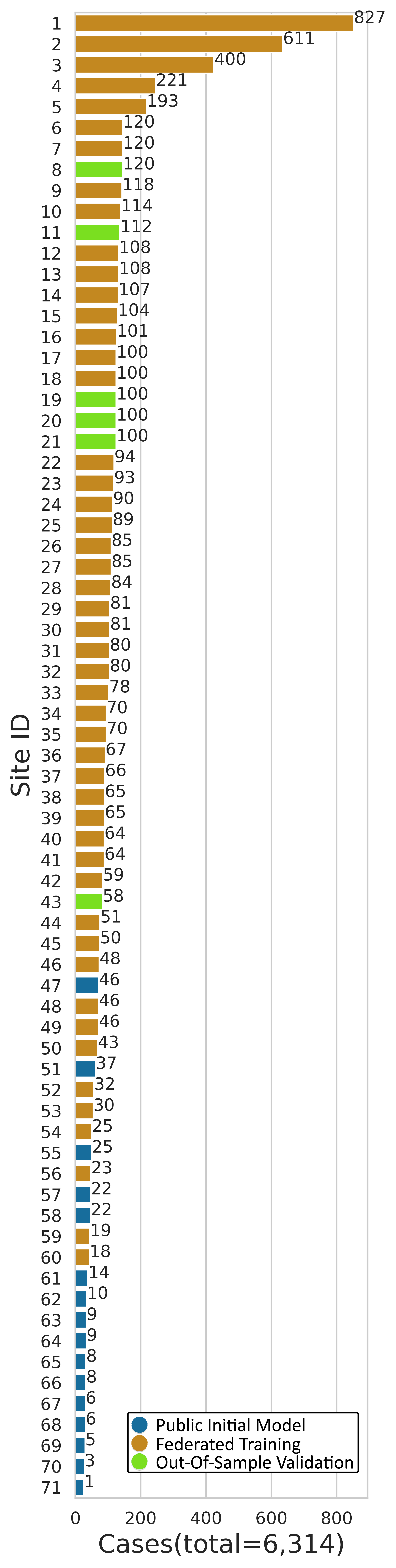
Co-authors in this study have previously introduced FL in healthcare in a simulated setting [29] and evaluated different FL approaches [25] for the same use case as the present study, i.e., detecting the boundaries of glioblastoma sub-compartments. Findings from these studies supported the superiority of the FL approach used in the present study (i.e., based on an aggregation server [28, 24]), which had almost identical performance to CL. The present study describes the largest to-date global FL effort to develop an accurate and generalizable ML model for detecting glioblastoma sub-compartment boundaries, based on MRI scans (over 5TB) of glioblastoma patients from geographically distinct sites, across continents (Fig. 2.a). Notably, this describes the largest and most diverse dataset of glioblastoma patients ever considered in the literature. It was the use of FL that successfully enabled our ML model to gain knowledge from such an unprecedented dataset. The extended global footprint and the task complexity is what sets this study apart from current literature, since it dealt with a multi-parametric multi-class problem with reference standards that require expert clinicians following an involved manual annotation protocol, rather than recording a categorical entry from medical records [51, 30]. Moreover, varying characteristics of the mpMRI data due to scanner hardware and acquisition protocol differences [52, 53] were handled at each collaborating site via established harmonized preprocessing pipelines [54, 55, 56, 57].
The scientific contributions of this manuscript can be summarized by i) the insights garnered during this work that can pave the way for more successful FL studies of increased scale and task complexity, ii) making a potential impact for the treatment of the rare disease of glioblastoma, by eventually publicly releasing an optimized trained consensus model for use in resource-constrained clinical settings, and iii) demonstrating the effectiveness of FL at such scale and task complexity as a paradigm shift redefining multi-site collaborations, while alleviating the need for data sharing.
Methods
Data
The data considered in this study described patient populations with adult-type diffuse glioma [44], and specifically displaying the radiological features of glioblastoma, scanned with mpMRI to characterize the anatomical tissue structure [39]. Each case is specifically described by i) native T1-weighted (T1), ii) Gadolinium-enhanced T1-weighted (T1Gd), iii) T2-weighted (T2), and iv) T2-weighted-Fluid-Attenuated-Inversion-Recovery (T2-FLAIR) MRI scans. Cases with any of these sequences missing were not included in the study. Note that no inclusion/exclusion criterion applied relating to the type of acquisition (i.e., both 2D axial and 3D acquisitions were included, with a preference for 3D if available), or the exact type of sequence (e.g., MP-RAGE vs SPGR). The only exclusion criterion was for T1-FLAIR scans that were intentionally excluded to avoid mixing varying tissue appearance due to the type of sequence, across native T1-weighted scans.
The eligibility of collaborating sites to participate in the federation was determined based on data availability, and approval by their respective institutional review board. sites participated as independent collaborators in the study defining a dataset of cases. The MRI scanners used for data acquisition were from multiple vendors (i.e., Siemens, GE, Philips, Hitachi, Toshiba), with magnetic field strength ranging from 1T to 3T. The data from all collaborating sites followed a male:female ratio of with age ranging between and years.
From all collaborating sites, were chosen to be part of the training phase, and sites were categorized as “out-of-sample”, i.e., none of these were part of the training stage. These specific out-of-sample sites (Site IDs: 8, 11, 19, 20, 21, 43) were allocated based on their availability, i.e., they have indicated an expected delayed participation rendering them optimal for model generalizability validation. One of these out-of-sample sites (Site 11) contributed aggregated a priori data from a multi-site randomized clinical trial for newly diagnosed glioblastoma (ClinicalTrials.gov Identifier: NCT00884741, RTOG0825 [58, 59], ACRIN6686 [60, 61]), with inherent diversity benefiting the intended generalizability validation purpose. The American College of Radiology (ACR - Site 11) serves as the custodian of this trial’s imaging data on behalf of ECOG-ACRIN, who made the data available for this study. Following screening for availability of the required mpMRI scans with sufficient signal-to-noise ratio judged by visual observation, a subset of cases from the original trial data were included in this study. The out-of-sample data totaled cases intentionally held-out of the federation, with the intention of validating the consensus model in completely unseen cases. To facilitate further such generalizability evaluation without burdening the collaborating sites, a subset consisting of cases (including the multi-site clinical data provided by ACR) from this out-of-sample data was aggregated, to serve as the “centralized out-of-sample” dataset. Furthermore, the sites participating in the training phase define a collective dataset of cases. The exact site IDs are: 1, 2, 3, 4, 5, 6, 7, 9, 10, 12, 13, 14, 15, 16, 17, 18, 22, 23, 24, 25, 26, 27, 28, 29, 30, 31, 32, 33, 34, 35, 36, 37, 38, 39, 40, 41, 42, 44, 45, 46, 48, 49, 50, 52, 53, 54, 56, 59, 60. These cases were automatically split at each site following a ratio between cases for training and local validation. During the federated training phase the data used for the public initial model were also included as a dataset from a separate node, such that the contribution of sites providing the publicly available data is not forgotten within the global consensus model. This results in the final consensus model being developed based on data from sites over a total dataset of cases.
Harmonized Data Preprocessing
Once each collaborating site identified their local data, they were asked to use the preprocessing functionality of the software platform we provided. This functionality follows the harmonized data preprocessing protocol defined by the BraTS challenge [54, 55, 56, 57], to account for inter-site acquisition protocol variations, e.g., 3D vs 2D axial plane acquisitions.
The neural network architecture
The trained model to delineate the different tumor sub-compartments was based on the popular 3D U-Net with residual connections (3D-ResUNet) [62, 63, 64, 65, 66]. The network had base filters, with a learning rate of optimized using the Adam optimizer [67]. For the loss function used in training, we used the generalized score [68, 69] (represented mathematically in Eq. 1) on the absolute complement of each tumor sub-compartment independently. Such mirrored loss has been shown to capture variations in smaller regions better [70]. No penalties were used in the loss function, due to our use of ‘mirrored’ loss [71, 72, 73]. The final layer of the model was a sigmoid layer, providing three channel outputs for each voxel in the input volume, one output channel per tumor sub-compartment. While the generalized score was calculated using a binarized version of the output (check sigmoid value against the threshold ) for the final prediction, we used the floating point [74] during the training process.
The Federation
The collaborative network of the present study spans continents (Fig. 2), with data from geographically distinct sites. The training process was initiated when each collaborator securely connected to a central aggregation server, which resided behind a firewall at the University of Pennsylvania. As soon as the secure connection was established, the public initial model was passed to the collaborating site. Using FL based on an aggregation server, collaborating sites then trained the same network architecture on their local data for one epoch, and shared model updates with the central aggregation server. The central aggregation server received model updates from all collaborators, combined them (by averaging model parameters) and sent the consensus model back to each collaborator to continue their local training. Each such iteration is called a “federated round”. After not observing any meaningful changes since round , we stopped the training after a total of federated rounds. Additionally, we performed all operations on the aggregator on secure hardware (by leveraging trusted execution environments [77]), in order to increase the trust by all parties in the confidentiality of the model updates being computed and shared, as well as to increase the confidence in the integrity of the computations being performed [78].
The federated training was initialized using a “public initial model” trained on cases from sites. This was done as opposed to a random initialization point to facilitate faster convergence of the model performance [79, 80]. After the training process was complete, the “final consensus model” was obtained after model selection from all the global consensus models obtained for each federated round. Thus, the final consensus model was developed on cases from sites. To quantitatively evaluate the performance of the trained models, of the total cases contributed by each participating site were excluded from the model training process and were used as “local validation data”. To further evaluate the generalizability of the models in unseen data, sites were not involved in any of the training stages to represent an unseen “out-of-sample” data population of cases. To facilitate further evaluation without burdening the collaborating sites, a subset () of these cases was aggregated to serve as a “centralized out-of-sample” dataset. Model performance was quantitatively evaluated here using the Dice Similarity Coefficient (), which assesses the spatial agreement between the model’s prediction and the reference standard for each of the tumor sub-compartments (ET, TC, WT).
Model Run-time in Clinical Environments
Clinical environments typically have constrained computational resources, such as the availability of specialized hardware (e.g., DL acceleration cards) and increased memory, which affect the run-time performance of DL inference workloads. Thus, taking into consideration the potential deployment of the final consensus model in such low-resource settings, we decided to proceed with a single 3D-ResUNet, rather than an ensemble of multiple models. This decision ensured a reduced computational burden, when compared with running multiple models, which is typically done in academic research projects [54, 55, 56, 57].
To further facilitate use in low-resource environments, we plan to publicly release run-time optimized [81] version of the final consensus model. For this model, graph level optimizations (i.e., operators fusion) were initially applied, followed by optimizations for low precision inference, i.e., converting the floating point single precision model to a fixed precision 8-bit integer model (a process known as “quantization” [82]). In particular, we used accuracy-aware quantization [83], where model layers were iteratively scaled to a lower precision format. These optimizations yielded several run-time performance benefits, such as lower inference latency (a platform-dependent average speedup and reduced memory requirement when compared with the original consensus model), and higher throughput (equal to the speedup improvement since the batch size used is equal to ), while the trade-off was an insignificant () drop in the average .
Code availability
Motivated by findability, accessibility, interoperability, and reusability (FAIR) criteria in scientific research [84], all the code used to design the Federated Tumor Segmentation (FeTS) platform for this study is available at https://github.com/FETS-AI/Front-End. The functionality related to preprocessing (i.e., DICOM to NIfTI conversion, population-based harmonized preprocessing, co-registration) and manual refinements of annotation is derived from the open-source Cancer Imaging Phenomics Toolkit (CaPTk, https://github.com/CBICA/CaPTk) [85, 86, 87]. The co-registration is performed using the Greedy framework [88], available via CaPTk [85, 86, 87], ITK-SNAP [89], and the FeTS tool. The brain extraction [90] is done using the BrainMaGe method [91], and is available in https://github.com/CBICA/BrainMaGe and via the Generally Nuanced Deep Learning Framework (GaNDLF) [92] at https://github.com/CBICA/GaNDLF. To generate automated annotations, DeepMedic’s [93] integration with CaPTk was used, and we used the model weights and inference mechanism provided by the other algorithm developers (DeepScan [94] and nnU-Net [70] (https://github.com/MIC-DKFZ/nnunet)). DeepMedic’s original implementation is available in https://github.com/deepmedic/deepmedic, whereas the one we used in this study can be found at https://github.com/CBICA/deepmedic. The fusion of the labels was done using the Label Fusion tool [95] available at https://github.com/FETS-AI/LabelFusion. The data loading pipeline and network architecture was developed using the GaNDLF framework [92] by using PyTorch [96]. The data augmentation was done via GaNDLF by leveraging TorchIO [97]. The FL backend developed for this project has been open-sourced as a separate software library, to encourage further research on FL [98] and is available at https://github.com/intel/openfl. The optimization of the consensus model inference workload was performed via OpenVINO [99] (https://github.com/openvinotoolkit/openvino/tree/2021.4.1), which is an open-source toolkit enabling acceleration of neural network models through various optimization techniques. The optimizations were evaluated on an Intel Core® i7-1185G7E CPU @ 2.80GHz with GB DDR4 3200MHz memory on Ubuntu 18.04.6 OS and Linux kernel version 5.9.0-050900-generic.
Results
At the time of initialization of the federation, the public initial model was evaluated against the local validation data of all sites, resulting in an average (across all cases of all sites) per sub-compartment, of: , , . To summarize the model performance with a single collective score, we then calculate the average (across all tumor sub-compartments per case, and then across all cases of all sites) as equal to .
Following model training across all sites, the final consensus model garnered significant performance improvements against the collaborators’ local validation data of (), (), and (), for ET, TC, and WT, respectively (Fig. 2.c).
To further evaluate the potential generalizability improvements of the final consensus model on unseen data, we compared it with the public initial model against the complete out-of-sample data, and noted significant performance improvements of (), % (), and (), for ET, TC, and WT, respectively (Fig. 2.d).
Notably, the only difference between the public initial model and the final consensus model, was that the latter gained knowledge during training from increased datasets contributed by the complete set of collaborators. The conclusion of these findings reinforces the importance of using large and diverse data for generalizable models to ultimately drive patient care.
Discussion
In this study, we have described the largest real-world FL effort to-date utilizing data of glioblastoma patients from geographically unique sites spread across continents, to develop an accurate and generalizable ML model for detecting glioblastoma sub-compartment boundaries. Notably, this extensive global footprint of the collaborating sites in this study, also yield the largest dataset ever reported in the literature assessing this rare disease. It is the use of FL that successfully enabled i) access to such an unprecedented dataset of the most common and fatal adult brain tumor, and ii) meaningful ML training to ensure generalizability of models across out-of-sample data. Since glioblastoma boundary detection is critical for treatment planning and the requisite first step for further quantitative analyses, the models generated during this study have the potential to make far-reaching clinical impact.
The large and diverse data that FL enabled, led to the final consensus model garnering significant performance improvements over the public initial model against both the collaborators’ local validation data and the complete out-of-sample data. The improved result is a clear indication of the benefit that can be afforded through access to more data. In comparison with the limited existing real-world FL studies [51, 30], our use case is larger in scale and substantially more complex, since it 1) addresses a multi-parametric multi-class problem, with reference standards that require expert collaborating clinicians to follow an involved manual annotation protocol, rather than simply recording a categorical entry from medical records, and 2) requires the data to be preprocessed in a harmonized manner to account for differences in MRI acquisition.
We have demonstrated the utility of an FL approach to develop an accurate and generalizable ML model for detecting glioblastoma sub-compartment boundaries, a finding which is of particular relevance for neurosurgical and radiotherapy planning in patients with this disease. This study is meant to be used as a blueprint for future FL studies that result in clinically deployable ML models. Building on this study, a continuous FL consortium would enable downstream quantitative analyses with implications for both routine practice and clinical trials, and most importantly, increase access to high-quality precision care worldwide. Furthermore, the lessons learned from this study with such a global footprint are invaluable and can be applied to a broad array of clinical scenarios with the potential for great impact to rare diseases and underrepresented populations.
Acknowledgments
Research and main methodological developments reported in this publication were partly supported by the National Institutes of Health (NIH) under award numbers NIH/NCI:U01CA242871 (S.Bakas), NIH/NINDS:R01NS042645 (C.Davatzikos), NIH/NCI:U24CA189523 (C.Davatzikos), NIH/NCI:U24CA215109 (J.Saltz), NIH/NCI:U01CA248226 (P.Tiwari), NIH/NCI:P30CA51008 (Y.Gusev), NIH:R50CA211270 (M.Muzi), NIH/NCATS:UL1TR001433 (Y.Yuan), NIH/NIBIB:R21EB030209 (Y.Yuan), NIH/NCI:R37CA214955 (A.Rao), and NIH:R01CA233888 (A.L.Simpson). The authors would also like to acknowledge the following NIH funded awards for the multi-site clinical trial (NCT00884741, RTOG0825/ACRIN6686): U10CA21661, U10CA37422, U10CA180820, U10CA180794, U01CA176110, R01CA082500, CA079778, CA080098, CA180794, CA180820, CA180822, CA180868. Research reported in this publication was also partly supported by the National Science Foundation, under award numbers 2040532 (S.Baek), and 2040462 (B.Landman). Research reported in this publication was also supported by i) a research grant from Varian Medical Systems (Palo Alto, CA USA) (Y.Yuan), ii) the Ministry of Health of the Czech Republic (Grant Nr. NU21-08-00359) (M.Keřkovský and M.Kozubek), iii) Deutsche Forschungsgemeinschaft (DFG, German Research Foundation) Project-ID 404521405, SFB 1389, Work Package C02, and Priority Programme 2177 “Radiomics: Next Generation of Biomedical Imaging” (KI 2410/1-1 — MA 6340/18-1) (P.Vollmuth), iv) DFG Project-ID B12, SFB 824 (B.Wiestler), v) the Helmholtz Association (funding number ZT-I-OO1 4) (K.Maier-Hein), vi) the Dutch Cancer Society (KWF project number EMCR 2015-7859) (S.R.van der Voort), vii) the Chilean National Agency for Research and Development (ANID-Basal FB0008 (AC3E) and FB210017 (CENIA)) (P.Guevara), viii) the Canada CIFAR AI Chairs Program (M.Vallières), ix) Leeds Hospital Charity (Ref: 9RO1/1403) (S.Currie), x) the Cancer Research UK funding for the Leeds Radiotherapy Research Centre of Excellence (RadNet) and the grant number C19942/A28832 (S.Currie), xi) Medical Research Council (MRC) Doctoral Training Programme in Precision Medicine (Award Reference No. 2096671) (J.Bernal), xii) The European Research Council (ERC) under the European Union’s Horizon 2020 research and innovation programme (Grant Agreement No. 757173) (B.Glocker), xiii) The UKRI London Medical Imaging & Artificial Intelligence Centre for Value Based Healthcare (K.Kamnitsas), xiv) Wellcome/Engineering and Physical Sciences Research Council (EPSRC) Center for Medical Engineering (WT 203148/Z/16/Z) (T.C.Booth), xv) American Cancer Society Research Scholar Grant RSG-16-005-01 (A.Rao), xvi) the Department of Defense (DOD) Peer Reviewed Cancer Research Program (PRCRP) W81XWH-18-1-0404, Dana Foundation David Mahoney Neuroimaging Program, the V Foundation Translational Research Award, Johnson & Johnson WiSTEM2D Award (P.Tiwari), xvii) RSNA Research & Education Foundation under grant number RR2011 (E.Calabrese), xviii) the National Research Fund of Luxembourg (FNR) (grant number: C20/BM/14646004/GLASS-LUX/Niclou) (S.P.Niclou), xix) EU Marie Curie FP7-PEOPLE-2012-ITN project TRANSACT (PITN-GA-2012-316679) and the Swiss National Science Foundation (project number 140958) (J.Slotboom), and xx) CNPq 303808/2018-7 and FAPESP 2014/12236-1 (A.Xavier Falcão). The content of this publication is solely the responsibility of the authors and does not represent the official views of the NIH, the NSF, the RSNA R&E Foundation, or any of the additional funding bodies.
Author Contributions
Study Conception: S.Pati, U.Baid, B.Edwards, M.Sheller, G.A.Reina, J.Martin, S.Bakas.
Development of software used in the study: S.Pati, B.Edwards, M.Sheller, S.Wang, G.A.Reina, P.Foley, A.Gruzdev, D.Karkada.
Data Acquisition: M.Bilello, S.Mohan, E.Calabrese, J.Rudie, J.Saini, R.Y.Huang, K.Chang, T.So, P.Heng, T.F.Cloughesy, C.Raymond, T.Oughourlian, A.Hagiwara, C.Wang, M.To, M.Keřkovský, T.Kopřivová, M.Dostál, V.Vybíhal, J.A.Maldjian, M.C.Pinho, D.Reddy, J.Holcomb, B.Wiestler, M.Metz, R.Jain, M.Lee, P.Tiwari, R.Verma, Y.Gusev, K.Bhuvaneshwar, C.Bencheqroun, A.Belouali, A.Abayazeed, A.Abbassy, S.Gamal, M.Qayati, M.Mekhaimar, M.Reyes, R.R.Colen, M.Ak, P.Vollmuth, G.Brugnara, F.Sahm, M.Bendszus, W.Wick, A.Mahajan, C.Balaña Quintero, J.Capellades, J.Puig, Y.Choi, M.Muzi, H.F.Shaykh, A.Herrera-Trujillo, W.Escobar, A.Abello, P.LaMontagne, B.Landman, K.Ramadass, K.Xu, S.Chotai, L.B.Chambless, A.Mistry, R.C.Thompson, J.Bapuraj, N.Wang, S.R.van der Voort, F.Incekara, M.M.J.Wijnenga, R.Gahrmann, J.W.Schouten, H.J.Dubbink, A.J.P.E.Vincent, M.J.van den Bent, H.I.Sair, C.K.Jones, A.Venkataraman, J.Garrett, M.Larson, B.Menze, T.Weiss, M.Weller, A.Bink, B.Pouymayou, Y.Yuan, S.Sharma, T.Tseng, B.C.A.Teixeira, F.Sprenger, S.P.Niclou, O.Keunen, L.V.M.Dixon, M.Williams, R.G.H.Beets-Tan, H.Franco-Maldonado, F.Loayza, J.Slotboom, P.Radojewski, R.Meier, R.Wiest, J.Trenkler, J.Pichler, G.Necker, S.Meckel, E.Torche, F.Vera, E.Lóópez, Y.Kim, H.Ismael, B.Allen, J.M.Buatti, J.Park, P.Zampakis, V.Panagiotopoulos, P.Tsiganos, E.Challiasos, D.M.Kardamakis, P.Prasanna, K.M.Mani, D.Payne, T.Kurc, L.Poisson, M.Vallières, D.Fortin, M.Lepage, F.Morón, J.Mandel, C.Badve, A.E.Sloan, J.S.Barnholtz-Sloan, K.Waite, G.Shukla, S.Liem, G.S.Alexandre, J.Lombardo, J.D.Palmer, A.E.Flanders, A.P.Dicker, G.Ogbole, M.Soneye, D.Oyekunle, O.Odafe-Oyibotha, B.Osobu, M.Shu’aibu, F.Dako, A.Dorcas, D.Murcia, R.Haas, J.Thompson, D.R.Ormond, S.Currie, K.Fatania, R.Frood, J.Mitchell, J.Farinhas, A.L.Simpson, J.J.Peoples, R.Hu, D.Cutler, F.Y.Moraes, A.Tran, M.Hamghalam, M.A.Boss, J.Gimpel, B.Bialecki, A.Chelliah.
Data Processing: C.Sako, S.Ghodasara, E.Calabrese, J.Rudie, M.Jadhav, U.Pandey, R.Y.Huang, M.Jiang, C.Chen, C.Raymond, S.Bhardwaj, C.Chong, M.Agzarian, M.Kozubek, F.Lux, J.Michálek, P.Matula, C.Bangalore Yogananda, D.Reddy, B.C.Wagner, I.Ezhov, M.Lee, Y.W.Lui, R.Verma, R.Bareja, I.Yadav, J.Chen, N.Kumar, K.Bhuvaneshwar, A.Sayah, C.Bencheqroun, K.Kolodziej, M.Hill, M.Reyes, L.Pei, M.Ak, A.Kotrotsou, P.Vollmuth, G.Brugnara, C.J.Preetha, M.Zenk, J.Puig, M.Muzi, H.F.Shaykh, A.Abello, J.Bernal, J.Gómez, P.LaMontagne, K.Ramadass, S.Chotai, N.Wang, M.Smits, S.R.van der Voort, A.Alafandi, F.Incekara, M.M.J.Wijnenga, G.Kapsas, R.Gahrmann, A.J.P.E.Vincent, P.J.French, S.Klein, H.I.Sair, C.K.Jones, J.Garrett, H.Li, F.Kofler, Y.Yuan, S.Adabi, A.Xavier Falcão, S.B.Martins, D.Menotti, D.R.Lucio, O.Keunen, A.Hau, K.Kamnitsas, L.Dixon, S.Benson, E.Pelaez, H.Franco-Maldonado, F.Loayza, S.Quevedo, R.McKinley, J.Trenkler, A.Haunschmidt, C.Mendoza, E.Ríos, J.Choi, S.Baek, J.Yun, P.Zampakis, V.Panagiotopoulos, P.Tsiganos, E.I.Zacharaki, C.Kalogeropoulou, P.Prasanna, S.Shreshtra, T.Kurc, B.Luo, N.Wen, M.Vallières, D.Fortin, F.Morón, C.Badve, V.Vadmal, G.Shukla, G.Ogbole, D.Oyekunle, F.Dako, D.Murcia, E.Fu, S.Currie, R.Frood, M.A.Vogelbaum, J.Mitchell, J.Farinhas, J.J.Peoples, M.Hamghalam, D.Kattil Veettil, K.Schmidt, B.Bialecki, S.Marella, T.C.Booth, A.Chelliah, M.Modat, C.Dragos, H.Shuaib.
Data Analysis & Interpretation: S.Pati, U.Baid, B.Edwards, M.Sheller, S.Bakas.
Site PI/Senior member (of each collaborating group): C.Davatzikos, J.Villanueva-Meyer, M.Ingalhalikar, R.Y.Huang, Q.Dou, B.M.Ellingson, M.To, M.Kozubek, J.A.Maldjian, B.Wiestler, R.Jain, P.Tiwari, Y.Gusev, A.Abayazeed, R.R.Colen, P.Vollmuth, A.Mahajan, C.Balaña Quintero, S.Lee, M.Muzi, H.F.Shaykh, M.Trujillo, D.Marcus, B.Landman, A.Rao, M.Smits, H.I.Sair, R.Jeraj, B.Menze, Y.Yuan, A.Xavier Falcão, S.P.Niclou, B.Glocker, J.Teuwen, E.Pelaez, R.Wiest, S.Meckel, P.Guevara, S.Baek, H.Kim, D.M.Kardamakis, J.Saltz, L.Poisson, M.Vallières, F.Morón, A.E.Sloan, A.E.Flanders, G.Ogbole, D.R.Ormond, S.Currie, J.Farinhas, A.L.Simpson, C.Apgar, T.C.Booth.
Writing the Original Manuscript: S.Pati, U.Baid, B.Edwards, M.Sheller, S.Bakas.
Review, Edit, & Approval of the Final Manuscript: All authors.
Competing Interest Declaration
The Intel affiliated authors (B.Edwards, M.Sheller, S.Wang, G.A.Reina, P.Foley, A.Gruzdev, D.Karkada, P.Shah, J.Martin) would like to disclose the following (potential) competing interest as Intel employees. Intel may develop proprietary software that is related in reputation to the OpenFL open source project highlighted in this work. In addition, the work demonstrates feasibility of federated learning for brain tumor boundary detection models. Intel may benefit by selling products to support an increase in demand for this use case.
References
- [1] Hamed Akbari et al. “Pattern analysis of dynamic susceptibility contrast-enhanced MR imaging demonstrates peritumoral tissue heterogeneity” In Radiology 273.2 Radiological Society of North America, 2014, pp. 502–510
- [2] Hamed Akbari et al. “Imaging surrogates of infiltration obtained via multiparametric imaging pattern analysis predict subsequent location of recurrence of glioblastoma” In Neurosurgery 78.4 Oxford University Press, 2016, pp. 572–580
- [3] Spyridon Bakas et al. “In vivo detection of EGFRvIII in glioblastoma via perfusion magnetic resonance imaging signature consistent with deep peritumoral infiltration: the -index” In Clinical Cancer Research 23.16 AACR, 2017, pp. 4724–4734
- [4] Hamed Akbari et al. “In vivo evaluation of EGFRvIII mutation in primary glioblastoma patients via complex multiparametric MRI signature” In Neuro-oncology 20.8 Oxford University Press US, 2018, pp. 1068–1079
- [5] Zev Binder et al. “Epidermal growth factor receptor extracellular domain mutations in glioblastoma present opportunities for clinical imaging and therapeutic development” In Cancer cell 34.1 Elsevier, 2018, pp. 163–177
- [6] Saima Rathore et al. “Deriving stable multi-parametric MRI radiomic signatures in the presence of inter-scanner variations: survival prediction of glioblastoma via imaging pattern analysis and machine learning techniques” In Medical Imaging 2018: Computer-Aided Diagnosis 10575, 2018, pp. 1057509 International Society for OpticsPhotonics
- [7] Spyridon Bakas et al. “NIMG-40. Non-invasive in vivo signature of idh1 mutational status in high grade glioma, from clinically-acquired multi-parametric magnetic resonance imaging, using multivariate machine learning” In Neuro-Oncology 20.suppl_6 Oxford University Press US, 2018, pp. vi184–vi185
- [8] Jared Pisapia et al. “Use of fetal magnetic resonance image analysis and machine learning to predict the need for postnatal cerebrospinal fluid diversion in fetal ventriculomegaly” In JAMA pediatrics 172.2 American Medical Association, 2018, pp. 128–135
- [9] Christos Davatzikos et al. “Precision diagnostics based on machine learning-derived imaging signatures” In Magnetic resonance imaging 64 Elsevier, 2019, pp. 49–61
- [10] Anahita Fathi, Spyridon Bakas, Hamidreza Saligheh and Christos Davatzikos “Imaging signatures of glioblastoma molecular characteristics: a radiogenomics review” In Journal of Magnetic Resonance Imaging 52.1 Wiley Online Library, 2020, pp. 54–69
- [11] Spyridon Bakas et al. “Overall survival prediction in glioblastoma patients using structural magnetic resonance imaging (MRI): advanced radiomic features may compensate for lack of advanced MRI modalities” In Journal of Medical Imaging 7.3 International Society for OpticsPhotonics, 2020, pp. 031505
- [12] Hamed Akbari et al. “Histopathology-validated machine learning radiographic biomarker for noninvasive discrimination between true progression and pseudo-progression in glioblastoma” In Cancer 126.11 Wiley Online Library, 2020, pp. 2625–2636
- [13] Andreas Mang et al. “Integrated biophysical modeling and image analysis: application to neuro-oncology” In Annual review of biomedical engineering 22 Annual Reviews, 2020, pp. 309–341
- [14] Bjoern Menze et al. “Analyzing magnetic resonance imaging data from glioma patients using deep learning” In Computerized medical imaging and graphics 88 Elsevier, 2021, pp. 101828
- [15] Gustav Mårtensson et al. “The reliability of a deep learning model in clinical out-of-distribution MRI data: a multicohort study” In Medical Image Analysis 66 Elsevier, 2020, pp. 101714
- [16] John Zech et al. “Variable generalization performance of a deep learning model to detect pneumonia in chest radiographs: a cross-sectional study” In PLoS medicine 15.11 Public Library of Science San Francisco, CA USA, 2018, pp. e1002683
- [17] Ziad Obermeyer and Ezekiel Emanuel “Predicting the future—big data, machine learning, and clinical medicine” In The New England journal of medicine 375.13 NIH Public Access, 2016, pp. 1216
- [18] Gary Marcus “Deep learning: A critical appraisal” In arXiv preprint arXiv:1801.00631, 2018
- [19] Charu Aggarwal “Neural networks and deep learning” In Springer 10 Springer, 2018, pp. 978–3
- [20] Paul Thompson et al. “The ENIGMA Consortium: large-scale collaborative analyses of neuroimaging and genetic data” In Brain imaging and behavior 8.2 Springer, 2014, pp. 153–182
- [21] The Consortium “Glioma through the looking GLASS: molecular evolution of diffuse gliomas and the Glioma Longitudinal Analysis Consortium” In Neuro-Oncology 20.7, 2018, pp. 873–884
- [22] Christos Davatzikos et al. “AI-based prognostic imaging biomarkers for precision neuro-oncology: the ReSPOND consortium” In Neuro-oncology 22.6 Oxford University Press US, 2020, pp. 886–888
- [23] Spyridon Bakas et al. “iGLASS: imaging integration into the Glioma Longitudinal Analysis Consortium” In Neuro-oncology 22.10 Oxford University Press US, 2020, pp. 1545–1546
- [24] Nicola Rieke et al. “The future of digital health with federated learning” In NPJ digital medicine 3.1 Nature Publishing Group, 2020, pp. 1–7
- [25] Micah Sheller et al. “Federated learning in medicine: facilitating multi-institutional collaborations without sharing patient data” In Scientific reports 10.1 Nature Publishing Group, 2020, pp. 1–12
- [26] George Annas “HIPAA regulations-a new era of medical-record privacy?” In New England Journal of Medicine 348.15 MEDICAL PUBLISHING GROUP-MASS MEDIC SOCIETY, 2003, pp. 1486–1490
- [27] Paul Voigt and Axel Von “The eu general data protection regulation (gdpr)” In A Practical Guide, 1st Ed., Cham: Springer International Publishing 10 Springer, 2017, pp. 3152676
- [28] Brendan McMahan et al. “Communication-efficient learning of deep networks from decentralized data” In Artificial intelligence and statistics, 2017, pp. 1273–1282 PMLR
- [29] Micah Sheller et al. “Multi-institutional deep learning modeling without sharing patient data: A feasibility study on brain tumor segmentation” In International MICCAI Brainlesion Workshop, 2018, pp. 92–104 Springer
- [30] Ittai Dayan et al. “Federated learning for predicting clinical outcomes in patients with COVID-19” In Nature medicine 27.10 Nature Publishing Group, 2021, pp. 1735–1743
- [31] Ken Chang et al. “Distributed deep learning networks among institutions for medical imaging” In Journal of the American Medical Informatics Association 25.8 Oxford University Press, 2018, pp. 945–954
- [32] Adrian Nilsson et al. “A performance evaluation of federated learning algorithms” In Proceedings of the Second Workshop on Distributed Infrastructures for Deep Learning, 2018, pp. 1–8
- [33] Karthik Sarma et al. “Federated learning improves site performance in multicenter deep learning without data sharing” In Journal of the American Medical Informatics Association 28.6 Oxford University Press, 2021, pp. 1259–1264
- [34] Chen Shen et al. “Multi-task Federated Learning for Heterogeneous Pancreas Segmentation” In Clinical Image-Based Procedures, Distributed and Collaborative Learning, Artificial Intelligence for Combating COVID-19 and Secure and Privacy-Preserving Machine Learning Springer, 2021, pp. 101–110
- [35] Dong Yang et al. “Federated semi-supervised learning for COVID region segmentation in chest CT using multi-national data from China, Italy, Japan” In Medical image analysis 70 Elsevier, 2021, pp. 101992
- [36] Jeffrey De et al. “Clinically applicable deep learning for diagnosis and referral in retinal disease” In Nature medicine 24.9 Nature Publishing Group, 2018, pp. 1342–1350
- [37] Awni Hannun et al. “Cardiologist-level arrhythmia detection and classification in ambulatory electrocardiograms using a deep neural network” In Nature medicine 25.1 Nature Publishing Group, 2019, pp. 65–69
- [38] Robert Griggs et al. “Clinical research for rare disease: opportunities, challenges, and solutions” In Molecular genetics and metabolism 96.1 Elsevier, 2009, pp. 20–26
- [39] Gaurav Shukla et al. “Advanced magnetic resonance imaging in glioblastoma: a review” In Chin Clin Oncol 6.4, 2017, pp. 40
- [40] Cameron Brennan et al. “The somatic genomic landscape of glioblastoma” In Cell 155.2 Elsevier, 2013, pp. 462–477
- [41] Roel Verhaak et al. “Integrated genomic analysis identifies clinically relevant subtypes of glioblastoma characterized by abnormalities in PDGFRA, IDH1, EGFR, and NF1” In Cancer cell 17.1 Elsevier, 2010, pp. 98–110
- [42] Andrea Sottoriva et al. “Intratumor heterogeneity in human glioblastoma reflects cancer evolutionary dynamics” In Proceedings of the National Academy of Sciences 110.10 National Acad Sciences, 2013, pp. 4009–4014
- [43] Quinn Ostrom et al. “CBTRUS statistical report: primary brain and other central nervous system tumors diagnosed in the United States in 2012–2016” In Neuro-oncology 21.Supplement_5 Oxford University Press US, 2019, pp. v1–v100
- [44] David Louis et al. “The 2021 WHO classification of tumors of the central nervous system: a summary” In Neuro-oncology 23.8 Oxford Academic, 2021, pp. 1231–1251
- [45] W Han et al. “Deep transfer learning and radiomics feature prediction of survival of patients with high-grade gliomas” In American Journal of Neuroradiology 41.1 Am Soc Neuroradiology, 2020, pp. 40–48
- [46] Spyridon Bakas et al. “GLISTRboost: combining multimodal MRI segmentation, registration, and biophysical tumor growth modeling with gradient boosting machines for glioma segmentation” In BrainLes 2015, 2015, pp. 144–155 Springer
- [47] Spyridon Bakas et al. “Segmentation of gliomas in multimodal magnetic resonance imaging volumes based on a hybrid generative-discriminative framework” In Proceeding of the Multimodal Brain Tumor Image Segmentation Challenge, 2015, pp. 5–12
- [48] Ke Zeng et al. “Segmentation of gliomas in pre-operative and post-operative multimodal magnetic resonance imaging volumes based on a hybrid generative-discriminative framework” In International Workshop on Brainlesion: Glioma, Multiple Sclerosis, Stroke and Traumatic Brain Injuries, 2016, pp. 184–194 Springer
- [49] Jeffrey Rudie et al. “Multi-disease segmentation of gliomas and white matter hyperintensities in the BraTS data using a 3D convolutional neural network” In Frontiers in Computational Neuroscience 13 Frontiers, 2019, pp. 84
- [50] Linmin Pei et al. “Longitudinal brain tumor segmentation prediction in MRI using feature and label fusion” In Biomedical signal processing and control 55 Elsevier, 2020, pp. 101648
- [51] Holger Roth et al. “Federated learning for breast density classification: A real-world implementation” In Domain Adaptation and Representation Transfer, and Distributed and Collaborative Learning Springer, 2020, pp. 181–191
- [52] Kaisorn Chaichana et al. “Multi-institutional validation of a preoperative scoring system which predicts survival for patients with glioblastoma” In Journal of Clinical Neuroscience 20.10 Elsevier, 2013, pp. 1422–1426
- [53] Anahita Fathi et al. “Cancer imaging phenomics via CaPTk: multi-institutional prediction of progression-free survival and pattern of recurrence in glioblastoma” In JCO clinical cancer informatics 4 American Society of Clinical Oncology, 2020, pp. 234–244
- [54] Bjoern Menze et al. “The multimodal brain tumor image segmentation benchmark (BRATS)” In IEEE transactions on medical imaging 34.10 IEEE, 2014, pp. 1993–2024
- [55] Spyridon Bakas et al. “Advancing the cancer genome atlas glioma MRI collections with expert segmentation labels and radiomic features” In Scientific data 4.1 Nature Publishing Group, 2017, pp. 1–13
- [56] Spyridon Bakas et al. “Identifying the best machine learning algorithms for brain tumor segmentation, progression assessment, and overall survival prediction in the BRATS challenge” In arXiv preprint arXiv:1811.02629, 2018
- [57] Ujjwal Baid et al. “The rsna-asnr-miccai brats 2021 benchmark on brain tumor segmentation and radiogenomic classification” In arXiv preprint arXiv:2107.02314, 2021
- [58] Mark Gilbert et al. “RTOG 0825: Phase III double-blind placebo-controlled trial evaluating bevacizumab (Bev) in patients (Pts) with newly diagnosed glioblastoma (GBM).” American Society of Clinical Oncology, 2013
- [59] Mark Gilbert et al. “A randomized trial of bevacizumab for newly diagnosed glioblastoma” In New England Journal of Medicine 370.8 Mass Medical Soc, 2014, pp. 699–708
- [60] Jerrold Boxerman et al. “Prognostic value of contrast enhancement and FLAIR for survival in newly diagnosed glioblastoma treated with and without bevacizumab: results from ACRIN 6686” In Neuro-oncology 20.10 Oxford University Press US, 2018, pp. 1400–1410
- [61] Kathleen Schmainda et al. “Value of dynamic contrast perfusion MRI to predict early response to bevacizumab in newly diagnosed glioblastoma: results from ACRIN 6686 multicenter trial” In Neuro-oncology 23.2 Oxford University Press US, 2021, pp. 314–323
- [62] Olaf Ronneberger, Philipp Fischer and Thomas Brox “U-net: Convolutional networks for biomedical image segmentation” In International Conference on Medical image computing and computer-assisted intervention, 2015, pp. 234–241 Springer
- [63] Özgün Çiçek et al. “3D U-Net: learning dense volumetric segmentation from sparse annotation” In International conference on medical image computing and computer-assisted intervention, 2016, pp. 424–432 Springer
- [64] Kaiming He, Xiangyu Zhang, Shaoqing Ren and Jian Sun “Deep residual learning for image recognition” In Proceedings of the IEEE conference on computer vision and pattern recognition, 2016, pp. 770–778
- [65] Michal Drozdzal et al. “The importance of skip connections in biomedical image segmentation” In Deep Learning and Data Labeling for Medical Applications Springer, 2016, pp. 179–187
- [66] Megh Bhalerao and Siddhesh Thakur “Brain tumor segmentation based on 3D residual U-Net” In International MICCAI Brainlesion Workshop, 2019, pp. 218–225 Springer
- [67] Diederik Kingma and Jimmy Ba “Adam: A method for stochastic optimization” In arXiv preprint arXiv:1412.6980, 2014
- [68] Carole Sudre et al. “Generalised dice overlap as a deep learning loss function for highly unbalanced segmentations” In Deep learning in medical image analysis and multimodal learning for clinical decision support Springer, 2017, pp. 240–248
- [69] Alex Zijdenbos, Benoit Dawant, Richard Margolin and Andrew Palmer “Morphometric analysis of white matter lesions in MR images: method and validation” In IEEE transactions on medical imaging 13.4 IEEE, 1994, pp. 716–724
- [70] Fabian Isensee et al. “nnU-Net: a self-configuring method for deep learning-based biomedical image segmentation” In Nature Methods 18.2 Nature Publishing Group, 2021, pp. 203–211
- [71] Liangjun Chen et al. “Efficient and robust deep learning with correntropy-induced loss function” In Neural Computing and Applications 27.4 Springer, 2016, pp. 1019–1031
- [72] Seyed Salehi, Deniz Erdogmus and Ali Gholipour “Tversky loss function for image segmentation using 3D fully convolutional deep networks” In International workshop on machine learning in medical imaging, 2017, pp. 379–387 Springer
- [73] Francesco Caliva et al. “Distance map loss penalty term for semantic segmentation” In arXiv preprint arXiv:1908.03679, 2019
- [74] Reuben Shamir et al. “Continuous dice coefficient: a method for evaluating probabilistic segmentations” In arXiv preprint arXiv:1906.11031, 2019
- [75] Roger Horn “The hadamard product” In Proc. Symp. Appl. Math 40, 1990, pp. 87–169
- [76] Ian Barrodale “L1 approximation and the analysis of data” In Journal of the Royal Statistical Society: Series C (Applied Statistics) 17.1 Wiley Online Library, 1968, pp. 51–57
- [77] Georgios Kaissis, Marcus Makowski, Daniel Rückert and Rickmer Braren “Secure, privacy-preserving and federated machine learning in medical imaging” In Nature Machine Intelligence 2.6 Nature Publishing Group, 2020, pp. 305–311
- [78] Jan-Erik Ekberg, Kari Kostiainen and N Asokan “The untapped potential of trusted execution environments on mobile devices” In IEEE Security & Privacy 12.4 IEEE, 2014, pp. 29–37
- [79] Maithra Raghu, Chiyuan Zhang, Jon Kleinberg and Samy Bengio “Transfusion: Understanding transfer learning for medical imaging” In Advances in neural information processing systems 32, 2019
- [80] Julio Young and Alethea Suryadibrata “Applicability of various pre-trained deep convolutional neural networks for pneumonia classification based on X-Ray Images” In International Journal of Advanced Trends in Computer Science and Engineering 9.3 Warse, 2020
- [81] Andres Rodriguez et al. “Lower numerical precision deep learning inference and training” In Intel White Paper 3, 2018, pp. 1–19
- [82] Darryl Lin, Sachin Talathi and Sreekanth Annapureddy “Fixed point quantization of deep convolutional networks” In International conference on machine learning, 2016, pp. 2849–2858 PMLR
- [83] Shervin Vakili, JM Langlois and Guy Bois “Enhanced precision analysis for accuracy-aware bit-width optimization using affine arithmetic” In IEEE Transactions on Computer-Aided Design of Integrated Circuits and Systems 32.12 IEEE, 2013, pp. 1853–1865
- [84] Mark Wilkinson et al. “The FAIR Guiding Principles for scientific data management and stewardship” In Scientific data 3.1 Nature Publishing Group, 2016, pp. 1–9
- [85] Christos Davatzikos et al. “Cancer imaging phenomics toolkit: quantitative imaging analytics for precision diagnostics and predictive modeling of clinical outcome” In Journal of medical imaging 5.1 International Society for OpticsPhotonics, 2018, pp. 011018
- [86] Saima Rathore et al. “Brain cancer imaging phenomics toolkit (brain-CaPTk): an interactive platform for quantitative analysis of glioblastoma” In International MICCAI Brainlesion Workshop, 2017, pp. 133–145 Springer
- [87] Sarthak Pati et al. “The cancer imaging phenomics toolkit (captk): Technical overview” In International MICCAI Brainlesion Workshop, 2019, pp. 380–394 Springer
- [88] Paul Yushkevich et al. “Fast automatic segmentation of hippocampal subfields and medial temporal lobe subregions in 3 Tesla and 7 Tesla T2-weighted MRI” In Alzheimer’s & Dementia: The Journal of the Alzheimer’s Association 12.7 Elsevier, 2016, pp. P126–P127
- [89] Paul. Yushkevich et al. “User-Guided 3D Active Contour Segmentation of Anatomical Structures: Significantly Improved Efficiency and Reliability” In Neuroimage 31.3, 2006, pp. 1116–1128
- [90] Xu Han et al. “Brain extraction from normal and pathological images: A joint PCA/image-reconstruction approach” In NeuroImage 176 Elsevier, 2018, pp. 431–445
- [91] Siddhesh Thakur et al. “Brain extraction on MRI scans in presence of diffuse glioma: Multi-institutional performance evaluation of deep learning methods and robust modality-agnostic training” In NeuroImage 220 Elsevier, 2020, pp. 117081
- [92] Sarthak Pati et al. “Gandlf: A generally nuanced deep learning framework for scalable end-to-end clinical workflows in medical imaging” In arXiv preprint arXiv:2103.01006, 2021
- [93] Konstantinos Kamnitsas et al. “Efficient multi-scale 3D CNN with fully connected CRF for accurate brain lesion segmentation” In Medical image analysis 36 Elsevier, 2017, pp. 61–78
- [94] Richard McKinley, Raphael Meier and Roland Wiest “Ensembles of densely-connected CNNs with label-uncertainty for brain tumor segmentation” In International MICCAI Brainlesion Workshop, 2018, pp. 456–465 Springer
- [95] Sarthak Pati and Spyridon Bakas “LabelFusion: Medical Image label fusion of segmentations” Zenodo, 2021
- [96] Adam Paszke et al. “Pytorch: An imperative style, high-performance deep learning library” In Advances in neural information processing systems, 2019, pp. 8026–8037
- [97] Fernando Pérez-García, Rachel Sparks and Sébastien Ourselin “TorchIO: a Python library for efficient loading, preprocessing, augmentation and patch-based sampling of medical images in deep learning” In Computer Methods and Programs in Biomedicine, 2021, pp. 106236
- [98] G Reina et al. “OpenFL: An open-source framework for Federated Learning” In arXiv preprint arXiv:2105.06413, 2021
- [99] Yury Gorbachev et al. “OpenVINO deep learning workbench: Comprehensive analysis and tuning of neural networks inference” In Proceedings of the IEEE/CVF International Conference on Computer Vision Workshops, 2019, pp. 783–787