Spectral Broadening and Ultrafast Dynamics of a Nitrogen-Vacancy Center Ensemble in Diamond
Abstract
Many applications of nitrogen-vacancy (NV) centers in diamond crucially rely on a spectrally narrow and stable optical zero-phonon line transition. Though many impressive proof-of-principle experiments have been demonstrated, much work remains in engineering NV centers with spectral properties that are sufficiently robust for practical implementation. To elucidate the mechanisms underlying their interactions with the environment, we apply multi-dimensional coherent spectroscopy to an NV center ensemble in bulk diamond at cryogenic temperatures. Our spectra reveal thermal dephasing due to quasi-localized vibrational modes as well as ultrafast spectral diffusion on the picosecond timescale. The intrinsic, ensemble-averaged homogeneous linewidth is found to be in the tens of GHz range by extrapolating to zero temperature. We also observe a temperature-dependent Stark splitting of the excited state manifold, relevant to NV sensing protocols.
Vacancy centers in diamond Aharonovich and Neu 2014; Schröder et al. 2016 have attracted sustained interest due to their exceptional optical properties. In particular, nitrogen-vacancy (NV) centers Doherty et al. 2013, the most well-studied of all defects in diamond, has found a variety of potential applications such as nanoscale sensors Schirhagl et al. 2014, solid-state qubits Dobrovitski et al. 2013, and single-photon emitters Aharonovich et al. 2016.
However, an obstacle to practical implementation of NV centers in devices is their non-zero electric dipole moment that leads to spectral instability and linewidth broadening Acosta et al. 2012. Other defects with inversion symmetry, most notably silicon-vacancy (SiV) centers, have proven less sensitive to their environment but possess their own disadvantages such as short spin lifetimes and low internal quantum efficiencies (i.e. up to around 10% for SiV centers Neu et al. 2012 compared to over 70% for NV centers Radko et al. 2016). There has thus been much recent effort towards engineering environmentally insensitive NV centers Chu et al. 2014; van Dam et al. 2019, but doing so rationally will require a greater understanding of the fundamental interactions between NV centers and their environment.
Specifically, the dynamics of NV centers at both ultrafast timescales ( ps) and low temperatures remains largely unexplored. In this regime, physical processes that lead to environmental perturbations of NV centers may be characterized by observing broadening of their optical transition linewidth Liu and Cundiff 2020. We further desire their ensemble-averaged properties, which inform us about the underlying physics without being biased by single exceptional centers. Unfortunately, the homogeneous properties of NV centers are obscured in ensemble measurements by inhomogeneous broadening that arises from locally varying electric field and strain.
We circumvent the inhomogeneous broadening of an NV center ensemble in bulk diamond by applying four-wave mixing (FWM) spectroscopy at cryogenic temperatures. First, multi-dimensional coherent spectroscopy (MDCS) is performed to extract the maximum amount of information from the system’s nonlinear optical response. The measured third-order optical response exhibits a broad inhomogeneous lineshape, twice the inhomogeneous width of linear ensemble spectra, comprised of two distinct orbital state distributions which exhibit dramatic shifts with changing temperature. Temperature- and waiting time-dependent measurements then determine the dominant linewidth-broadening mechanisms to be quasi-localized phonon-induced dephasing and spectral diffusion on ultrafast timescales. Complementary time-domain integrated FWM measurements are performed, which reveal non-Markovian dephasing dynamics on the picosecond timescale. These results provide crucial insight into the decoherence processes in NV centers that limit quantum technologies.
I Materials and Methods
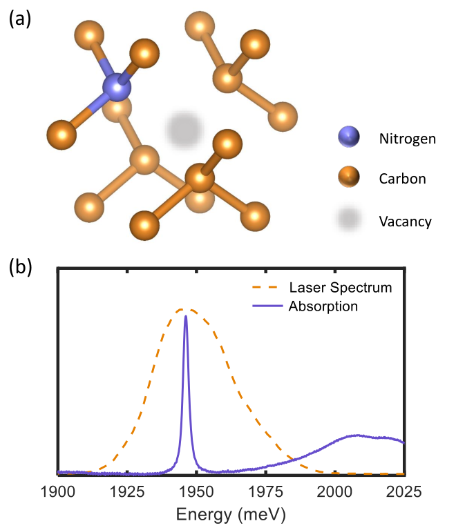
The sample studied is type Ib bulk monocrystalline diamond, where vacancy centers were introduced by irradiation with 1 MeV electrons and subsequent annealing, resulting in an NV- density of 1-2 ppm. The atomic structure of such a center is shown in Fig. 1a, which consists of a vacancy point-defect and an adjacent nitrogen substitution. This configuration gives rise to a cryogenic absorption spectrum consisting of a narrow zero-phonon line (ZPL) and an adjacent phonon sideband, as shown in Fig. 1b. We center our excitation laser spectrum on the ZPL, and our narrow pulse bandwidth (compared to the known discrete vibrational modes Huxter et al. 2013; Ulbricht et al. 2018) means that we predominantly measure the ZPL resonance of NV- without other phonon-assisted transitions. The ZPL center energy is known to shift with temperature Davies 1974; Doherty et al. 2014 due to lattice interactions Liu et al. 2019a, but is relatively stationary in our measurements performed below 150 K. NV centers in their neutral charge state (NV0) have a higher energy transition energy of 2156 meV (575 nm) and are not excited in our experiment.
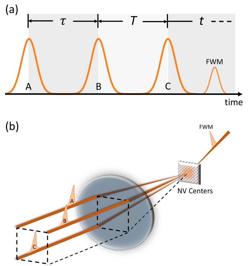
Three resonant laser pulses are used to generate a FWM signal as shown in Fig. 2a. To perform MDCS, the FWM signal is recorded as a function of three time-delays and subsequent Fourier transforms along two or all three variables returns a multi-dimensional spectrum. Here we Fourier transform an emitted photon echo along delays and , which returns a so-called one-quantum spectrum Liu et al. 2019b; Liu et al. 2019c. By correlating absorption and emission dynamics, a one-quantum spectrum unambiguously separates homogeneous and inhomogeneous broadening in orthogonal directions Siemens et al. 2010; Liu et al. 2019b; Liu et al. 2019c.
A schematic of our MDCS experiment is shown in Fig. 2b, in which three pulses are focused onto our sample in the box geometry to generate a photon echo FWM signal that is heterodyne detected with a separate local-oscillator pulse routed around the sample. The pulses used are approximately 90 fs in duration and generated by an optical parametric amplifier at a repetition rate of 250 kHz. The laser excitation density used to obtain the data is 1 W/cm2 except for that in Fig. 4 where it is 3.3 W/cm2 (both verified to generate predominately third-order responses via power-dependence measurements). The sample is mounted to a cold-finger cryostat and cooled to cryogenic temperatures.
II Results and Discussion
II.1 Multi-Dimensional Coherent Spectroscopy
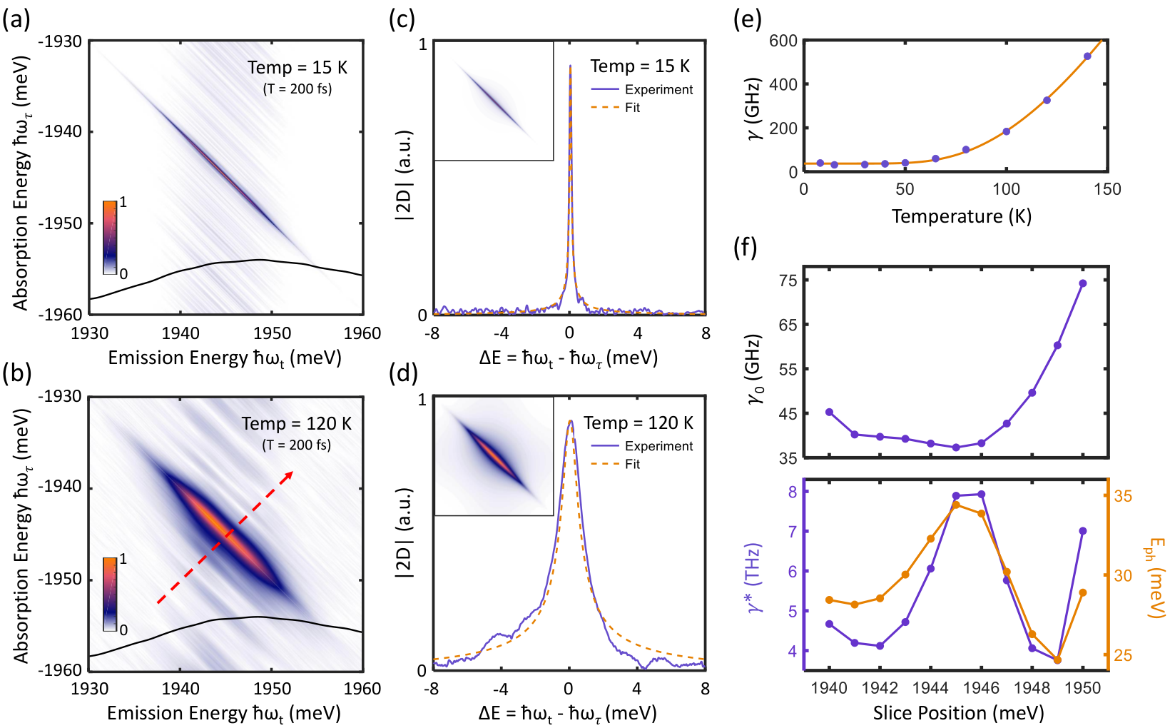
We examine the dephasing dynamics by resolving homogeneous lineshapes in one-quantum spectra. One-quantum spectra at temperatures 15 K and 120 K are shown in Fig. 3a and 3b, in which the vertical axes are opposite in sign to the horizontal axes due to inverse phase-evolution during delays and . Strong inhomogeneous broadening of the ZPL transition manifests as peak elongation along the diagonal () direction Siemens et al. 2010, while lineshapes in the orthogonal cross-diagonal direction reflect homogeneous broadening. However, homogeneous () and inhomogeneous (standard deviation of a Gaussian transition frequency distribution) broadening are completely separated in each direction only for systems dominated by inhomogeneous broadening (). To extract and in the presence of moderate inhomogeneous broadening, as in NV centers, lineshapes in the diagonal and cross-diagonal directions must be simultaneously fit Siemens et al. 2010. The experimental and fitted cross-diagonal lineshapes for each temperature are shown in Fig. 3c and 3d, with the resultant one-quantum spectra plotted inset.
A temperature dependence of measured for a resonance energy of 1945 meV is plotted in Fig. 3e, where the observed exponential increase in dephasing rate with temperature is characteristic of pure dephasing due to elastic interactions with discrete phonons. The thermal dephasing may be modeled by a linear dependence on the phonon mode occupation Singh et al. 2013:
| (1) |
where is the intrinsic zero-temperature dephasing rate, is the thermal dephasing amplitude, is the sample temperature, and is the energy of the involved phonon mode. A least-squares fit to our data is plotted as a solid line in Fig. 3e, with fitted parameters GHz ( ps), GHz, and meV. The phonon energy is in very good agreement with previous experimental results on the activation energy for ZPL dephasing from single-defect linewidth measurements Fu et al. 2009 and transient absorption spectroscopy Ulbricht et al. 2016. This phonon mode was calculated to be a vibronic mode originating from the Jahn-Teller distortion in the electronic, doubly-degenerate excited state Abtew et al. 2011. Another simulation found a quasi-localized vibrational mode of similar energy that involves vibrations of carbon atoms almost exclusively Zhang et al. 2011.
We also repeat our linewidth temperature dependence analysis for cross-diagonal lineshapes taken at positions ranging from meV to 1950 meV, corresponding to varying resonance energy. The slice position dependence of the three thermal broadening parameters , , and are plotted in Fig. 3f. We find that the zero-temperature linewidth sharply increase for energies higher than 1946 meV, and that both and vary strongly throughout the inhomogeneous distribution. These observations provide crucial information concerning the underlying mechanism of inhomogeneous broadening in this system.
We note that an activation energy of meV was previously reported by integrated FWM measurements Lenef et al. 1996 which, measured at a long waiting time of ns, is inconsistent with our results likely due to spectral diffusion. Femtosecond polarization anisotropy measurements in Ulbricht et al. 2016, which probe the depolarization of the excitation-induced coherence between the two excited state orbitals, reported a decoherence time at the low temperature limit of = 14.4 ps. This perfectly matches the values reported here. Note that the significant spectral bandwidth of the femtosecond pulses used in both studies necessarily excites both orbital states. This differs from cryogenic PLE experiments on single NV centers Shen et al. 2008; Fu et al. 2009, in which an optical coherence is induced between the ground state and only a single orbital by means of narrowband laser excitation. The discrepancy between time-resolved dephasing lifetime measurements (here and elsewhere Lenef et al. 1996) and PLE measurements that report radiatively-limited optical coherence times may thus originate from modification of orbital relaxation due to simultaneous excitation of the entire excited state manifold.
II.2 Ultrafast Spectral Diffusion
MDCS also enables the powerful capability to probe ultrafast spectral diffusion. While pulses and probe absorption and emission dynamics of the sample, the second pulse may be thought to induce a population state whose energy will vary in time. As the waiting time between pulses and increases, absorption and emission energies of a resonance become less correlated and broadening in the cross-diagonal direction occurs. For systems with strong spectral diffusion, simultaneous broadening and distortion of the cross-diagonal lineshape occurs, which allows extraction of the frequency-frequency correlation function Singh et al. 2016 that quantifies the resonance energy fluctuations. However, we find that broadening of NV center lineshapes is too weak (within our experimental delay limit) to perform such an analysis. We therefore plot a dependence of on waiting time in Fig. 4, which provides an effective measure of spectral diffusion at ultrafast timescales. From measurements taken at two different locations on our sample, as is increased from 1 ps to 2 ns the fitted effective dephasing rate increases at 1.98 MHz/ps and 1.59 MHz/ps. In a previous photon correlation study of NV centers in nanodiamonds Wolters et al. 2013 ultrafast spectral diffusion was found to occur only with simultaneous 532 nm excitation, which implicated impurity photo-ionization and subsequent charge trapping as the underlying mechanism. Our measurements of spectral diffusion, which involve only resonant excitation of the ZPL, indicate that spectral diffusion of NV centers in bulk diamond may occur due to other mechanisms such as reorganization of the surrounding diamond lattice following optical excitation.
II.3 Integrated Four-Wave Mixing
The ultrafast spectral diffusion observed comprises a microscopic mechanism of pure dephasing Mukamel 1999. For excitations in crystals (such as the case for NV centers), resonance energy fluctuations primarily arise from frequency-dependent interactions with their thermal environment, which often gives rise to memory-effects in decoherence Carmele and Reitzenstein 2019. While these memory-effects are difficult to observe in the frequency-domain, they clearly manifest in the time-domain as non-exponential (non-Markovian) dephasing Lorenz and Cundiff 2005; Liu et al. 2019b.
We therefore measure the integrated photon-echo field as a function of delay . In the case of memoryless (Markovian) dephasing, the photon-echo field decays exponentially in with a time-constant identical to twice the dephasing time of an inhomogeneously-broadened optical transition Lorenz and Cundiff 2005. We note however, that this dephasing time is an effective value averaged over the inhomogeneous resonance frequency distribution. Measurements performed at representative temperatures are shown in Fig. 5a, which exhibit an increase in dephasing rate with temperature. Unexpectedly, the dephasing dynamics exhibit two distinct time constants with more rapid dephasing occuring at longer . Such behavior is reminiscent of cross-over between Markovian and non-Markovian dephasing, previously observed in atomic vapors Lorenz and Cundiff 2005. Separate fitting of each segment yields two dephasing times, shown in Fig. 5b, that differ by over 10 ps at the lowest temperatures but converge as temperature increases. The longer fitted dephasing times in Fig. 5b at low temperatures agree well with the intrinsic dephasing rates extracted from our 2D measurements shown in Fig. 3. Interestingly, the values and temperature dependence of the fast decay component perfectly match the dephasing times determined through previous transient polarization anisotropy measurements Ulbricht et al. 2016.
II.4 Bimodal Inhomogeneous Distribution
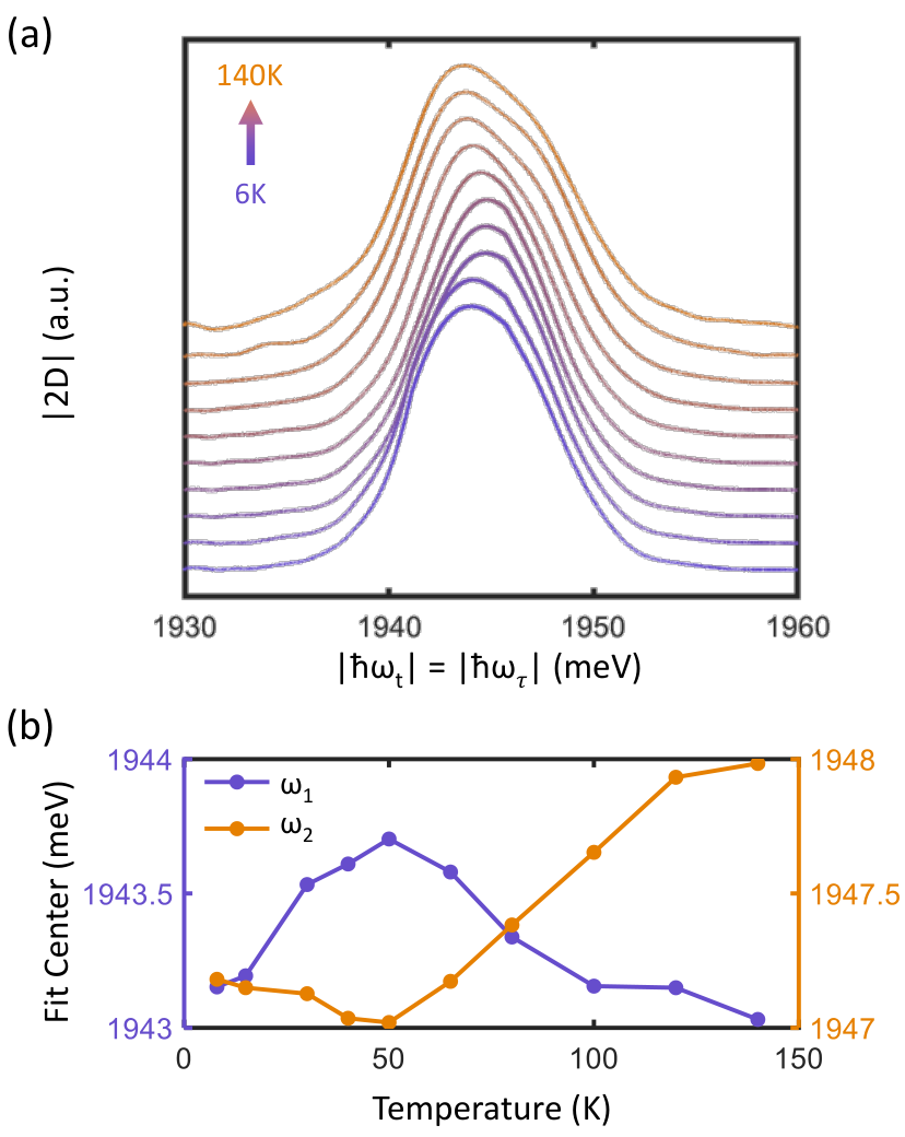
We next examine diagonal slices of the 2-D spectra taken along (plotted in Fig. 6a as a function of temperature). Interestingly, increasing temperature results in the formation of two distinct lobes in the diagonal slice lineshape, not visible in the linear absorption spectra (see Supplemental Info), which we attribute to electric field-induced splitting Tamarat et al. 2006; Bassett et al. 2011 of the manifold. An absence of sidebands in the one-quantum spectra also indicates that the two families observed are not coherently coupled. We thus modeled this system as two independent Gaussian distributions, each corresponding to an excited orbital state. The two Gaussians are of fixed widths meV (627.8 GHz) and meV (555.4 GHz), with only center frequencies allowed to vary, which reproduced the data well (see Supplemental Info). The fitted center frequencies as a function of temperature are plotted in Fig. 6b, which vary in the single meV range and appear anti-correlated. Indeed, symmetric splitting of the excited orbital states has recently been predicted and observed in NV center ensembles in response to perpendicularly-oriented electric fields Block et al. 2020. Remarkably, our observed splitting of 5 meV (40 cm-1) perfectly matches the values reported from photon echo Lenef et al. 1996 and ODMR van Oort et al. 1991 experiments, where the former report Lenef et al. 1996 proposed a Jahn-Teller splitting in the excited state manifold that has since been disputed Goss et al. 1997. According to the transverse electric-field susceptibility of MHz/(V/cm) reported by Block et al. Block et al. 2020, the splittings we observe correspond to fields ranging from 0.29 MV/cm at 50 K to 0.43 MV/cm at 140 K. These internal fields are possibly generated by positively charged single substitutional nitrogen defects in close proximity to NV centers Manson et al. 2018.
III Conclusion
In conclusion, we have measured both integrated FWM and one-quantum spectra of NV centers in diamond, which simultaneously provide both the homogeneous dephasing rate and inhomogeneous resonance frequency distribution. The primary ZPL dephasing mechanism is found to be a Jahn-Teller-induced vibronic state, in accordance with previous studies. In addition, waiting time-dependent measurements reveal another source of extrinsic broadening due to ultrafast spectral diffusion that occurs solely from resonant photo-excitation. Knowledge of these broadening mechanisms provide crucial, fundamental knowledge for engineering spectrally stable NV centers with narrow optical linewidths, with applications toward quantum information protocols Sipahigil et al. 2012; Bernien et al. 2013. Lastly we observe Stark-splitting of excited orbital states that is not visible in linear spectroscopy and forms a bimodal structure in the inhomogeneous optical response, and whose center frequencies show distinct temperature dependencies. With recent advances in the rapid acquisition of multi-dimensional spectra Lomsadze and Cundiff 2017; Lomsadze et al. 2018, our results suggest a microwave-free, all-optical analogue of a recently proposed electric-field sensing protocol Block et al. 2020 in NV center ensembles.
We acknowledge N. Manson for fruitful discussions. This work was supported by the Department of Energy grant number DE-SC0015782. D.B.A. acknowledges support by a fellowship from the Brazilian National Council for Scientific and Technological Development (CNPq). R.U. acknowledges support by a DFG fellowship (UL 474/1-1).
References
- Aharonovich and Neu (2014) I. Aharonovich and E. Neu, Diamond nanophotonics, Adv. Opt. Mater. 2, 911 (2014).
- Schröder et al. (2016) T. Schröder, S. L. Mouradian, J. Zheng, M. E. Trusheim, M. Walsh, E. H. Chen, L. Li, I. Bayn, and D. Englund, Quantum nanophotonics in diamond [invited], Journal of the Optical Society of America B 33, B65 (2016).
- Doherty et al. (2013) M. W. Doherty, N. B. Manson, P. Delaney, F. Jelezko, J. Wrachtrup, and L. C. L. Hollenberg, The nitrogen-vacancy colour centre in diamond, Phys. Rep. 528, 1 (2013).
- Schirhagl et al. (2014) R. Schirhagl, K. Chang, M. Loretz, and C. L. Degen, Nitrogen-vacancy centers in diamond: Nanoscale sensors for physics and biology, Annu. Rev. Phys. Chem. 65, 83 (2014).
- Dobrovitski et al. (2013) V. V. Dobrovitski, G. D. Fuchs, A. L. Falk, C. Santori, and D. D. Awschalom, Quantum control over single spins in diamond, Annual Review of Condensed Matter Physics 4, 23 (2013).
- Aharonovich et al. (2016) I. Aharonovich, D. Englund, and M. Toth, Solid-state single-photon emitters, Nat. Photonics 10, 631 EP (2016), review Article.
- Acosta et al. (2012) V. M. Acosta, C. Santori, A. Faraon, Z. Huang, K.-M. C. Fu, A. Stacey, D. A. Simpson, K. Ganesan, S. Tomljenovic-Hanic, A. D. Greentree, S. Prawer, and R. G. Beausoleil, Dynamic stabilization of the optical resonances of single nitrogen-vacancy centers in diamond, Phys. Rev. Lett. 108, 206401 (2012).
- Neu et al. (2012) E. Neu, M. Agio, and C. Becher, Photophysics of single silicon vacancy centers in diamond: implications for single photon emission, Opt. Express 20, 19956 (2012).
- Radko et al. (2016) I. P. Radko, M. Boll, N. M. Israelsen, N. Raatz, J. Meijer, F. Jelezko, U. L. Andersen, and A. Huck, Determining the internal quantum efficiency of shallow-implanted nitrogen-vacancy defects in bulk diamond, Opt. Express 24, 27715 (2016).
- Chu et al. (2014) Y. Chu, N. P. de Leon, B. J. Shields, B. Hausmann, R. Evans, E. Togan, M. J. Burek, M. Markham, A. Stacey, A. S. Zibrov, A. Yacoby, D. J. Twitchen, M. Loncar, H. Park, P. Maletinsky, and M. D. Lukin, Coherent optical transitions in implanted nitrogen vacancy centers, Nano Lett. 14, 1982 (2014).
- van Dam et al. (2019) S. B. van Dam, M. Walsh, M. J. Degen, E. Bersin, S. L. Mouradian, A. Galiullin, M. Ruf, M. IJspeert, T. H. Taminiau, R. Hanson, and D. R. Englund, Optical coherence of diamond nitrogen-vacancy centers formed by ion implantation and annealing, Phys. Rev. B 99, 161203 (2019).
- Liu and Cundiff (2020) A. Liu and S. T. Cundiff, Spectroscopic signatures of electron-phonon coupling in silicon-vacancy centers in diamond, Phys. Rev. Mater. 4, 055202 (2020).
- Huxter et al. (2013) V. M. Huxter, T. A. A. Oliver, D. Budker, and G. R. Fleming, Vibrational and electronic dynamics of nitrogen-vacancy centres in diamond revealed by two-dimensional ultrafast spectroscopy, Nat. Phys. 9, 744 EP (2013), article.
- Ulbricht et al. (2018) R. Ulbricht, S. Dong, A. Gali, S. Meng, and Z.-H. Loh, Vibrational relaxation dynamics of the nitrogen-vacancy center in diamond, Phys. Rev. B 97, 220302 (2018).
- Davies (1974) G. Davies, Vibronic spectra in diamond, 7, 3797 (1974).
- Doherty et al. (2014) M. W. Doherty, V. M. Acosta, A. Jarmola, M. S. J. Barson, N. B. Manson, D. Budker, and L. C. L. Hollenberg, Temperature shifts of the resonances of the nv- center in diamond, Phys. Rev. B 90, 041201 (2014).
- Liu et al. (2019a) A. Liu, L. G. Bonato, F. Sessa, D. B. Almeida, E. Isele, G. Nagamine, L. F. Zagonel, A. F. Nogueira, L. A. Padilha, and S. T. Cundiff, Effect of dimensionality on the optical absorption properties of cspbi3 perovskite nanocrystals, J. Chem. Phys. 151, 191103 (2019a).
- Liu et al. (2019b) A. Liu, D. B. Almeida, W. K. Bae, L. A. Padilha, and S. T. Cundiff, Non-markovian exciton-phonon interactions in core-shell colloidal quantum dots at femtosecond timescales, Phys. Rev. Lett. 123, 057403 (2019b).
- Liu et al. (2019c) A. Liu, D. B. Almeida, W.-K. Bae, L. A. Padilha, and S. T. Cundiff, Simultaneous existence of confined and delocalized vibrational modes in colloidal quantum dots, J. Phys. Chem. Lett. , 6144 (2019c).
- Siemens et al. (2010) M. E. Siemens, G. Moody, H. Li, A. D. Bristow, and S. T. Cundiff, Resonance lineshapes in two-dimensional fourier transform spectroscopy, Opt. Express 18, 17699 (2010).
- Singh et al. (2013) R. Singh, T. M. Autry, G. Nardin, G. Moody, H. Li, K. Pierz, M. Bieler, and S. T. Cundiff, Anisotropic homogeneous linewidth of the heavy-hole exciton in (110)-oriented gaas quantum wells, Phys. Rev. B 88, 045304 (2013).
- Fu et al. (2009) K.-M. C. Fu, C. Santori, P. E. Barclay, L. J. Rogers, N. B. Manson, and R. G. Beausoleil, Observation of the dynamic jahn-teller effect in the excited states of nitrogen-vacancy centers in diamond, Phys. Rev. Lett. 103, 256404 (2009).
- Ulbricht et al. (2016) R. Ulbricht, S. Dong, I.-Y. Chang, B. M. K. Mariserla, K. M. Dani, K. Hyeon-Deuk, and Z.-H. Loh, Jahn-teller-induced femtosecond electronic depolarization dynamics of the nitrogen-vacancy defect in diamond, Nat. Commun. 7, 13510 EP (2016), article.
- Abtew et al. (2011) T. A. Abtew, Y. Y. Sun, B.-C. Shih, P. Dev, S. B. Zhang, and P. Zhang, Dynamic jahn-teller effect in the nv- center in diamond, Phys. Rev. Lett. 107, 146403 (2011).
- Zhang et al. (2011) J. Zhang, C.-Z. Wang, Z. Z. Zhu, and V. V. Dobrovitski, Vibrational modes and lattice distortion of a nitrogen-vacancy center in diamond from first-principles calculations, Phys. Rev. B 84, 035211 (2011).
- Lenef et al. (1996) A. Lenef, S. W. Brown, D. A. Redman, S. C. Rand, J. Shigley, and E. Fritsch, Electronic structure of the n-v center in diamond: Experiments, Phys. Rev. B 53, 13427 (1996).
- Shen et al. (2008) Y. Shen, T. M. Sweeney, and H. Wang, Zero-phonon linewidth of single nitrogen vacancy centers in diamond nanocrystals, Phys. Rev. B 77, 033201 (2008).
- Singh et al. (2016) R. Singh, G. Moody, M. E. Siemens, H. Li, and S. T. Cundiff, Quantifying spectral diffusion by the direct measurement of the correlation function for excitons in semiconductor quantum wells, Journal of the Optical Society of America B 33, C137 (2016).
- Wolters et al. (2013) J. Wolters, N. Sadzak, A. W. Schell, T. Schröder, and O. Benson, Measurement of the ultrafast spectral diffusion of the optical transition of nitrogen vacancy centers in nano-size diamond using correlation interferometry, Phys. Rev. Lett. 110, 027401 (2013).
- Mukamel (1999) S. Mukamel, Principles of Nonlinear Optical Spectroscopy (Oxford University Press, 1999).
- Carmele and Reitzenstein (2019) A. Carmele and S. Reitzenstein, Non-markovian features in semiconductor quantum optics: quantifying the role of phonons in experiment and theory, Nanophotonics 8, 655 (2019).
- Lorenz and Cundiff (2005) V. O. Lorenz and S. T. Cundiff, Non-markovian dynamics in a dense potassium vapor, Phys. Rev. Lett. 95, 163601 (2005).
- Tamarat et al. (2006) P. Tamarat, T. Gaebel, J. R. Rabeau, M. Khan, A. D. Greentree, H. Wilson, L. C. L. Hollenberg, S. Prawer, P. Hemmer, F. Jelezko, and J. Wrachtrup, Stark shift control of single optical centers in diamond, Phys. Rev. Lett. 97, 083002 (2006).
- Bassett et al. (2011) L. C. Bassett, F. J. Heremans, C. G. Yale, B. B. Buckley, and D. D. Awschalom, Electrical tuning of single nitrogen-vacancy center optical transitions enhanced by photoinduced fields, Phys. Rev. Lett. 107, 266403 (2011).
- Block et al. (2020) M. Block, B. Kobrin, A. Jarmola, S. Hsieh, C. Zu, N. L. Figueroa, V. M. Acosta, J. Minguzzi, J. R. Maze, D. Budker, and N. Y. Yao, Optically enhanced electric field sensing using nitrogen-vacancy ensembles (2020), arXiv:2004.02886 [quant-ph] .
- van Oort et al. (1991) E. van Oort, B. van der Kamp, R. Sitter, and M. Glasbeek, Microwave-induced line-narrowing of the n-v defect absorption in diamond, J. Lumin. 48-49, 803 (1991).
- Goss et al. (1997) J. P. Goss, R. Jones, P. R. Briddon, G. Davies, A. T. Collins, A. Mainwood, J. A. van Wyk, J. M. Baker, M. E. Newton, A. M. Stoneham, and S. C. Lawson, Comment on “electronic structure of the n-$v$ center in diamond: Theory”, Phys. Rev. B 56, 16031 (1997).
- Manson et al. (2018) N. B. Manson, M. Hedges, M. S. J. Barson, R. Ahlefeldt, M. W. Doherty, H. Abe, T. Ohshima, and M. J. Sellars, Nv–n+ pair centre in 1b diamond, New J. Phys. 20, 113037 (2018).
- Sipahigil et al. (2012) A. Sipahigil, M. L. Goldman, E. Togan, Y. Chu, M. Markham, D. J. Twitchen, A. S. Zibrov, A. Kubanek, and M. D. Lukin, Quantum interference of single photons from remote nitrogen-vacancy centers in diamond, Phys. Rev. Lett. 108, 143601 (2012).
- Bernien et al. (2013) H. Bernien, B. Hensen, W. Pfaff, G. Koolstra, M. S. Blok, L. Robledo, T. H. Taminiau, M. Markham, D. J. Twitchen, L. Childress, and R. Hanson, Heralded entanglement between solid-state qubits separated by three metres, Nature 497, 86 (2013).
- Lomsadze and Cundiff (2017) B. Lomsadze and S. T. Cundiff, Frequency combs enable rapid and high-resolution multidimensional coherent spectroscopy, Science 357, 1389 (2017).
- Lomsadze et al. (2018) B. Lomsadze, B. C. Smith, and S. T. Cundiff, Tri-comb spectroscopy, Nat. Photonics 12, 676 (2018).