User-Guided Domain Adaptation for Rapid Annotation from User Interactions: A Study on Pathological Liver Segmentation
Abstract
Mask-based annotation of medical images, especially for D data, is a bottleneck in developing reliable machine learning models. Using minimal-labor user interactions to guide the annotation is promising, but challenges remain on best harmonizing the mask prediction with the user interactions. To address this, we propose the user-guided domain adaptation framework, which uses prediction-based adversarial domain adaptation to model the combined distribution of user interactions and mask predictions. The user interactions are then used as anchors to guide and align the mask prediction. Importantly, user-guided domain adaptation can both learn from unlabelled data and also model the high-level semantic meaning behind different user interactions. We test user-guided domain adaptation on annotating pathological livers using a clinically comprehensive dataset of patient studies. Using only extreme-point user interactions, we achieve a mean (worst-case) performance of (), compared to () for deep extreme points. Furthermore, we also show user-guided domain adaptation can retain this state-of-the-art performance even when only seeing a fraction of available user interactions, demonstrating an ability for robust and reliable user interaction-guided segmentation with extremely minimal labor demands.
Keywords:
Liver Segmentation Interactive Segmentation User-guided Domain Adaptation1 Introduction
Reliable computer-assisted segmentation of anatomical structures from medical images can allow for quantitative biomarkers for disease diagnosis, prognosis, and progression. Given the extreme labor to fully annotate data, especially for 3D volumes, a considerable body of work focuses on weakly-supervised segmentation solutions [24]. Solutions that can leverage user interactions, e.g., extreme-points, scribbles, and boundary marks, are an important such category.
The main challenge is effectively leveraging user interactions to constrain or guide the mask generation. Classic approaches, like the random walker algorithm [5], do so via propagating seed regions using intensity similarities. Later approaches add additional constraints, e.g., based on presegmentations [6] or learned probabilities [7]. With the advent of deep-learning, harmonizing mask predictions with the user interactions continues to be a challenge. Deep extreme points [17], which requires the user to click on the extreme boundary points of an object, is a popular and effective approach. But deep extreme points only adds the extreme point annotations as an additional channel when training the segmentor, meaning the predicted mask may not agree with the user interactions. Later work uses expectation-maximization strategies that alternate between network training and then regularization via random walker [22] or dense conditional random fields [20, 27, 2]. However, over and above their computational demands, the intensity-based random walker or conditional random field regularization may not capture high-level semantics and any guidance on the mask predictions still remains highly indirect. DeepIGeoS [28] offers an alternative that uses a deep-conditional random field optimizer to propagate scribbles. However, DeepIGeos would only allow boundary annotations or extreme points to be treated as simple seed regions, neglecting their rich semantic meaning.
To address these issues, we propose user-guided domain adaptation. Our new method uses prediction-based adversarial domain adaptation [26] to guide mask predictions by the user interactions. user-guided domain adaptation’s advantage is that it is equipped to model the high-level meaning behind different types of user interactions and how they should impact the ultimate mask prediction. Importantly, the user interactions are used as anchors when adapting the mask. Another advantage is that, like prediction-based adversarial domain adaptation, user-guided domain adaptation can learn from and exploit completely unlabelled data, in addition to those accompanied by user interactions. Without loss of generality, we focus on using deep extreme points-style extreme point user interactions because of their intuitiveness and effectiveness [19, 22]. But other types, e.g., boundary corrections, are equally possible in addition to, or instead of, extreme points. The only constraint is that we assume a fully-supervised dataset is available in order to model the interplay between mask and user interactions. But such data can originate from sources other than the target dataset, e.g., from public data. To the best of our knowledge, we are the first to use domain adaptation as a mechanism to drive user interaction-based segmentation.
We test user-guided domain adaptation on an extremely challenging pathological liver segmentation dataset collected directly from the picture archiving and communication system of Chang Gung Memorial hospital. This dataset comprises patient studies, all with hepatocellular carcinoma, intrahepatic cholangiocellular carcinoma, metastasized, or benign lesions and is one of the most challenging datasets to date for pathological liver segmentation. Liver volumetry analysis is a crucial pre-requisite prior to many hepatic procedures, such as liver transplantation [18, 25] or resection [15, 14]. Despite success with fully-automated solutions, modern deep solutions [29, 12, 11] are often trained only on public datasets, e.g., LiTS [1], which only represents hepatocellular carcinoma and metastasized lesions and does not fully represent patient/co-morbidity distributions. Thus, they may not generalize to all datasets, such as ours. Using only extreme-point user interactions, we achieve state-of-the-art mean (worst-case) Dice-Sørensen coefficient scores of () on our dataset, compared to () and () for a strong a fully-supervised baseline and DEXTR [17], respectively. We also show that user-guided domain adaptation can improve over prediction-based adversarial domain adaptation [26] by Dice-Sørensen coefficient and that user-guided domain adaptation can even perform robustly when only shown incomplete sets of user interactions. Finally, we demonstrate predicted masks align extraordinarily well with user interactions, allowing the user to interact with confidence and with minimal frustration.
2 Methods
We aim to produce reliable mask predictions on a target dataset or deployment scenario, given only minimal user interactions. More formally, we assume we are given a dataset composed of both user interaction-labelled and completely unlabelled volumes, , with and denoting the images and extreme points user interactions, respectively. In addition, we also assume a fully-supervised source dataset with masks is also available, . As long as the masks and extreme points describe the same anatomical structure, may originate from entirely different sources, e.g., public data. The goal is to use only the extreme point user interactions to robustly annotate . Fig. 1 outlines our workflow, which uses user-guided domain adaptation (UGDA) to efficiently and effectively exploit the extreme-point user interactions.

2.1 Supervised Workflow
The backbone of user-guided domain adaptation is two 3D fully-convolutional networks chained together, where the first fully-convolutional network predicts extreme-points while the second predicts a full mask. Working backward, the second fully-convolutional network acts very similarly to DEXTR [17], where the latter predicts a mask given an input image along with the extreme-point user interactions:
| (1) |
where we have used to represent the segmentation fully-convolutional network. Each of the extreme points are represented by a 3D Gaussian heat map centered on the user clicks and rendered into an additional input channel, . We find the method is not sensitive to the size of the Gaussian heat maps, and we use a kernel with -pixel standard deviation. However, unlike DEXTR, we do not assume all training volumes come with extreme-points, as only some target volumes in may have user interactions.
To loosen these restrictions, we use the first fully-convolutional network to predict extreme-point heatmaps for each volume. Following heatmap regression conventions [30], the extreme-point fully-convolutional network, , outputs 3D Gaussian heatmaps, each corresponding to one extreme point. These are then summed together into one channel prior to being inputted into our segmentation fully-convolutional network:
| (2) | ||||
| (3) |
where for convenience we have skipped showing the summation of heatmaps into one channel. By relying on predictions, this allows our system to operate even with unlabelled data. Ground-truth extreme-points can be generated from any user interactions performed on . Extreme-points for can be deterministically generated from the full masks, simulating the user interactions.
In terms of loss, if we use to denote any input volume associated with extreme-point user interactions, whether from or , then a supervised loss can be formulated:
| (4) | ||||
| (5) | ||||
| (6) |
where denotes the cardinality of . Following heat-map regression practices [30], we implement using mean-squared error. For we use a summation of cross entropy and Dice-Sørensen coefficient losses, which has experienced success in segmentation tasks [11]. While we focus on extreme points for this work, other types of user interactions, such as boundary corrections, can be readily incorporated in this framework.
2.2 User-Guided Domain Adaptation
Similar to DEXTR [17], (6) indirectly guides mask predictions by using extreme-point heat maps as an additional input channel for the segmentor. Additionally, for user interaction-labelled volumes in , the supervised loss in (5) encourages the extreme-point predictions to actually match the user interactions. However, the mask prediction may contradict the user interactions because there is no penalty for disagreement between the two. Thus, an additional mechanism is needed to align the mask with user interactions. We opt for an adversarial domain adaption approach to penalize discordant mask predictions. While image translation-based adversarial domain adaptation methods show excellent results [10, 13], these are unsuited to our task because we are concerned with adapting the prediction-space to produce a mask well-aligned with the user interactions. Thus, we use prediction-based adversarial domain adaptation (PADA) [26].
More specifically, we use a discriminator, , to learn the distribution and interplay between masks and extreme-points. Treating samples from as the “correct” distribution, the discriminator loss can be expressed as
| (7) |
where denotes the cross-entropy loss. Importantly, to model their combined distribution, the discriminator accepts both the user interaction and mask predictions. Following standard adversarial training, here gradients only flow through the discriminator. user-guided domain adaptation then attempts to fool the discriminator by predicting extreme-point/mask pairs for that match ’s distribution. More formally, an adversarial loss is set up for volumes in :
| (8) |
Note that compared to (7), the “label” for has been switched from to . Like standard prediction-based adversarial domain adaptation setups, gradients do not flow through the discriminator weights in (8). Importantly, gradients also do not flow through the extreme-point predictions when the user interactions are present. Consequently, when user interactions are available, extreme-point predictions are only influenced by the supervised loss in (5) to match the user interactions. While there is no strict guarantee, our results demonstrate almost perfect matching. Thus, the extreme-point predictions act as anchors, while the adversarial loss in (8) guides the mask predictions to properly align with the user interactions. This alignment is more than simply making mask predictions agree with the user interactions, as by modelling their interplay, prediction-based adversarial domain adaptation also guides mask regions far away from user interactions. Finally, the use of prediction-based adversarial domain adaptation provides another important benefit, as completely unlabelled volumes in can seamlessly contribute to the learning process in (7) and (8). In fact, user-guided domain adaptation can be seen as integrating domain adaptation learning processes [26] in addition to DEXTR-style guidance [17] from user interactions. Thus, the overall training objective for user-guided domain adaptation is to minimize the following total loss:
| (9) |
where we have purposely kept loss weighting to only the adversarial component to reduce hyper-parameter tuning.
3 Experiments

Dataset description. We test user-guided domain adaptation on segmenting pathological livers, using a target dataset of venous-phase computed tomography studies from the picture archiving and communication system of Chang Gung Memorial hospital. The only selection criterion was of patients with biospied or resecuted liver lesions, with computed tomography scans taken within one month before the procedure. Patients directly reflect clinical distributions and represent intrahepatic cholangiocellular carcinoma, hepatocellular carcinoma, benign or metastasized lesions, along with co-occuring maladies, such as liver fibrosis, splenomegaly, or embolized lesions. This serves as . From these, we selected and studies as validation and test sets, respectively, and delineated the patient livers. These computed tomographies are called evaluation volumes. We annotated the remainder using only extreme point user interactions. For we collected fully-labelled venous-phase computed tomography studies from public datasets [1, 4, 9, 3], which, unlike , comprises both healthy and pathological livers and only represents hepatocellular carcinoma and metastasized tumors. Corresponding extreme-point “user interactions” were generated from the full masks following [17]. For internal validation, we also split into , , and for training, testing and validation, respectively.
Implementation Details. For user-guided domain adaptation’s two fully-convolutional network architectures, we use a D version of the deeply-supervised progressive holistically nested network [8], which provides an efficient and decoder-free pipeline. As backbone, we use a D generalization of VGG-16 [23]. We first train a fully-supervised baseline on using (4), then we finetune after convergence using (9). This dual-PHNN baseline is very strong on our public data, achieving a Dice-Sørensen coefficient score of on the test set. For discriminator, we use a D version of a popular architecture [16] using atrous convolution, which has proved a useful discriminator for liver masks [21]. We do not use the multi-level discriminator variant proposed by Tsai et al. [26], as its added complexity does not seem to result in noticeable improvements for liver-based prediction-based adversarial domain adaptation [21]. Specific details on the choice of hyper parameters are listed in the supplementary material.
Evaluation protocols. We evaluate user-guided domain adaptation on how well it can annotate using only extreme-point user interactions. To do this, we include the evaluation volumes and their extreme point user interactions within the training procedure, but hide their masks. For evaluation, we measure Dice-Sørensen coefficient scores and also the mask-extreme-point agreement. The latter measures the average distance between all six of a predicted mask’s extreme-points vs. the ground-truth extreme points. This directly measures how well the model can produce a mask prediction that actually matches the extreme-point user interactions.
Comparisons. We test against the user-interactive DEXTR [17] using the same progressive holistically nested network backbone. DEXTR can only be trained on because it requires fully-labelled training data, but during inference it sees ’s extreme-point user interactions. The DEXTR authors claim their approach can generalize well to new data. We also test against two mask-based prediction-based adversarial domain adaptation [26] variants. The first variant, called “mask-based prediction-based adversarial domain adaptation (no user interactions)”, matches published practices [26] and just uses a single network (progressive holistically nested network in our case) to directly predict masks, using a discriminator to penalize ill-behaving mask predictions on unlabeled data. It does not incorporate user interactions. The second variant, simply called “mask-based prediction-based adversarial domain adaptation (w user interactions)”, is almost identical to user-guided domain adaptation, meaning the mask and extreme-point predictions are still trained with the supervised losses of (6) and (5), respectively, but the discriminator in (7) and (8) only models mask predictions without seeing the extreme-point predictions. Comparing this variant to user-guided domain adaptation reveals the impact of using extreme-points as anchors to guide the domain adaptation of the masks.
Finally, we also compare against user-guided domain adaptation variants trained with only a fraction of the user interactions available in , with the remainder being left unlabelled. This reveals how well user-guided domain adaptation can operate in scenarios when only a fraction of target volumes have user interaction-labels. When doing so, we remove the same percentage of user interaction-labels from both non-evaluation and evaluation volumes.
Results. Tab. 2 outlines the performance of all variants in annotating . As can be seen, compared to its performance on , the fully-supervised dual progressive holistically nested network’s performance drops from to due to the major differences between public liver datasets and our picture archiving and communication system-based clinical target dataset. This suggests that other strategies are required, e.g., exploiting minimal-labor user interactions.
| Model | % user interactions | Mean DSC | Mean MXA (mm) |
|---|---|---|---|
| Dual PHNN [8] | n/a | ||
| DEXTR [17] | |||
| Mask-based PADA (no user interactions) [26] | |||
| Mask-based PADA (w user interactions) [26] | |||
| user-guided domain adaptation | |||
| user-guided domain adaptation | |||
| user-guided domain adaptation |
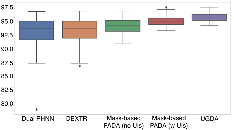
As the table also demonstrates, by exploiting user interactions, DEXTR can significantly boost the mask-extreme-point agreement, but its lackluster Dice-Sørensen coefficient scores suggest that the resulting masks, while aligning better with the extreme-points, still do not properly capture the liver extent. On the other hand, both mask-based prediction-based adversarial domain adaptation variants perform better, indicating that modelling mask distributions on top of deep extreme points-style user interaction guidance can more robustly annotate . Finally, user-guided domain adaptation performs best, demonstrating that modelling the interplay between the user interactions and mask predictions can boost performance even further. Importantly, user-guided domain adaptation’s mask-extreme-point agreement is extremely good, which shows that the mask predictions well match the user interactions. These mean scores are bolstered by Fig. 3’s box-and-whisker plots, which demonstrate that user-guided domain adaptation provides important boosts in reliability, with an extremely robust worst-case performance of Dice-Sørensen coefficient, compared to for the mask-based prediction-based adversarial domain adaptation (w user interactions) variant.
Viewing the qualitative examples in Fig. 2 reinforces these quantitative improvements. In particular, user-guided domain adaptation is able to ensure that masks both agree with extreme-points and provide robust predictions away from the user interactions.
Finally, as Tab. 2 also demonstrates, user-guided domain adaptation can perform almost as well when only a fraction of is user interaction-labelled, outperforming both deep extreme points and the mask-based prediction-based adversarial domain adaptation, both of which see all of the user interactions. These results indicate that user-guided domain adaptation can operate well even in scenarios with extremely minimal user interaction annotation. providing further evidence of its very high versatility.
4 Conclusion
We presented user-guided domain adaptation (UGDA), an effective approach to use user interactions to rapidly annotate 3D medical volumes. We tested user-guided domain adaptation on arguably the most challenging and comprehensive pathological liver segmentation dataset to date, demonstrating that by only using extreme-point user interactions we can achieve Dice-Sørensen coefficient scores of , outperforming both deep extreme points [17] and conventional mask-based prediction-based adversarial domain adaptation [26]. Future work includes testing on other anatomical structures, incorporating additional types of user interactions, and adapting user-guided domain adaptation for real-time interaction.
Supplementary material
Hyper parameters
We set to . The dual-progressive holistically nested network learning rates was set to and reduced by a factor of when validation Dice-Sørensen coefficients do not improve after epochs, whereas the discriminator has a constant learning rate of . We use the Adam optimizer [Kingma_2015] for both.
| Input volume resolution | |
|---|---|
| Standard deviation for D Gaussian map | 5 |
| GPU system | Quadro RTX 8000 |
| Cuda version | 10.2 |
| Number of GPUs and memory | |
| Deep learning framework and version | pytorch 1.4.0 |
References
- [1] Bilic, P., et al.: The liver tumor segmentation benchmark (lits). arXiv preprint arXiv:1901.04056 (2019)
- [2] Can, Y.B., Chaitanya, K., Mustafa, B., Koch, L.M., Konukoglu, E., Baumgartner, C.F.: Learning to segment medical images with scribble-supervision alone. In: Deep Learning in Medical Image Analysis and Multimodal Learning for Clinical Decision Support. pp. 236–244. Springer International Publishing, Cham (2018)
- [3] chaos: Chaos - combined (ct-mr) healthy abdominal organ segmentation (2019), https://chaos.grand-challenge.org/Combined_Healthy_Abdominal_Organ_Segmentation
- [4] Gibson, E., Giganti, F., Hu, Y., Bonmati, E., Bandula, S., Gurusamy, K., Davidson, B., Pereira, S.P., Clarkson, M.J., Barratt, D.C.: Multi-organ Abdominal CT Reference Standard Segmentations (Feb 2018), https://doi.org/10.5281/zenodo.1169361
- [5] Grady, L.: Random walks for image segmentation. IEEE Transactions on Pattern Analysis and Machine Intelligence 28(11), 1768–1783 (Nov 2006)
- [6] Grady, L., Funka-Lea, G.: An energy minimization approach to the data driven editing of presegmented images/volumes. In: Medical Image Computing and Computer-Assisted Intervention – MICCAI 2006. pp. 888–895. Springer Berlin Heidelberg, Berlin, Heidelberg (2006)
- [7] Harrison, A.P., Birkbeck, N., Sofka, M.: Intelledits: Intelligent learning-based editor of segmentations. In: Mori, K., Sakuma, I., Sato, Y., Barillot, C., Navab, N. (eds.) Medical Image Computing and Computer-Assisted Intervention – MICCAI 2013. pp. 235–242. Springer Berlin Heidelberg, Berlin, Heidelberg (2013)
- [8] Harrison, A.P., Xu, Z., George, K., Lu, L., Summers, R.M., Mollura, D.J.: Progressive and multi-path holistically nested neural networks for pathological lung segmentation from ct images. In: International conference on medical image computing and computer-assisted intervention. pp. 621–629. Springer (2017)
- [9] Heimann, T., et al.: Comparison and evaluation of methods for liver segmentation from ct datasets. IEEE Transactions on Medical Imaging 28(8), 1251–1265 (Aug 2009)
- [10] Hoffman, J., Tzeng, E., Park, T., Zhu, J.Y., Isola, P., Saenko, K., Efros, A., Darrell, T.: CyCADA: Cycle-consistent adversarial domain adaptation. In: Dy, J., Krause, A. (eds.) Proceedings of the 35th International Conference on Machine Learning. Proceedings of Machine Learning Research, vol. 80, pp. 1989–1998. PMLR, Stockholmsmässan, Stockholm Sweden (10–15 Jul 2018)
- [11] Isensee, F., Petersen, J., Klein, A., Zimmerer, D., Jaeger, P.F., Kohl, S., Wasserthal, J., Koehler, G., Norajitra, T., Wirkert, S., et al.: nnu-net: Self-adapting framework for u-net-based medical image segmentation. arXiv preprint arXiv:1809.10486 (2018)
- [12] Li, X., Chen, H., Qi, X., Dou, Q., Fu, C.W., Heng, P.A.: H-denseunet: hybrid densely connected unet for liver and tumor segmentation from ct volumes. IEEE transactions on medical imaging 37(12), 2663–2674 (2018)
- [13] Li, Y., Yuan, L., Vasconcelos, N.: Bidirectional learning for domain adaptation of semantic segmentation. In: IEEE Conference on Computer Vision and Pattern Recognition, CVPR 2019, Long Beach, CA, USA, June 16-20, 2019. pp. 6936–6945. Computer Vision Foundation / IEEE (2019). https://doi.org/10.1109/CVPR.2019.00710
- [14] Lim, M., Tan, C., Cai, J., Zheng, J., Kow, A.: Ct volumetry of the liver: where does it stand in clinical practice? Clinical radiology 69(9), 887–895 (2014)
- [15] Lodewick, T.M., Arnoldussen, C.W., Lahaye, M.J., van Mierlo, K.M., Neumann, U.P., Beets-Tan, R.G., Dejong, C.H., van Dam, R.M.: Fast and accurate liver volumetry prior to hepatectomy. HPB 18(9), 764–772 (2016)
- [16] Lv, F., Lian, Q., Yang, G., Lin, G., Jialin Pan, S., Duan, L.: Domain adaptive semantic segmentation through structure enhancement. In: The European Conference on Computer Vision (ECCV) Workshops (September 2018)
- [17] Maninis, K.., Caelles, S., Pont-Tuset, J., Van Gool, L.: Deep extreme cut: From extreme points to object segmentation. In: 2018 IEEE/CVF Conference on Computer Vision and Pattern Recognition. pp. 616–625 (June 2018)
- [18] Nakayama, Y., Li, Q., Katsuragawa, S., Ikeda, R., Hiai, Y., Awai, K., Kusunoki, S., Yamashita, Y., Okajima, H., Inomata, Y., et al.: Automated hepatic volumetry for living related liver transplantation at multisection ct. Radiology 240(3), 743–748 (2006)
- [19] Papadopoulos, D.P., Uijlings, J.R.R., Keller, F., Ferrari, V.: Extreme clicking for efficient object annotation. In: 2017 IEEE International Conference on Computer Vision (ICCV). pp. 4940–4949 (Oct 2017)
- [20] Rajchl, M., Lee, M.C.H., Oktay, O., Kamnitsas, K., Passerat-Palmbach, J., Bai, W., Damodaram, M., Rutherford, M.A., Hajnal, J.V., Kainz, B., Rueckert, D.: Deepcut: Object segmentation from bounding box annotations using convolutional neural networks. IEEE Transactions on Medical Imaging 36(2), 674–683 (Feb 2017)
- [21] Raju, A., Cheng, C.T., Huo, Y., Cai, J., Huang, J., Xiao, J., Lu, L., Liao, C., Harrison, A.P.: Co-heterogeneous and adaptive segmentation from multi-source and multi-phase ct imaging data: A study on pathological liver and lesion segmentation. In: ECCV 2020 (2020)
- [22] Roth, H., Zhang, L., Yang, D., Milletari, F., Xu, Z., Wang, X., Xu, D.: Weakly supervised segmentation from extreme points. In: Large-Scale Annotation of Biomedical Data and Expert Label Synthesis and Hardware Aware Learning for Medical Imaging and Computer Assisted Intervention, pp. 42–50. Springer (2019)
- [23] Simonyan, K., Zisserman, A.: Very deep convolutional networks for large-scale image recognition. In: International Conference on Learning Representations (2015)
- [24] Tajbakhsh, N., Jeyaseelan, L., Li, Q., Chiang, J., Wu, Z., Ding, X.: Embracing imperfect datasets: A review of deep learning solutions for medical image segmentation (2019)
- [25] Taner, C.B., Dayangac, M., Akin, B., Balci, D., Uraz, S., Duran, C., Killi, R., Ayanoglu, O., Yuzer, Y., Tokat, Y.: Donor safety and remnant liver volume in living donor liver transplantation. Liver transplantation 14(8), 1174–1179 (2008)
- [26] Tsai, Y.H., Hung, W.C., Schulter, S., Sohn, K., Yang, M.H., Chandraker, M.: Learning to adapt structured output space for semantic segmentation. In: Proceedings of the IEEE Conference on Computer Vision and Pattern Recognition. pp. 7472–7481 (2018)
- [27] Wang, G., Li, W., Zuluaga, M.A., Pratt, R., Patel, P.A., Aertsen, M., Doel, T., David, A.L., Deprest, J., Ourselin, S., Vercauteren, T.: Interactive medical image segmentation using deep learning with image-specific fine tuning. IEEE Transactions on Medical Imaging 37(7), 1562–1573 (July 2018)
- [28] Wang, G., Zuluaga, M.A., Li, W., Pratt, R., Patel, P.A., Aertsen, M., Doel, T., David, A.L., Deprest, J., Ourselin, S., Vercauteren, T.: Deepigeos: A deep interactive geodesic framework for medical image segmentation. IEEE Transactions on Pattern Analysis and Machine Intelligence 41(7), 1559–1572 (July 2019)
- [29] Zhang, J., Xie, Y., Zhang, P., Chen, H., Xia, Y., Shen, C.: Light-weight hybrid convolutional network for liver tumour segmentation. In: Proceedings of the 28th International Joint Conference on Artificial Intelligence, Macao, China. pp. 10–16 (2019)
- [30] Zhou, X., Zhuo, J., Krähenbühl, P.: Bottom-up object detection by grouping extreme and center points. In: CVPR (2019)