Spectroscopic Signature of Chemical Bond Dissociation Revealed by Calculated Core-Electron Spectra
Abstract
The advent of ultrashort soft x-ray pulse sources permits the use of established gas phase spectroscopy methods to investigate ultrafast photochemistry in isolated molecules with element and site specificity. In the present study, we simulate excited state wavepacket dynamics of a prototypical process, the ultrafast photodissociation of methyl iodide. Based on the simulation, we calculate time-dependent excited state carbon edge photoelectron and Auger electron spectra. We observe distinct signatures in both types of spectra and show their direct connection to C – I bond dissociation and charge rearrangement processes in the molecule. We demonstrate at the molecule that the observed signatures allow us to map the time-dependent dynamics of ultrafast photo-induced bond breaking with unprecedented details.
Some of the most important photochemical processes in nature take place in the excited states of organic molecules on ultrafast timescalesCheng and Fleming 2009; Polli et al. 2010. Thorough understanding of ultrafast photochemistry can be achieved by gas phase spectroscopy in combination with high-level ab initio excited state dynamics simulations. Such combined approaches work best, if the experimental and calculated observables can be directly compared as demonstrated for time-resolved valence photoelectron spectroscopyWolf et al. 2014; Horton et al. 2019; Coates et al. 2018; MacDonell et al. 2016; Schalk et al. 2016; Warne et al. 2019. Time-resolved spectroscopy of the valence electrons, however, is not able to produce site-specific information about photochemistry, e.g. the dissociation of a specific chemical bond. This can be achieved by soft x-ray spectroscopy of the strongly localized inner electrons. With femtosecond soft x-ray pulses from free-electron laser (FEL) and high-harmonic generation (HHG) sources now becoming availableEmma et al. 2010; Pertot et al. 2017; Attar et al. 2017; Teichmann et al. 2016; Johnson et al. 2018; Allaria et al. 2012; Ackermann et al. 2007, well established steady-state techniques like x-ray absorption spectroscopy, x-ray photoelectron spectroscopy (XPS), and Auger electron spectroscopy (AES) can be employed to follow ultrafast dynamics of moleculesMcFarland et al. 2014; Wolf et al. 2017; Wolf et al. 2017; Brauße et al. 2018; Siefermann et al. 2014; Sann et al. 2016.
Both steady-state XPS and AES are element specific, since ionization edges of different elements lie hundreds of eV apart. The core electron binding energies, which are measured in XPS, additionally, show a high sensitivity to the local bonding environment of a specific atom in a moleculeSiegbahn et al. 1969; Bagus et al. 1999. Thus, time-resolved XPS can be expected to be sensitive to changes in the bonding environment induced by ultrafast photochemistry. Photoionization of a electron as in XPS leaves the molecule with a strongly localized electron hole that typically decays within few femtoseconds via Auger decay creating two holes in the valence. Because of the Coulomb repulsion of the two valence holes, also time-resolved AES can be expected to be sensitive to structural changes in the molecule Inhester et al. 2012; McFarland et al. 2014; Marchenko et al. 2018. The interplay of molecular effects accompanying core ionization give rise to valence rearrangement between the different atoms that are of strong fundamental interestErk et al. 2014; Boll et al. 2016; Rudenko et al. 2017; Hao et al. 2019. Investigating the transition from an intact molecule to a dissociated fragment via femtosecond-resolved XPS and AES holds the potential for elucidating important aspects of these mechanisms.
Here, we present a theoretical investigation of the TR-XPS and TR-AES signatures of a prototypical process, the ultrafast photodissociation of the carbon-iodine bond in isolated methyl iodide. This reaction has been experimentally investigated in several studiesde Nalda et al. 2008; Corrales et al. 2014; Erk et al. 2014; Boll et al. 2016; Brauße et al. 2018; Tehlar and Wörner 2013; Warne et al. 2019. The molecule is therefore an ideal benchmark for the capabilities of time-resolved XPS and AES. Photoexcitation of methyl iodide in the ultraviolet (UV) spectral regime leads to a single electron transition from an iodine-centered molecular orbital (MO) to a carbon-iodine antibonding MO ( excitation). The dipole-allowed spin-orbit coupled and states contribute dominantly to the UV photoabsorption. For the region (absorption maximum for state), the triplet state accounts for 94% of the absorption intensityGedanken and Rowe 1975. Both states are repulsive and C – I dissociation is known to take place on a sub- timescalede Nalda et al. 2008; Corrales et al. 2014; Hammerich et al. 1994; Erk et al. 2014; Attar et al. 2015; Drescher et al. 2016; Boll et al. 2016; Baumann et al. 2018. The photodissociation dynamics of methyl iodide have been experimentally investigated by XPS in a seminal study at the iodine edge Brauße et al. 2018. The study identifies a time-dependent shift of the iodine binding energy as the XPS signature of C-I bond dissociation. With our present study, we theoretically investigate the XPS and AES during photochemical C-I bond breaking in using ab initio wavepacket simulations combined with a complete description of the electronic structure in the photoionization and subsequent Auger decay process employing the XMOLECULE toolkitHao et al. 2015; Inhester et al. 2016; Inhester et al. 2018. We demonstrate that the transient XPS signatures of bond dissociation can be considerably richer than the so far experimentally observed linear shift in binding energy. As opposed to the aforementioned experimental study, we are simulating carbon K-edge XPS and AES signatures. Other than the iodine edge, the carbon K-edge features do not exhibit extensive fine structure from spin-orbit coupling. This facilitates the interpretation of time-dependent spectroscopic signatures. We show that TR-XPS is highly sensitive to the immediate electron density redistribution during bond dissociation dynamics and electron rearrangement upon core-shell ionization. The sensitivity to electron density redistribution is enabled by dramatic changes in the ionization transition strength to different core-ionized states during the photodissociation, which has been to the best of our knowledge never been considered before. Thus, both time-resolved XPS and AES exhibit a distinct signature, when the redistribution of bonding electron density is completed. These signals can be utilized as a probe for molecular valence electron rearrangement accompanying core-shell ionization.
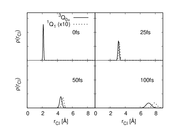

The evolution of the molecule in the transient excited state is shown in Fig. 1(a-b). Snapshots of the reduced density of the wave packet along the C – I bond distance that is obtained by projecting the wave packet onto one of the electronic states and tracing out all other nuclear degrees of freedom are shown in Fig. 1(a). Within , the bond distance is increased to two times the ground state equilibrium value. Due to the strongly repulsive potential, the wave packet stays relatively compact throughout the first after UV excitation. Additionally, the umbrella motion (not shown) brings the fragment from the tetrahedron structure to a closely planar geometry as the C – I bond dissociates. Some of the electronic state population is transferred to the lower state through non-adiabatic effects, but the molecule remains almost completely in the stateHammerich et al. 1994 (see Fig. 1(b)). For all considered bond lengths, the spin-orbit coupled state is dominated by the spin configuration. While for the considered UV wavelength spin-orbit effects are relevant for the photoexcitationGedanken and Rowe 1975 and for the iodine state into which the molecule dissociates, they play a minor role for the electronic state of the corresponding methyl fragmentde Nalda et al. 2008; Baumann et al. 2018; Kamiya and Taketsugu 2019; Corrales et al. 2014. For the XPS and AES due to carbon 1s ionization, spin-orbit splitting effects are smaller than Cutler et al. 1992 and thus play a minor role. Accordingly, we neglect in the following discussion spin orbit-coupling and discuss the time dependent spectra for the non-spin-orbit coupled state.


In Fig. 2(a), we present the calculated XPS for the photo-excited molecule as a function of bond-length assuming a photon energy of convoluted with a fixed-width Gaussian of full-width-at-half-maximum (FWHM). At the equilibrium geometry of the ground state, the C binding energy is for the singlet ground state. After excitation into the state, the C binding energy lowers to . The center of the XPS band observed at increases slightly with bond length up to an internuclear distance of . A satellite feature that can be attributed to valence shake-up is seen very weakly at equilibrium bond distance () at photoelectron energy . For larger bond distances this satellite feature becomes stronger and its binding energy decreases. For interatomic separations larger than , the initial weak satellite feature, which is at this bond distance at , becomes dominant and the original photoelectron line vanishes. While for the equilibrium bond distances very small splitting between doublet and quadruplet final electronic states is seen, at bond distances , distinct quadruplet and doublet features with an energy separation of occur. Due to their larger multiplicity, the two final quadruplet states (solid lines in Fig. 2(a)) dominante the TR-XPS with a relative strength twice as large as the corresponding doublet states. The TR-XPS spectrum resulting from the time-dependent wave packet (Fig. 1) is shown in Fig. 2(b). In this time-resolved spectrum, we have neglected the effect of the relatively small hydrogen motion that has little impact on the resulting x-ray spectra. The similarity of time and distance-dependent XPS showcases direct mapping onto the dissociation reaction coordinate. As can be seen, at a time delay of the position of the dominant photoelectron spectral line shows a pronounced jump that is connected with the wave packet being located at bond distances around .
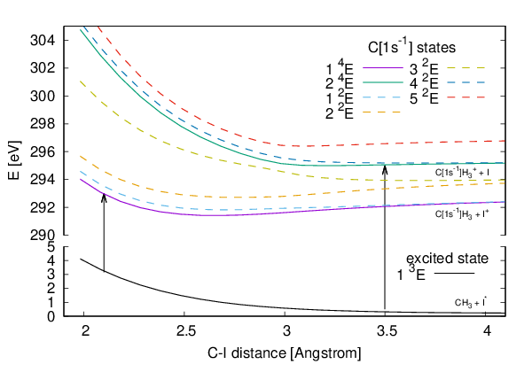
The spectral position of the photoelectron lines follow the difference of the potential energy curves of the excited and core ionized potential energy curves shown in Fig. 3. The jump in the XPS at a bond length is a consequence of the bond-length dependency of the core ionization cross-section to the different core-hole states.

This is clearly illustrated in Fig. 4, which shows the swap in the individual photoionization cross sections for the two involved final core-hole states of quadruplet spin multiplicity. The lower core-ionized state , which is dominantly populated through core shell ionization at equilibrium bond length, describes a redistribution of the valence electron density that screens the created core hole. It is instructive to realize that this lower carbon-core ionized state, which can be seen as a satellite for photoionization at large interatomic distance, corresponds to an asymptotic dissociation into I+ and a core-excited radical. The same counter-intuitive dissociation character of core hole states, where the charge is located on the atom that has not been core-ionized, has also been reported for COCorral et al. 2017. On the contrary, the upper core-ionized quadruplet state corresponds to an asymptotic dissociation into neutral I and a core-ionized cation. The electron density change accompanying core ionization of the initial photoexcited state is illustrated in the iso-surface plots in Fig. 4 for selected C – I distances. The blue iso-surface (electron density increase) around the carbon atom and the red iso-surfaces (electron density decrease) covering the hydrogen atoms clearly reflect the contraction of the valence electron density in the methyl moiety illustrating the screening of the core-hole. Additionally, one can see a shift of valence electron density involving the iodine atom that is qualitatively different for the two considered core-ionized states. The transition tends to shift electron density from the iodine towards the methyl moiety. In contrast, density change due to the transition is for small bond lengths characterized by a reshuffeling of density. For larger bond lengths, it shows almost no impact on the iodine atom. We note that the here discussed core-ionized or core-excited fragments are in transient states that will further charge up via Auger decay.
The characteristic valence electron changes following core ionization can be directly understood from the configurational mixing in the valence electronic structure as a function of interatomic separation. While the photoexcited state is for all bond distances characterized by a excitation, the two core hole states and show a mixing of core-hole configurations involving an additional excitation that reflects the tendency in the core ionized states to screen the core hole via shifting valence electrons from the iodine atom towards the methyl moiety. At equilibrium bond length, the state is dominated by the core-ionized configuration. The core hole leads to a somewhat stronger bond and, thus, to a slightly less dissociative potential energy curve compared to the initial valence excited state . Accordingly, the core-electron binding energy slightly increases with C – I distance (see Fig. 2(a)). This observation of strengthening of a bond through a core hole has been observed for a number of other core ionized moleculesCorral et al. 2017. With increasing bond length, the configurational mixing of the core-hole state is shifted more and more towards an additional excitation, which becomes dominant for bond length . Due to this change in configurational character the photoionization from the initial excited state is depleted. The higher-lying core-ionized state is for small bond distances dominated by the additional excitation resulting in a potential energy curve that is even more repulsive than the initial excited state (see Fig. 3). This is also indicated by the lowering of the respective photoelectron binding energy with inter-atomic distance (see Fig. 2(a)). The configurational character of this state changes with bond length in the opposite way: At the Franck-Condon region it describes a shake-up and for bond length it is characterized by the main configuration (i.e. without additional excitation). Accordingly, photoionization from the initial excited state increases with bond length.
From this finding we can directly connect the jump in the XPS with the suppression of the inter-atomic valence electron rearrangement in the context of core-hole screening. These dynamics can also be understood with the classical over-the-barrier model of charge migrationErk et al. 2014; Ryufuku et al. 1980 (see the supplementary material for details). According to this model, the electron transfer from I to is forbidden, when the internuclear distance is larger than the critical distance , which is consistent with the observed critical distance of in the XPS (Fig. 2(a)).
Strong sensitivity with respect to bond dissociation is also seen in the TR-AES that is shown in Fig. 5(a+b). Because of the multitude of different pathways resulting in densely lying Auger lines, a complete interpretation of all the lines and their bond distance dependence is tedious. There is a general trend for most of the Auger lines to show a shift to higher energy with increased inter-atomic separation. This effect can be attributed to the possible larger spatial separation and thus smaller Coulomb repulsion of the two valence holes in the final dicationic states that dissociate into two ion pairs. Around , many lines show a discontinuity that is a consequence of the discussed swap in the photoionization cross sections (Fig. 4). Above , the Auger lines seem to vary very weakly with interatomic distance. At these bond-lengths, the Auger decay in the fragment seems to be almost unaffected by the iodine fragment.


In the following we focus on the Auger line resulting in the fastest Auger electrons. Typically, these fast Auger electrons are rather isolated from other Auger lines, because they represent low energetic dicationic states, where the density of states of the dication is rather low compared to higher excited dicationic states. In general, fast Auger electrons are therefore easier to identify with specific dicationic electronic states. As shown in Fig. 5(a+b), the Auger spectrum exhibits a satellite feature above that becomes stronger around a C – I distance of and vanishes again for larger bond lengths. At equilibrium bond length , this Auger line is found at energies and its energetic position lowers with increasing C – I distance. Our calculation indicates that this Auger transition can be attributed to the decay of the higher-lying core-ionized quadruplet state into the dicationic state. The change of energetic position of this Auger line is dominated by the strongly repulsive curve of the core-ionized state. The dicationic state associated with this Auger channel dissociates into and . Thus, this Auger channel is accompanied with a transfer of a valence electron to the iodine atom. Analogous to the blocking of charge transfer in the XPS the intensity of this Auger line becomes therefore weaker with increasing bond length.
Via ab-initio modelling of XPS and AES, we have demonstrated here the power of element and site-specific soft x-ray spectroscopy for investigation of photoinduced bond dissociation, a fundamental process in photochemistry. We show at the example of methyl iodide that carbon time-resolved x-ray photoelectron spectra exhibit a distinct jump in between the initial core ionization potential of the molecule and of the reaction product . As we discuss in the Supp. Information, the jump is clearly preserved when finite time resolution of the experiment is taken into account. We connect this jump to the point in time and space ( and 20 fs), at which molecular charge redistribution upon carbon core ionization is blocked. The large sensitivity of XPS can be understood from the fact that the creation of a core hole is accompanied with screening effects in the valence shell, which lead to rearrangement of valence electrons from the electron-rich iodine atom to the moiety. The reported behavior also reveals that the production of fragments should show a strong sensitivity with delay time resulting from the different degree to populate the two different states through carbon core ionization. Thereby, our calculation present an interesting explanation of the bond-distance dependence of the molecular charge rearrangement mechanism upon x-ray ionizationErk et al. 2014; Boll et al. 2016. In comparison to our simulated XPS, the information content from time-resolved valence photoelectron spectroscopy is fundamentally different. The only such study on in the literature employs insufficient probe photon energies to follow the bond dissociation to the photoproducts. Warne et al. 2019. With sufficient probe photon energy, we can, however, expect to observe features, which correlate with the lowest cationic states of the photofragments and , to smoothly evolve on the dissociation coordinate from their binding energies in towards their binding energies in the fragmentsBerkowitz et al. 1981; Berkowitz et al. 1994. Instead, our simulated XPS provides exclusive information on the evolution of the valence electronic structure of the emerging fragment. Moreover, the valence photoelectron spectrum can not be expected to exhibit such a drastic change in the partial ionization cross-sections as we observe in the XPS. The TR-AES shows a remarkable sensitivity to C – I bond length. Although the spectrum is overall very crowded, particular features shift by more than , which should result in a clearly observable signal in a time-resolved AES experiment. It is remarkable that in such a pump-probe experiment, specific transient features are only visible in a region of intermediate C – I bond distance. Within a UV-pump-x-ray-probe scheme, this feature could be used as a sensitive indicator for a specific dissociation step.
While the predicted phenomena have not yet been fully resolved experimentally, Brauße et al. 2018 a confirmation of our predictions is well within the range of capabilities at current free electron laser facilities. We, furthermore, do not see any reason why they should be confined to our specific benchmark molecule and expect them to be general signatures of valence electron rearrangement during bond dissociation. With the help of the here reported spectral fingerprints of bond-breaking and new achievements in providing short and accurately controlled x-ray pulses, we expect time-resolved x-ray photoelectron spectroscopy and time-resolved Auger electron spectroscopy to become an enormously useful tool to investigate ultrafast photochemistry.
1 Methods
1.1 Quantum dynamics of photodissociation
In order to describe the non-Born-Oppenheimer nuclear dynamics triggered by photoexcitation of methyl iodide, we performed a five-dimensional quantum dynamics simulation using the multi-configuration time-dependent Hartree (MCTDH) methodBeck et al. 2000 reproducing the results of Ref. 34. In this work, the numerical procedure demonstrated to faithfully reproduce the branching ratio of I/I* and quantum states distribution of dissociation products. Since it is well described elsewhereBeck et al. 2000; Hammerich et al. 1994, we here only give a brief overview.
A set of coordinates is used to describe the processes depicted in Fig. 6, the C – I dissociation, C – H vibration, bending, umbrella motion of and wagging of I atom. In the chosen coordinate space, is the major coordinate characterizing the dissociation process. Motion of and are driven naturally by the recoil of C-I dissociation. is crucial to break the symmetry of the molecule to enable the non-Born-Oppenheimer coupling between the valence excited states, which leads to the formation of excited and ground state iodine atoms, and plays minor role. The initial state of the wave packet of the UV excited molecule is prepared by sudden excitation approximation following the Franck-Condon principle Hammerich et al. 1994. The employed potential energy surfaces for the and state are based on ab initio spin-orbit configuration interaction calculations Amatatsu et al. 1991; Hammerich et al. 1994.

The time-dependent wave function of the molecule is represented as
| (1) | |||||
for our case , where and denotes the discrete basis of the electronic degrees of freedom. The function are the time-dependent single particle function (SPF) basis that forms a co-moving cover of the wave packet. The equation of motion used to propagate the SPF and the expansion coefficients in time follow from the Dirac-Frenkel variation principle as
| (2) |
and
| (3) |
where the vector notation has been used. Further details on the MCTDH method and the the definitions of the mean field , the density matrix and the projector can be found in Ref. 47.
1.2 Femtosecond XPS and AES of the photodissociating molecule
For simplicity, we discuss the resulting XPS and AES without the effect of spin-orbit interaction. As discussed in the result section, the most dominantly populated state can be associated almost purely with a non-spin-orbit coupled state. The resulting XPS might be blurred due to splitting of the final, core-ionized states. We estimate the effect of splitting due to spin-orbit splitting to be smaller than Cutler et al. 1992. Given the additional spreading of the spectra due to the spatial extend of the vibrational wave-packet on the dissociating potential energy curves, spin-orbit splitting details will most-likely be hidden within a broad peak of the individual photo- and Auger electrons. The spin-orbit coupling therefore does not involve any characteristic changes in the XPS and AES and for our discussion it is therefore sufficient to neglect spin-orbit interaction.
For a given molecular geometry, the photoionization cross section of the initial electronic state can be calculated as
| (4) |
where is the fine structure constant, and are the initial and final molecular eigenstate with energy and , respectively, is the photon energy, and a function that determines the shape and position of the line. The many-electron dipole-matrix element in Eq. (4) is given by
| (5) |
where is the dipole moment operator, and are fermionic operators that annihilate and create an electron, respectively, and the sum over and runs over a complete set of one-particle basis functions.
To account for the finite width of the vibrational wave-packet evolving in time and following the arguments in Ref. 25, we employ
| (6) |
where is the time-dependent vibrational wavepacket density projected along the coordinate and a parameter that accounts for additional broadening effects, e.g. finite lifetime and spectral bandwidth of the x-ray pulse. In the following we choose .
For the calculation of the XPS, we employ the XMOLECULE toolkitHao et al. 2015; Inhester et al. 2016; Inhester et al. 2018. We evaluate the transition dipole matrix element in Eq. (5) on the basis of the one-center approximation. To that end, the final electronic state, , is separated into a continuum wave function and a bound, core-ionized multielectron part . The respective continuum wave function describing the photoelectron with kinetic energy is approximated with the continuum wave function from an atomic carbon Auger decay calculation. We describe the bound electronic structure with a CASCI(6,4)/6-311G calculation, where for the final electronic state , the carbon core orbital is kept singly occupied. This is done employing an orbital basis obtained from a ROHF calculation for the neutral, excited state and the carbon-core-ionized configuration, respectively. Because, we use different sets of orbitals for the initial and final states, evaluation of the transition dipole moment (Eq. (5)) requires the calculation of overlap matrices of the involved spin configurationsInhester et al. 2018. We calculate the XPS for all the accessible core ionized states of irreducible representation that result from the full diagonalization of the complete active space. This results in the core ionized states , whose potential energy curves are depicted in Fig. 3 together with the potential energy curve of the initial UV-excited state .
For the calculation of the AES, we employ the methods described in Ref. 40. The rate for the auto-ionization of an initial core ionized state with a hole in orbital to a final state with reoccupied core hole, two holes in the valence, and an Auger electron is given by
| (7) |
where is the Coulomb matrix element for the interaction of two valence electrons and with the continuum electron and the core electron . In Eq. (7) the sum over runs over all valence electron pairs and . We evaluate the two-electron integral matrix elements by employing the one-center approximationInhester et al. 2016. In this approximation the wave function for the Auger electron, , is approximated with the wave function of the atomic Auger electron. The initial state is described the same way as the bound part of the final electronic states in the XPS calculation. The bound electronic structure of the final states is described with a multi-reference CI calculation involving all two-valence-hole configurations out of the CASCI(6,4) reference space employing an orbital set obtained from a ROHF/6-311G calculation for the dicationic triplet ground state (). We assume that the molecular geometry is static during the short lifetime of the core hole (). To account for the fact that we have multiple core hole states, the resulting Auger spectrum is constructed from the product of the partial core-photoionization cross section and respective Auger decay rate summed over all intermediate core ionized states. We convolute the resulting Auger spectrum with Gaussian of full-width-at-half-maximum to account for vibrational and finite lifetime broadening effects.
omparison of calculated XPS and AES data with available experimental data, discussion on the impact of finite-time resolution in realistic experimental setups, complementary analysis of the charge transfer upon core shell ionization employing the classical over-the-barrier model and the MCTDH simulation.
References
- Cheng and Fleming (2009) Cheng, Y.-C.; Fleming, G. R. Dynamics of Light Harvesting in Photosynthesis. Ann. Rev. Phys. Chem. 2009, 60, 241–262.
- Polli et al. (2010) Polli, D.; Altoè, P.; Weingart, O.; Spillane, K. M.; Manzoni, C.; Brida, D.; Tomasello, G.; Orlandi, G.; Kukura, P.; Mathies, R. A. et al. Conical Intersection Dynamics of the Primary Photoisomerization Event in Vision. Nature 2010, 467, 440–443.
- Wolf et al. (2014) Wolf, T.; Kuhlman, T. S.; Schalk, O.; Martinez, T.; Møller, K. B.; Stolow, A.; Unterreiner, A.-N. Hexamethylcyclopentadiene: Time-Resolved Photoelectron Spectroscopy and Ab Initio Multiple Spawning Simulations. Phys. Chem. Chem. Phys. 2014, 16, 11770–11779.
- Horton et al. (2019) Horton, S. L.; Liu, Y.; Forbes, R.; Makhija, V.; Lausten, R.; Stolow, A.; Hockett, P.; Marquetand, P.; Rozgonyi, T.; Weinacht, T. Excited State Dynamics of CH2I2 and CH2BrI Studied with UV Pump VUV Probe Photoelectron Spectroscopy. J. Chem. Phys. 2019, 150, 174201.
- Coates et al. (2018) Coates, M. R.; Larsen, M. A. B.; Forbes, R.; Neville, S. P.; Boguslavskiy, A. E.; Wilkinson, I.; Sølling, T. I.; Lausten, R.; Stolow, A.; Schuurman, M. S. Vacuum Ultraviolet Excited State Dynamics of the Smallest Ring, Cyclopropane. II. Time-Resolved Photoelectron Spectroscopy and Ab Initio Dynamics. J. Chem. Phys. 2018, 149, 144311.
- MacDonell et al. (2016) MacDonell, R. J.; Schalk, O.; Geng, T.; Thomas, R. D.; Feifel, R.; Hansson, T.; Schuurman, M. S. Excited State Dynamics of Acrylonitrile: Substituent Effects at Conical Intersections Interrogated via Time-Resolved Photoelectron Spectroscopy and Ab Initio Simulation. J. Chem. Phys. 2016, 145, 114306.
- Schalk et al. (2016) Schalk, O.; Geng, T.; Thompson, T.; Baluyot, N.; Thomas, R. D.; Tapavicza, E.; Hansson, T. Cyclohexadiene Revisited: A Time-Resolved Photoelectron Spectroscopy and Ab Initio Study. J. Phys. Chem. A 2016, 120, 2320–2329.
- Warne et al. (2019) Warne, E. M.; Downes-Ward, B.; Woodhouse, J.; Parkes, M. A.; Bellshaw, D.; Springate, E.; Majchrzak, P.; Zhang, Y.; Karras, G.; Wyatt, A. S. et al. Photodissociation Dynamics of CH3I Probed via Multiphoton Ionisation Photoelectron Spectroscopy. Phys. Chem. Chem. Phys. 2019, 21, 11142–11149.
- Emma et al. (2010) Emma, P.; Akre, R.; Arthur, J.; Bionta, R.; Bostedt, C.; Bozek, J.; Brachmann, A.; Bucksbaum, P.; Coffee, R.; Decker, F.-J. et al. First Lasing and Operation of an Ångstrom-Wavelength Free-Electron Laser. Nat. Photonics 2010, 4, 641–647.
- Pertot et al. (2017) Pertot, Y.; Schmidt, C.; Matthews, M.; Chauvet, A.; Huppert, M.; Svoboda, V.; von Conta, A.; Tehlar, A.; Baykusheva, D.; Wolf, J.-P. et al. Time-Resolved x-Ray Absorption Spectroscopy with a Water Window High-Harmonic Source. Science 2017, 355, 264–267.
- Attar et al. (2017) Attar, A. R.; Bhattacherjee, A.; Pemmaraju, C. D.; Schnorr, K.; Closser, K. D.; Prendergast, D.; Leone, S. R. Femtosecond X-Ray Spectroscopy of an Electrocyclic Ring-Opening Reaction. Science 2017, 356, 54–59.
- Teichmann et al. (2016) Teichmann, S. M.; Silva, F.; Cousin, S. L.; Hemmer, M.; Biegert, J. 0.5-keV Soft X-Ray Attosecond Continua. Nat. Commun. 2016, 7, 11493.
- Johnson et al. (2018) Johnson, A. S.; Austin, D. R.; Wood, D. A.; Brahms, C.; Gregory, A.; Holzner, K. B.; Jarosch, S.; Larsen, E. W.; Parker, S.; Strüber, C. S. et al. High-Flux Soft x-Ray Harmonic Generation from Ionization-Shaped Few-Cycle Laser Pulses. Sci. Adv. 2018, 4, eaar3761.
- Allaria et al. (2012) Allaria, E.; Appio, R.; Badano, L.; Barletta, W. A.; Bassanese, S.; Biedron, S. G.; Borga, A.; Busetto, E.; Castronovo, D.; Cinquegrana, P. et al. Highly Coherent and Stable Pulses from the FERMI Seeded Free-Electron Laser in the Extreme Ultraviolet. Nat. Photonics 2012, 6, 699–704.
- Ackermann et al. (2007) Ackermann, W.; Asova, G.; Ayvazyan, V.; Azima, A.; Baboi, N.; Bähr, J.; Balandin, V.; Beutner, B.; Brandt, A.; Bolzmann, A. et al. Operation of a Free-Electron Laser from the Extreme Ultraviolet to the Water Window. Nat. Photonics 2007, 1, 336–342.
- McFarland et al. (2014) McFarland, B. K.; Farrell, J. P.; Miyabe, S.; Tarantelli, F.; Aguilar, A.; Berrah, N.; Bostedt, C.; Bozek, J. D.; Bucksbaum, P. H.; Castagna, J. C. et al. Ultrafast X-Ray Auger Probing of Photoexcited Molecular Dynamics. Nat. Commun. 2014, 5, 4235.
- Wolf et al. (2017) Wolf, T. J. A.; Myhre, R. H.; Cryan, J. P.; Coriani, S.; Squibb, R. J.; Battistoni, A.; Berrah, N.; Bostedt, C.; Bucksbaum, P.; Coslovich, G. et al. Probing Ultrafast */n* Internal Conversion in Organic Chromophores via K-Edge Resonant Absorption. Nat. Commun. 2017, 8, 29.
- Wolf et al. (2017) Wolf, T. J. A.; Holzmeier, F.; Wagner, I.; Berrah, N.; Bostedt, C.; Bozek, J.; Bucksbaum, P.; Coffee, R.; Cryan, J.; Farrell, J. et al. Observing Femtosecond Fragmentation Using Ultrafast X-Ray-Induced Auger Spectra. Appl. Sci. 2017, 7, 681.
- Brauße et al. (2018) Brauße, F.; Goldsztejn, G.; Amini, K.; Boll, R.; Bari, S.; Bomme, C.; Brouard, M.; Burt, M.; de Miranda, B. C.; Düsterer, S. et al. Time-Resolved Inner-Shell Photoelectron Spectroscopy: From a Bound Molecule to an Isolated Atom. Phys. Rev. A 2018, 97, 043429.
- Siefermann et al. (2014) Siefermann, K. R.; Pemmaraju, C. D.; Neppl, S.; Shavorskiy, A.; Cordones, A. A.; Vura-Weis, J.; Slaughter, D. S.; Sturm, F. P.; Weise, F.; Bluhm, H. et al. Atomic-Scale Perspective of Ultrafast Charge Transfer at a Dye–Semiconductor Interface. J. Phys. Chem. Lett. 2014, 5, 2753–2759.
- Sann et al. (2016) Sann, H.; Havermeier, T.; Müller, C.; Kim, H.-K.; Trinter, F.; Waitz, M.; Voigtsberger, J.; Sturm, F.; Bauer, T.; Wallauer, R. et al. Imaging the Temporal Evolution of Molecular Orbitals during Ultrafast Dissociation. Phys. Rev. Lett. 2016, 117, 243002.
- Siegbahn et al. (1969) Siegbahn, K.; Nordling, C.; Fallmann, C. ESCA Applied to Free Molecules; North-Holland Publishing Co. Amsterdam, 1969.
- Bagus et al. (1999) Bagus, P. S.; Illas, F.; Pacchioni, G.; Parmigiani, F. Mechanisms Responsible for Chemical Shifts of Core-Level Binding Energies and Their Relationship to Chemical Bonding. J. Electron. Spectrosc. Relat. Phenom. 1999, 100, 215–236.
- Inhester et al. (2012) Inhester, L.; Burmeister, C. F.; Groenhof, G.; Grubmüller, H. Auger Spectrum of a Water Molecule after Single and Double Core Ionization. J. Chem. Phys. 2012, 136, 144304.
- Marchenko et al. (2018) Marchenko, T.; Inhester, L.; Goldsztejn, G.; Travnikova, O.; Journel, L.; Guillemin, R.; Ismail, I.; Koulentianos, D.; Ceolin, D.; Püttner, R. et al. Ultrafast Nuclear Dynamics in the Double-Core Ionized Water Molecule Observed via Auger Spectroscopy. Phys. Rev. A 2018,
- Erk et al. (2014) Erk, B.; Boll, R.; Trippel, S.; Anielski, D.; Foucar, L.; Rudek, B.; Epp, S. W.; Coffee, R.; Carron, S.; Schorb, S. et al. Imaging Charge Transfer in Iodomethane upon X-Ray Photoabsorption. Science 2014, 345, 288–291.
- Boll et al. (2016) Boll, R.; Erk, B.; Coffee, R.; Trippel, S.; Kierspel, T.; Bomme, C.; Bozek, J. D.; Burkett, M.; Carron, S.; Ferguson, K. R. et al. Charge Transfer in Dissociating Iodomethane and Fluoromethane Molecules Ionized by Intense Femtosecond X-Ray Pulses. Struct. Dyn. 2016, 3, 043207.
- Rudenko et al. (2017) Rudenko, A.; Inhester, L.; Hanasaki, K.; Li, X.; Robatjazi, S. J.; Erk, B.; Boll, R.; Toyota, K.; Hao, Y.; Vendrell, O. et al. Femtosecond Response of Polyatomic Molecules to Ultra-Intense Hard X-Rays. Nature 2017, 546, 129–132.
- Hao et al. (2019) Hao, Y.; Inhester, L.; Son, S.-K.; Santra, R. Theoretical Evidence for the Sensitivity of Charge-Rearrangement-Enhanced x-Ray Ionization to Molecular Size. Phys. Rev. A 2019, 100, 013402.
- de Nalda et al. (2008) de Nalda, R.; Durá, J.; García-Vela, A.; Izquierdo, J. G.; González-Vázquez, J.; Banares, L. A Detailed Experimental and Theoretical Study of the Femtosecond A-Band Photodissociation of CH3I. J. Chem. Phys. 2008, 128, 244309.
- Corrales et al. (2014) Corrales, M.; González-Vázquez, J.; Balerdi, G.; Solá, I.; De Nalda, R.; Bañares, L. Control of Ultrafast Molecular Photodissociation by Laser-Field-Induced Potentials. Nat. Chem. 2014, 6, 785.
- Tehlar and Wörner (2013) Tehlar, A.; Wörner, H. J. Time-Resolved High-Harmonic Spectroscopy of the Photodissociation of CH3I and CF3I. Mol. Phys. 2013, 111, 2057–2067.
- Gedanken and Rowe (1975) Gedanken, A.; Rowe, M. D. Magnetic Circular Dichroism Spectra of the Methyl Halides. Resolution of the n* Continuum. Chem. Phys. Lett. 1975, 34, 39–43.
- Hammerich et al. (1994) Hammerich, A. D.; Manthe, U.; Kosloff, R.; Meyer, H.-D.; Cederbaum, L. S. Time-Dependent Photodissociation of Methyl Iodide with Five Active Modes. J. Chem. Phys. 1994, 101, 5623–5646.
- Attar et al. (2015) Attar, A. R.; Bhattacherjee, A.; Leone, S. R. Direct Observation of the Transition-State Region in the Photodissociation of CH3I by Femtosecond Extreme Ultraviolet Transient Absorption Spectroscopy. J. Phys. Chem. Lett. 2015, 6, 5072–5077.
- Drescher et al. (2016) Drescher, L.; Galbraith, M. C. E.; Reitsma, G.; Dura, J.; Zhavoronkov, N.; Patchkovskii, S.; Vrakking, M. J. J.; Mikosch, J. Communication: XUV Transient Absorption Spectroscopy of Iodomethane and Iodobenzene Photodissociation. J. Chem. Phys. 2016, 145, 011101.
- Baumann et al. (2018) Baumann, A.; Rompotis, D.; Schepp, O.; Wieland, M.; Drescher, M. Time-Resolved Dissociation Dynamics of Iodomethane Resulting from Rydberg and Valence Excitation. J. Phys. Chem. A 2018, 122, 4779–4784.
- Hao et al. (2015) Hao, Y.; Inhester, L.; Hanasaki, K.; Son, S.-K.; Santra, R. Efficient Electronic Structure Calculation for Molecular Ionization Dynamics at High X-Ray Intensity. Struct. Dyn. 2015, 2, 041707.
- Inhester et al. (2016) Inhester, L.; Hanasaki, K.; Hao, Y.; Son, S.-K.; Santra, R. X-Ray Multiphoton Ionization Dynamics of a Water Molecule Irradiated by an x-Ray Free-Electron Laser Pulse. Phys. Rev. A 2016, 94, 023422.
- Inhester et al. (2018) Inhester, L.; Oostenrijk, B.; Patanen, M.; Kokkonen, E.; Southworth, S. H.; Bostedt, C.; Travnikova, O.; Marchenko, T.; Son, S.-K.; Santra, R. et al. Chemical Understanding of the Limited Site-Specificity in Molecular Inner-Shell Photofragmentation. J. Phys. Chem. Lett. 2018, 9, 1156–1163.
- Kamiya and Taketsugu (2019) Kamiya, M.; Taketsugu, T. Ab Initio Surface Hopping Excited-State Molecular Dynamics Approach on the Basis of Spin–Orbit Coupled States: An Application to the A-Band Photodissociation of CH3I. J. Comput. Chem. 2019, 40, 456–463.
- Cutler et al. (1992) Cutler, J. N.; Bancroft, G. M.; Tan, K. H. Ligand-field Splittings and Core-level Linewidths in I 4d Photoelectron Spectra of Iodine Molecules. J. Chem. Phys. 1992, 97, 7932–7943.
- Corral et al. (2017) Corral, I.; González-Vázquez, J.; Martín, F. Potential Energy Surfaces of Core-Hole and Shake-Up States for Dissociative Ionization Studies. J. Chem. Theory Comput. 2017, 13, 1723–1736.
- Ryufuku et al. (1980) Ryufuku, H.; Sasaki, K.; Watanabe, T. Oscillatory Behavior of Charge Transfer Cross Sections as a Function of the Charge of Projectiles in Low-Energy Collisions. Phys. Rev. A 1980, 21, 745–750.
- Berkowitz et al. (1981) Berkowitz, J.; Batson, C. H.; Goodman, G. L. Photoionization of Atomic Iodine and Atomic Tellurium. Phys. Rev. A 1981, 24, 149–160.
- Berkowitz et al. (1994) Berkowitz, J.; Ellison, G. B.; Gutman, D. Three Methods to Measure RH Bond Energies. J. Phys. Chem. 1994, 98, 2744–2765.
- Beck et al. (2000) Beck, M. H.; Jäckle, A.; Worth, G. A.; Meyer, H.-D. The Multiconfiguration Time-Dependent Hartree (MCTDH) Method. a Highly Efficient Algorithm for Propagating Wavepackets. Phys. Rep. 2000, 324, 1–105.
- Amatatsu et al. (1991) Amatatsu, Y.; Morokuma, K.; Yabushita, S. Ab Initio Potential Energy Surfaces and Trajectory Studies of A-band Photodissociation Dynamics: CH3I*CH3+I and CH3+I*. J. Chem. Phys. 1991, 94, 4858–4876.