1 TU Wien,
Gusshausstrasse 25/E389,
Vienna,
Austria
2 ÉTS,
1100 Notre Dame St W,
Montreal,
Canada
3 CentraleSupélec-CNRS-Université Paris Sud,
3 rue Joliot-Curie,
Gif-sur-Yvette,
France
ubfigurelabel[3]#1\subfigurelabel#2
myCSVstyleseparator=semicolon, respect all, head to column names=true
[myCSVstyle, filter strcmp=\csvcoliavg]csv-data/compare-dc.csv
On Direct Distribution Matching for Adapting Segmentation Networks
Abstract
Minimization of distribution matching losses is a principled approach to domain adaptation in the context of image classification. However, it is largely overlooked in adapting segmentation networks, which is currently dominated by adversarial models. We propose a class of loss functions, which encourage direct kernel density matching in the network-output space, up to some geometric transformations computed from unlabeled inputs. Rather than using an intermediate domain discriminator, our direct approach unifies distribution matching and segmentation in a single loss. Therefore, it simplifies segmentation adaptation by avoiding extra adversarial steps, while improving quality, stability and efficiency of training. We juxtapose our approach to state-of-the-art segmentation adaptation via adversarial training in the network-output space. In the challenging task of adapting brain segmentation across different magnetic resonance imaging (MRI) modalities, our approach achieves significantly better results both in terms of accuracy and stability.
keywords
domain adaptation, unsupervised domain adaptation, semantic segmentation, direct distribution matching1 Introduction
Semantic segmentation is of pivotal importance towards high-level understanding of image content, which is useful in a breadth of application areas, from autonomous driving to health care, for instance. Particularly, in medical imaging, segmentation facilitates clinical tasks, including disease diagnosis, treatment and follow-up, among others. Modern medical segmentation approaches rely on deep learning techniques, which have demonstrated outstanding performance in a breadth of applications Dolz et al. 2018a; Dolz et al. 2018b; Litjens et al. 2017. Despite their success, generalization of trained models to new scenarios is hampered if the gap between data distributions across domains is large. A trivial solution to address this issue would be to re-annotate images from different domains and re-train or fine-tune the deep models. Nevertheless, obtaining such massive amounts of labeled data is a cumbersome process which, for some applications, may require user expertise, resulting in a prohibitive and unrealistic approach.
To tackle this problem, uda (uda) techniques have been widely investigated. These methods aim at learning robust classifiersin the presence of a shift between source and target distributions when the target data is unlabeled. In this scenario, the goal is typically to minimize the discrepancy between distributions across domains at the input Bousmalis et al. 2017; Chen et al. 2018a; Hoffman et al. 2018; Russo et al. 2018; Sankaranarayanan and Balaji 2018; Wu et al. 2018 or intermediate-feature level Ganin and Lempitsky 2015; Ghifary et al. 2016; Kamnitsas et al. 2017; Liu et al. 2018; Long et al. 2015; Long et al. 2016; Tzeng et al. 2017, while leveraging labeled source examples to retain discriminative power on the feature space. Generative techniques either operate on a pixel-level Bousmalis et al. 2017; Chen et al. 2018a; Russo et al. 2018; Shrivastava et al. 2017; Zhang et al. 2018a or in feature space Dou et al. 2018b; Ganin and Lempitsky 2015; Kamnitsas et al. 2017; Long et al. 2015; Tzeng et al. 2017 and align the image appearance between domains, so that the target data “style” is transferred to source data, or vice-versa. Then, supervised learning is performed with the newly generated synthetic data. A downside of these approaches is that they perform satisfactorily only for small images and narrow domain shifts, which limits their applicability. Within the current paradigm of learning domain-invariant representations, domain adversarial training Ganin and Lempitsky 2015; Tzeng et al. 2017 and mmd (mmd) Long et al. 2015; Sun and Saenko 2016; Yan et al. 2017 have become very popular choices.
For semantic segmentation problems, adversarial training models Goodfellow et al. 2014 are currently dominating the literature Chen et al. 2017; Chen et al. 2018b; Dou et al. 2018a; Kamnitsas et al. 2017; Hoffman et al. 2016; Hong et al. 2018; Saito et al. 2018; Tsai et al. 2018; Vu et al. 2019. Such models alternate the training of two networks: a discriminator that learns a decision boundary between source and target features and a segmentation network that uses the learned decision boundary to match the feature distributions across domains. Some other approaches rely on generative networks, which yield target images conditioned on the source, or vice-versa, aligning both domains at the pixel level Cai et al. 2019; Huo et al. 2018; Murez et al. 2018; Sankaranarayanan and Balaji 2018; Zhang et al. 2018b; Zhao et al. 2019.
While adversarial training achieved outstanding performances in image classification, our numerical evidence and intuition suggest that it may not be suitable for segmentation tasks to the same degree. First, learning a discriminator boundary for a segmentation task is much more complex as the label space is exponentially large. Intuitively, a high dimensional label space implies that the discriminator boundary can be very complicated and thus hard to learn. Therefore, as we will see later in our experiments, alternating both adversarial and prediction tasks in segmentation might cause more significant training instabilities than in image classification tasks. Moreover, it is more unlikely that source and target domain share the same multi-level feature representations if the label space is high dimensional.
While the inputs can differ significantly from one domain to another, the output (label) space in semantic segmentation conveys very rich information related to the spatial layout and local context, which is shared across domains. Inspired by this observation, Tsai et al.Tsai et al. 2018 proposed adversarial training in the output (softmax segmentation) space, achieving better performance than features-matching approaches on the Cityscapes dataset. Leveraging this information is even more meaningful in medical images, where label (output) statistics remain domain-independent, despite significant differences in image inputs across domains. Nevertheless, following the trend in uda approaches for natural image segmentation, adversarial learning has become the de facto choice in medical image segmentation Chen et al. 2019; Dou et al. 2018a; Gholami et al. 2018; Javanmardi and Tasdizen 2018; Kamnitsas et al. 2017; Zhang et al. 2018a; Zhao et al. 2019. It is worth mentioning that some recent natural image segmentation works Zhang et al. 2017; Zou et al. 2018 pointed out that adversarial models for classification do not translate well to segmentation. These studies showed that similar or better performances can be achieved by other alternatives.
Here, we propose a simple, easily trainable approach to uda, that can be applied in cases where the underlying (latent) ground truth is identical for source and target domains, up to some geometric transformations of unlabeled images. While unrealistic for natural images, this can easily be achieved in medical imaging, e.g., by obtaining separate scans of one patient with different imaging methods or by applying multi-modal registration algorithms to unlabeled image pairs. The class of loss functions we propose encourages direct density matching in the network’s output space. It follows the principle of Minimization of distribution matching losses, a principled approach to da (da) in the context of image classification, e.g., mmd Long et al. 2015; Sun and Saenko 2016; Yan et al. 2017. Rather than using an intermediate domain discriminator, our direct approach unifies distribution matching and segmentation in a single loss. Therefore, it simplifies segmentation adaptation by avoiding extra adversarial steps, while improving quality, stability and efficiency of training. We compare our approach to the state-of-art segmentation method in Tsai et al. 2018. In the challenging task of adapting brain segmentation across different mri (mri) modalities, our approach achieves significantly better performance than adversarial output adaption, both in terms of accuracy and stability. We also investigate experimentally the sensitivity of our approach to the alignment of unlabeled image pairs.
2 Formulation
Consider an unsupervised domain-adaptation setting with two distinct subsets: contains labeled source-domain images and the corresponding ground-truth segmentations , and contains unlabeled image pairs, each involving a source image and a target image . For each labeled source image , , the ground-truth labeling is a matrix whose columns are binary vectors, encoding the assignment of pixel to one of classes (segmentation regions): , where if and only if label is assigned to pixel of the -th image. For any image , let denote the probability vector of softmax outputs for pixel , with the trainable parameters of the network. For the sake of simplicity, we will omit the subscript in the following.
We propose to minimize the following loss function:
| (1) |
where
- •
evaluates the discrepancy between two probability distributions and , e.g., kl (kl) divergence , where superscript denotes transposition.
- •
denotes standard cross-entropy loss for labeled source-domain images: .
- •
is a non-negative multiplier.
- •
could be simply identity if unlabeled images and are aligned, e.g., by acquisition11 1 In some practical scenarios, images from different modalities are aligned when acquired at the same time.. Also, could be a geometric transformation, which aligns pairs of unlabeled images, for instance, using a standard automatic cross-modality registration algorithm Oliveira and Tavares 2014.
The first term in our model (1) is the usual cross-entropy loss of a semantic segmentation problem on the source domain, while the second term, which is based on unlabeled image pairs, encourages the network outputs (softmax segmentations) in the target domain to closely match those in the source domain. In fact, when corresponds to some kernel function, i.e., , the summation over pixels in the second term of (1) can be expressed in terms of a kernel between two softmax segmentations in :
| (2) |
with denoting the matrix whose columns are the softmax outputs at each pixel, i.e., probability vectors . Now, notice that the kernel density estimate (KDE)22 2 KDEs are also commonly referred to as Parzen window estimates. of the distribution of source-domain softmax segmentations, i.e., the network outputs in , can be written as follows: . Therefore, by maximizing these source density estimates at target-domain segmentations, we directly match the distributions of the source and target domains in the network-output space. This amounts to minimizing the following direct distribution-matching loss:
| (3) |
Clearly, from the expression of kernel in (2), the second term in our loss in (1) can be viewed as an approximation of (3) based on a subset of pairwise matching kernels. Therefore, our loss in (1) encourages direct density matching in the network-output space.
Fig. 1 highlights the conceptual differences between our direct matching (Fig. 1) and the state-of-art adversarial method in Tsai et al. 2018, which pursues a two-step adversarial learning in the network-output space (Fig. 1 and 1), so as to achieve the same goal as our loss: matching the source and target distributions of label predictions.
[Adversarial (Discriminator)]
 \subfigure[Adversarial (Segmenter)]
 \subfigure[Direct Distribution Matching]
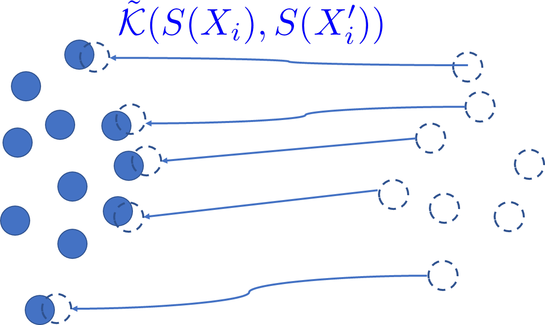
The model in Tsai et al. 2018 alternates the training of two networks: a discriminator, which learns to distinguish between source and target outputs; and a segmentation network, which is trained using the discriminator. The discriminator is used to encourage the target outputs to be similar to those of the source domain. Rather than using an intermediate domain discriminator, our direct method unifies distribution matching and segmentation in a single loss. Therefore, it simplifies segmentation adaptation by avoiding extra adversarial steps, while improving both the quality, stability and efficiency of training. While adversarial training achieved outstanding performances in image classification, our numerical evidence and intuition suggest that it may not be suitable for segmentation, in which case learning a discriminator boundary is much more complex as it solves for predictions in an exponentially large label space. In fact, intuitively, a large label space implies large spaces of possible solutions for discriminator boundaries and target predictions, both of which are latent; see dashed boundaries and data points in Fig. 1. Alternating both adversarial and prediction tasks in segmentation can cause more significant instabilities than in image classification tasks, as we will see later in our experiments.
Another important difference between our approach and adversarial training is that we account for the fact that target and source data have a common ground truth in the label space, up to some geometric transformation. Such prior information is very common and useful in medical imaging problems, but adversarial approaches do not have mechanisms to take advantage of it.
3 Experiments
We evaluated our approach extensively on the challenging task of brain tissue segmentation in mri scans, and compared the performances to the state-of-the-art method in Tsai et al. 2018.
3.1 Experimental details
Datasets: We performed numerical studies on two public segmentation benchmarks: MRBrainS2013 Mendrik et al. 2015 and iSEG2017 Wang et al. 2019. The MRBrainS dataset contains 5 labeled and 15 unlabeled scans of adult brains. The iSEG dataset is composed of 10 labeled and 13 unlabeled infant brain scans. We tested our domain adaptation on the T1 and T2-FLAIR modalities of MRBrainS and the T1 and T2 modalities of iSEG. The task consists of segmenting the wm (wm), gm (gm) and csf (csf). The original T2 images from iSEG were resampled into an isotropic resolution, and then aligned onto their corresponding T1 images with a simple affine registration method. The sequences from the MRBrainS Challenge were aligned by rigid registration, using Elastix Klein et al. 2010.
Training: The data consists of two distinct subsets. is the labeled subset, which contains images from the source domain with their corresponding ground truth . Unlabeled subset contains pairs of aligned source and target data, respectively, without a ground-truth. In our experiments, we found that the choice of distance functions does not significantly alter performances. If not mentioned otherwise, we used kl divergence and the multiplier .
Due to the limited size of the training set, we employed a leave-one-out-cross-validation strategy, where only one image was used for testing/evaluation, leaving the remaining images for training. We used four of the five labeled scans in the MRBrainS2013 dataset as samples in . The one remaining scan was used for evaluation. As the iSEG dataset contains more labeled scans, we opted to use 8 scans for training in and one scan for testing and evaluation, respectively. Furthermore, all unlabeled scans, i.e., 15 and 13 in the MRBrainS2013 and iSEG datasets, respectively, are used in to compute the unsupervised term in (1). Each experiment was performed three times with different evaluation/testing data splits and the average as well as the empirical standard deviation reported subsequently were computed over these three runs.
Baselines and comparisons: In order to evaluate the impact of the adaptation approaches, we trained the segmentation network in a supervised manner on the source and target data, providing a lower and upper bound for the uda results. While the network trained on source images is referred to as no adaptation, the network trained on the target domain is referred to as the oracle. In addition, we compare the proposed approach with the adversarial method proposed in Tsai et al. 2018. For a fair comparison, we used the same segmentation network for the proposed and the adversarial approach. For simplicity, we chose the “single-level” strategy, performing da only on the output layer. We used the same discriminator model as Tsai et al. 2018. The Lagrange multiplier for training the segmentation network was chosen to be . Although AdaptSegNet does not utilize the fact that source and target data are aligned in , we nevertheless trained the discriminator with these aligned pairs. Subsequent runs indeed revealed that this does not have an impact on the performance of AdaptSegNet.
Implementation details: We used a slightly modified U-Net Ronneberger et al. 2015 for the segmentation task, operating on 2D slices. Particularly, the employed network follows the original implementation Ronneberger et al. 2015, but the depth is reduced by one, i.e., max-pool is performed only three times instead of four. We used ReLU activation functions and did not include dropout, to avoid any regularization that does not originate from our proposed da strategy. To obtain 2D input, the 3D images are sliced along the z-axis. However, Dice coefficients are computed on the 3D scans. The implementation was done in TensorFlow, and the experiments were run on a server equipped with a NVidia Titan V GPU with 12 GB memory. For all networks, we employed the Adam Kingma and Ba 2015 optimizer with learning rate and a batch size of . We performed fully supervised pre-training for 200 epochs on the source domain data. Subsequently, we trained for 800 epochs with the full loss (1), totaling 1000 training epochs. The code is publicly available at https://github.com/g-pichler/DDMSegNet.
Evaluation: We resorted to the common Dice coefficient, widely employed in medical image segmentation, to compare quantitatively the performances of the different methods. When using the iSEG dataset, the mean Dice coefficient on the test scan was used to determine the best model during training. We then report the performance of this model on the evaluation sample. Due to the limited size of the MRBrainS2013 dataset, here, the testing and evaluation sets are identical.
We report Dice coefficients in percent and when comparing the performance of two models, we refer to the absolute difference in pp (pp).
Mean Dice Oracle No adaptation AdaptSegNet Proposed Source Target TargetTarget SourceTarget SourceTarget SourceTarget MRB (T1) MRB (T2-FLAIR) GM WM CSF Mean MRB (T2-FLAIR) MRB (T1) GM WM CSF Mean iSEG (T1) iSEG (T2) GM WM CSF Mean iSEG (T2) iSEG (T1) GM WM CSF Mean
3.2 Results
Table 1 reports the class-specific and mean Dice coefficients in percent. Looking at the results achieved by the oracle, one can observe that, without adaptation, the performance drops dramatically, particularly for wm. The adversarial adaptation strategy proposed in Tsai et al. 2018, AdaptSegNet, is able to infer target domain information during learning and to recovers segmentation performance. For example, when shifting from T1 to T2, AdaptSegNet improves the mean performance by at least in comparison to no adaptation, in both MRBrainS and iSEG images. Despite this improvement, there is still a considerable gap of at least compared to the oracle. On the other hand, the increased performance achieved by our method is more pronounced, getting closer to the performance of the oracle. Particularly, in all the four settings, differences with respect to training the network on target images and our method are in the range between . Furthermore, in most cases, the standard deviation is largely decreased by employing the proposed approach rather than the adversarial method. Another interesting finding when independently analyzing the class-specific results is that the proposed method reliably follows the behavior of the oracle. For each of the four analyzed settings, the class segmentation rank for both oracle and proposed approach remains the same.
Qualitative results of these models are depicted in Figure 2. Specifically, cross-sectional 2D mri scans of two given patients are shown, for both source and target domains, along with the corresponding ground truth and segmentation masks obtained by the different models. We can observe that if no adaptation method is applied, the model trained on the source domain completely fails to segment the target image. Including an adaptation adversarial module visually improves the segmentation, which aligns with the numerical values reported in Table 1. Having a closer look to the AdaptSegNet segmentation, we observe that while the csf (in brown) seems to correlate with the ground truth, both wm and gm (in yellow and green, respectively) only capture global information, being imprecise in local details. This can be due to the fact that appearance of this particular structure remains similar across domains, whereas intensity distribution of white and gm highly differ between source and target domains. Indeed, this observation also holds for the no adaptation setting, where csf segmentation obtains the best performance for da on MRBrainS. Contrary, the proposed direct distribution matching method is able to correctly capture differences between images, satisfactorily adapting both domains.

3.2.1 Sensitivity to image disalignment.
Our proposed method assumes perfectly aligned images between the source and the target domain in the unlabeled training set . In order to test the sensitivity of our approach to a violation of this assumption of alignment between and , we should pair scans of different individuals in . As the datasets are small, instead, we deliberately shuffled the unsupervised pairs using a cyclic shift, and then performed our experiments with the modified unlabeled training data . However, in order to avoid a misalignment due to the imaging procedure, we did perform a 3D affine registration using the SimpleITK software package, registering to using mutual information Mattes et al. 2003 as optimization metric.
The results are detailed in Table 2. When adapting from T1 to T2, the proposed approach achieves similar results than the adversarial method, even offering a slight increase of and in the iSEG and MRBrainS datasets, respectively. On the iSEG dataset neither method substantially outperforms the no adaptation strategy, both AdaptSegNet and the proposed method being within . Only when adapting from T2 to T1 on the MRBrainS dataset, while still improving substantially upon no adaptation, AdaptSegNet outperforms the proposed approach by .
While AdaptSegNet does not leverage the alignment between images, while our proposed approach is built upon the assumption of perfect image alignment. However, the data in Table 2 suggest that yet the proposed approach might sill be useful if the alignment between the domains is not perfect and, e.g., achieved by a pre-registration step.
Oracle No adaptation AdaptSegNet Proposed MRBrainS, T1 T2 MRBrainS, T2 T1 iSEG, T1 T2 iSEG, T2 T1
Training stability: In addition to segmentation performance, we juxtaposed our method to the adversarial approach in terms of learning stability. Fig. 3 depicts the testing evolution of the mean 3D Dice for AdaptSegNet and our approach, evaluated every 5 epochs. In both datasets, training is very unstable for the adversarial approach. As a consequence, the performance can differ drastically depending on the number of training epochs and the stopping criterion. On the other hand, the proposed method shows a significantly better stability, smoothly converging during training.
[MRBrainS: T1T2-FLAIR] \subfigure[iSEG: T1T2]
Kernel choice: In addition to kl divergence, we conducted experiments with the squared Euclidean distance and the negative Bhattacharyya kernel , on both datasets. As shown in Table 3, the kernel choice has a negligible impact on the performances.
| Sq. Euclidean | Bhattacharyya | kl divergence | |
|---|---|---|---|
| iSEG, T1 T2 | |||
| MRBrainS, T1 T2 |
Impact of parameter : We experimented with different value of parameter to examine the sensitivity of the method with respect to the choice of this parameter. The results are reported in Table 4.
0.1 0.05 0.01 0.005 0.001 iSEG, T1 T2 iSEG, T2 T1 MRBrainS, T1 T2 MRBrainS, T2 T1
4 Conclusions
In this paper, we proposed a direct distribution matching approach for uda in the context of semantic segmentation of medical images. Unlike adversarial approaches, our method matches the distributions from both domains with a single network, avoiding complex and unstable adversarial steps. It also leverages the contextual similarities of the output (label) spaces corresponding to pairs of images from different modalities but depicting the same structures, up to some geometric transformations, as is very common in medical imaging. Unlike natural images, this property is specific to multi-modal medical images and provides a very important structure prior for uda. Adversarial approaches do not have a mechanism to account for such an important prior. As demonstrated in our experiments, directly matching output distributions has several benefits compared to adversarial learning: significantly superior performances and better training stability.
Acknowledgments
Dr. Georg Pichler and Prof. Pablo Piantanida would like to acknowledge support for this project from the CNRS via the International Associated Laboratory (LIA) on Information, Learning and Control. This project has received funding from the European Union’s Horizon 2020 research and innovation programme under the Marie Skłodowska-Curie grant agreement No 792464.
Prof. Jose Dolz would like to thank NVIDIA for the donation of one TITAN V to support his research.
Some computations were made on the supercomputer “Helios” from Laval University, managed by Calcul Québec and Compute Canada. The operation of this supercomputer is funded by the Canada Foundation for Innovation (CFI), the ministère de l’Économie, de la science et de l’innovation du Québec (MESI) and the Fonds de recherche du Québec - Nature et technologies (FRQ-NT).
Part of this work was performed using HPC resources from the Mésocentre computing center of CentraleSupélec and École Normale Supérieure Paris-Saclay supported by CNRS and Région Île-de-France.
Appendix A Evolution of DICE coefficients
Fig. 4 shows the evolution of the individual DICE coefficients, for gray matter (GM), white matter (WM) and cerebrospinal fluid (CSF), as well as their mean dice, extending Fig. 3. Where available, we also included the proposed algorithm with the Battacharyya kernel.
[MRBrainS: T1T2-FLAIR; GM] \subfigure[MRBrainS: T1T2-FLAIR; WM] \subfigure[MRBrainS: T1T2-FLAIR; CSF] \subfigure[MRBrainS: T1T2-FLAIR; Mean]
[MRBrainS: T2-FLAIRT1; GM] \subfigure[MRBrainS: T2-FLAIRT1; WM] \subfigure[MRBrainS: T2-FLAIRT1; CSF] \subfigure[MRBrainS: T2-FLAIRT1; Mean]
[iSEG: T1T2; GM] \subfigure[iSEG: T1T2; WM] \subfigure[iSEG: T1T2; CSF] \subfigure[iSEG: T1T2; Mean]
[iSEG: T2T1; GM] \subfigure[iSEG: T2T1; WM] \subfigure[iSEG: T2T1; CSF] \subfigure[iSEG: T2T1; Mean]
References
- Bousmalis et al. (2017) Konstantinos Bousmalis, Nathan Silberman, David Dohan, Dumitru Erhan, and Dilip Krishnan. Unsupervised pixel-level domain adaptation with generative adversarial networks. In Proc. IEEE Conference on Computer Vision and Pattern Recognition, pages 3722–3731, 2017.
- Cai et al. (2019) Jinzheng Cai, Zizhao Zhang, Lei Cui, Yefeng Zheng, and Lin Yang. Towards cross-modal organ translation and segmentation: A cycle-and shape-consistent generative adversarial network. Medical Image Analysis, 52:174–184, 2019.
- Chen et al. (2018a) Cheng Chen, Qi Dou, Hao Chen, and Pheng-Ann Heng. Semantic-aware generative adversarial nets for unsupervised domain adaptation in chest x-ray segmentation. In Proc. International Workshop on Machine Learning in Medical Imaging, pages 143–151. Springer, 2018a.
- Chen et al. (2019) Cheng Chen, Qi Dou, Hao Chen, Jing Qin, and Pheng-Ann Heng. Synergistic image and feature adaptation: Towards cross-modality domain adaptation for medical image segmentation. In Proc. AAAI Conference on Artificial Intelligence, volume 33, pages 865–872, 2019.
- Chen et al. (2017) Yi-Hsin Chen, Wei-Yu Chen, Yu-Ting Chen, Bo-Cheng Tsai, Yu-Chiang Frank Wang, and Min Sun. No more discrimination: Cross city adaptation of road scene segmenters. In Proc. IEEE International Conference on Computer Vision, pages 1992–2001, 2017.
- Chen et al. (2018b) Yuhua Chen, Wen Li, and Luc Van Gool. Road: Reality oriented adaptation for semantic segmentation of urban scenes. In Proc. IEEE Conference on Computer Vision and Pattern Recognition, pages 7892–7901, 2018b.
- Dolz et al. (2018a) Jose Dolz, Christian Desrosiers, and Ismail Ben Ayed. 3D fully convolutional networks for subcortical segmentation in MRI: A large-scale study. NeuroImage, 170:456–470, 2018a.
- Dolz et al. (2018b) Jose Dolz, Karthik Gopinath, Jing Yuan, Herve Lombaert, Christian Desrosiers, and Ismail Ben Ayed. Hyperdense-net: A hyper-densely connected CNN for multi-modal image segmentation. IEEE Transactions on Medical Imaging, 2018b.
- Dou et al. (2018a) Qi Dou, Cheng Ouyang, Cheng Chen, Hao Chen, Ben Glocker, Xiahai Zhuang, and Pheng-Ann Heng. PnP-AdaNet: Plug-and-play adversarial domain adaptation network with a benchmark at cross-modality cardiac segmentation. arXiv preprint arXiv:1812.07907, 2018a.
- Dou et al. (2018b) Qi Dou, Cheng Ouyang, Cheng Chen, Hao Chen, and Pheng-Ann Heng. Unsupervised cross-modality domain adaptation of convnets for biomedical image segmentations with adversarial loss. In Proc. International Joint Conference on Intelligence, pages 691–697. International Joint Conferences on Artificial Intelligence Organization, 7 2018b. 10.24963/ijcai.2018/96. URL https://doi.org/10.24963/ijcai.2018/96.
- Ganin and Lempitsky (2015) Yaroslav Ganin and Victor Lempitsky. Unsupervised domain adaptation by backpropagation. In International Conference on Machine Learning, pages 1180–1189, 2015.
- Ghifary et al. (2016) Muhammad Ghifary, W Bastiaan Kleijn, Mengjie Zhang, David Balduzzi, and Wen Li. Deep reconstruction-classification networks for unsupervised domain adaptation. In European Conference on Computer Vision, pages 597–613. Springer, 2016.
- Gholami et al. (2018) Amir Gholami, Shashank Subramanian, Varun Shenoy, Naveen Himthani, Xiangyu Yue, Sicheng Zhao, Peter Jin, George Biros, and Kurt Keutzer. A novel domain adaptation framework for medical image segmentation. In International MICCAI Brainlesion Workshop, pages 289–298. Springer, 2018.
- Goodfellow et al. (2014) Ian Goodfellow, Jean Pouget-Abadie, Mehdi Mirza, Bing Xu, David Warde-Farley, Sherjil Ozair, Aaron Courville, and Yoshua Bengio. Generative adversarial nets. In Advances in neural information processing systems, pages 2672–2680, 2014.
- Hoffman et al. (2016) Judy Hoffman, Dequan Wang, Fisher Yu, and Trevor Darrell. FCNs in the wild: Pixel-level adversarial and constraint-based adaptation. arXiv preprint arXiv:1612.02649, 2016.
- Hoffman et al. (2018) Judy Hoffman, Eric Tzeng, Taesung Park, Jun-Yan Zhu, Phillip Isola, Kate Saenko, Alexei A Efros, and Trevor Darrell. Cycada: Cycle-consistent adversarial domain adaptation. In International Conference on Machine Learning, pages 1989–1998, 2018.
- Hong et al. (2018) Weixiang Hong, Zhenzhen Wang, Ming Yang, and Junsong Yuan. Conditional generative adversarial network for structured domain adaptation. In Proc. IEEE Conference on Computer Vision and Pattern Recognition, pages 1335–1344, 2018.
- Huo et al. (2018) Yuankai Huo, Zhoubing Xu, Hyeonsoo Moon, Shunxing Bao, Albert Assad, Tamara K Moyo, Michael R Savona, Richard G Abramson, and Bennett A Landman. Synseg-net: Synthetic segmentation without target modality ground truth. IEEE Transactions on Medical Imaging, 2018.
- Javanmardi and Tasdizen (2018) Mehran Javanmardi and Tolga Tasdizen. Domain adaptation for biomedical image segmentation using adversarial training. In 2018 IEEE 15th International Symposium on Biomedical Imaging, pages 554–558. IEEE, 2018.
- Kamnitsas et al. (2017) Konstantinos Kamnitsas, Christian Baumgartner, Christian Ledig, Virginia Newcombe, Joanna Simpson, Andrew Kane, David Menon, Aditya Nori, Antonio Criminisi, Daniel Rueckert, et al. Unsupervised domain adaptation in brain lesion segmentation with adversarial networks. In International Conference on Information Processing in Medical Imaging, pages 597–609. Springer, 2017.
- Kingma and Ba (2015) Diederik P. Kingma and Jimmy Ba. Adam: A method for stochastic optimization. Proc. International Conference on Learning Representations, 2015.
- Klein et al. (2010) Stefan Klein, Marius Staring, Keelin Murphy, Max A Viergever, and Josien PW Pluim. Elastix: a toolbox for intensity-based medical image registration. IEEE Transactions on Medical Imaging, 29(1):196–205, 2010.
- Litjens et al. (2017) Geert Litjens, Thijs Kooi, Babak Ehteshami Bejnordi, Arnaud Arindra Adiyoso Setio, Francesco Ciompi, Mohsen Ghafoorian, Jeroen Awm Van Der Laak, Bram Van Ginneken, and Clara I Sánchez. A survey on deep learning in medical image analysis. Medical Image Analysis, 42:60–88, 2017.
- Liu et al. (2018) Yen-Cheng Liu, Yu-Ying Yeh, Tzu-Chien Fu, Sheng-De Wang, Wei-Chen Chiu, and Yu-Chiang Frank Wang. Detach and adapt: Learning cross-domain disentangled deep representation. In Proc. IEEE Conference on Computer Vision and Pattern Recognition, pages 8867–8876, 2018.
- Long et al. (2015) Mingsheng Long, Yue Cao, Jianmin Wang, and Michael I Jordan. Learning transferable features with deep adaptation networks. In International Conference on Machine Learning, 2015.
- Long et al. (2016) Mingsheng Long, Han Zhu, Jianmin Wang, and Michael I Jordan. Unsupervised domain adaptation with residual transfer networks. In Advances in Neural Information Processing Systems, pages 136–144, 2016.
- Mattes et al. (2003) David Mattes, David R. Haynor, Hubert Vesselle, Thomas K. Lewellen, and William Eubank. PET-CT image registration in the chest using free-form deformations. IEEE Transactions on Medical Imaging, 22(1):120–128, 2003.
- Mendrik et al. (2015) Adriënne M Mendrik, Koen L Vincken, Hugo J Kuijf, Marcel Breeuwer, Willem H Bouvy, Jeroen De Bresser, Amir Alansary, Marleen De Bruijne, Aaron Carass, Ayman El-Baz, et al. Mrbrains challenge: online evaluation framework for brain image segmentation in 3t mri scans. Computational intelligence and neuroscience, 2015:1, 2015.
- Murez et al. (2018) Zak Murez, Soheil Kolouri, David Kriegman, Ravi Ramamoorthi, and Kyungnam Kim. Image to image translation for domain adaptation. In Proc. IEEE Conference on Computer Vision and Pattern Recognition, pages 4500–4509, 2018.
- Oliveira and Tavares (2014) Francisco P.M. Oliveira and Joao Manuel R.S. Tavares. Medical image registration: a review. Computer Methods in Biomechanics and Biomedical Engineering, 17(2):73–93, 2014.
- Ronneberger et al. (2015) Olaf Ronneberger, Philipp Fischer, and Thomas Brox. U-Net: Convolutional networks for biomedical image segmentation. In International Conference on Medical image computing and computer-assisted intervention, pages 234–241. Springer, 2015.
- Russo et al. (2018) Paolo Russo, Fabio M Carlucci, Tatiana Tommasi, and Barbara Caputo. From source to target and back: symmetric bi-directional adaptive GAN. In Proc. IEEE Conference on Computer Vision and Pattern Recognition, pages 8099–8108, 2018.
- Saito et al. (2018) Kuniaki Saito, Kohei Watanabe, Yoshitaka Ushiku, and Tatsuya Harada. Maximum classifier discrepancy for unsupervised domain adaptation. In Proc. IEEE Conference on Computer Vision and Pattern Recognition, pages 3723–3732, 2018.
- Sankaranarayanan and Balaji (2018) Swami Sankaranarayanan and Yogesh Balaji. Generate to adapt: Aligning domains using generative adversarial networks. In Computer Vision and Pattern Recognition, 2018.
- Shrivastava et al. (2017) Ashish Shrivastava, Tomas Pfister, Oncel Tuzel, Joshua Susskind, Wenda Wang, and Russell Webb. Learning from simulated and unsupervised images through adversarial training. In Proc. IEEE Conference on Computer Vision and Pattern Recognition, pages 2107–2116, 2017.
- Sun and Saenko (2016) Baochen Sun and Kate Saenko. Deep CORAL: Correlation alignment for deep domain adaptation. In European Conference on Computer Vision, pages 443–450. Springer, 2016.
- Tsai et al. (2018) Yi-Hsuan Tsai, Wei-Chih Hung, Samuel Schulter, Kihyuk Sohn, Ming-Hsuan Yang, and Manmohan Chandraker. Learning to adapt structured output space for semantic segmentation. In Proc. Computer Vision and Pattern Recognition, 2018.
- Tzeng et al. (2017) Eric Tzeng, Judy Hoffman, Kate Saenko, and Trevor Darrell. Adversarial discriminative domain adaptation. In Proc. IEEE Conference on Computer Vision and Pattern Recognition, pages 2962–2971, July 2017. 10.1109/CVPR.2017.316.
- Vu et al. (2019) Tuan-Hung Vu, Himalaya Jain, Maxime Bucher, Mathieu Cord, and Patrick Pérez. Advent: Adversarial entropy minimization for domain adaptation in semantic segmentation. In Proc. IEEE Conference on Computer Vision and Pattern Recognition, pages 2517–2526, 2019.
- Wang et al. (2019) L. Wang, D. Nie, G. Li, É. Puybareau, J. Dolz, Q. Zhang, F. Wang, J. Xia, Z. Wu, J. Chen, K. Thung, T. D. Bui, J. Shin, G. Zeng, G. Zheng, V. S. Fonov, A. Doyle, Y. Xu, P. Moeskops, J. P. W. Pluim, C. Desrosiers, I. Ben Ayed, G. Sanroma, O. M. Benkarim, A. Casamitjana, V. Vilaplana, W. Lin, G. Li, and D. Shen. Benchmark on automatic 6-month-old infant brain segmentation algorithms: The iseg-2017 challenge. IEEE Transactions on Medical Imaging, pages 1–1, 2019. ISSN 0278-0062. 10.1109/TMI.2019.2901712.
- Wu et al. (2018) Zuxuan Wu, Xintong Han, Yen-Liang Lin, Mustafa Gokhan Uzunbas, Tom Goldstein, Ser Nam Lim, and Larry S Davis. DCAN: Dual channel-wise alignment networks for unsupervised scene adaptation. In Proc. European Conference on Computer Vision, pages 518–534, 2018.
- Yan et al. (2017) Hongliang Yan, Yukang Ding, Peihua Li, Qilong Wang, Yong Xu, and Wangmeng Zuo. Mind the class weight bias: Weighted maximum mean discrepancy for unsupervised domain adaptation. In Proc. IEEE Conference on Computer Vision and Pattern Recognition, pages 2272–2281, 2017.
- Zhang et al. (2017) Yang Zhang, Philip David, and Boqing Gong. Curriculum domain adaptation for semantic segmentation of urban scenes. In Proc. IEEE International Conference on Computer Vision, pages 2020–2030, 2017.
- Zhang et al. (2018a) Yue Zhang, Shun Miao, Tommaso Mansi, and Rui Liao. Task driven generative modeling for unsupervised domain adaptation: Application to x-ray image segmentation. In International Conference on Medical Image Computing and Computer-Assisted Intervention, pages 599–607. Springer, 2018a.
- Zhang et al. (2018b) Zizhao Zhang, Lin Yang, and Yefeng Zheng. Translating and segmenting multimodal medical volumes with cycle-and shape-consistency generative adversarial network. In Proc. IEEE Conference on Computer Vision and Pattern Recognition, pages 9242–9251, 2018b.
- Zhao et al. (2019) He Zhao, Huiqi Li, Sebastian Maurer-Stroh, Yuhong Guo, Qiuju Deng, and Li Cheng. Supervised segmentation of un-annotated retinal fundus images by synthesis. IEEE Transactions on Medical Imaging, 38(1):46–56, 2019.
- Zou et al. (2018) Yang Zou, Zhiding Yu, BVK Vijaya Kumar, and Jinsong Wang. Unsupervised domain adaptation for semantic segmentation via class-balanced self-training. In Proc. European Conference on Computer Vision, pages 289–305, 2018.