Thermodynamics of force-induced B-DNA melting: single-strand discreteness matters
Abstract
Overstretching of B-DNA is currently understood as force-induced melting. Depending on the geometry of the stretching experiment, the force threshold for the overstretching transition is around 65 or 110 pN. Although the mechanisms behind force-induced melting have been correctly described by Rouzina and Bloomfield RouzinaBloomfield2001a, neither force threshold has been exactly calculated by theory. In this work, a detailed analysis of the force-extension curve is presented, based on a description of single-stranded DNA in terms of the discrete Kratky-Porod model, consistent with (i) the contour length expected from the crystallographically determined monomer distance, and (ii) a high value of the elastic stretch modulus arising from covalent bonding. The value estimated for the ss-DNA persistence length, nm, is at the low end of currently known estimates and reflects the intrinsic stiffness of the partially, or fully stretched state, where electrostatic repulsion effects are expected to be minimal. A detailed analysis of single- and double-stranded DNA free energies provides estimates of the overstretching force thresholds. In the unconstrained geometry, the predicted threshold is 64 pN. In the constrained geometry, after allowing for the entropic penalty of the plectonemic topology of the molten state, the predicted threshold is 111 pN.
pacs
36.20.-r, 87.14.Gg, 87.15.AaI Introduction
Understanding the elastic properties of single-stranded (ss) DNA is an important problem in a variety of biophysical contexts, e.g. molecular recognition, folding into hairpin-like structures, unzipping and/or overstretching of the double helix. The latter process, where an applied force of about 65 pN (or 110 pN, depending on the boundary conditions) destroys the B-DNA structure Busta1996 is - following some speculation over a possible distinct double-stranded (ds) intermediate state - currently understood as force-induced melting vanMam2009; RouzinaBloomfield2001a.
Current estimates of ss-DNA flexibility, as expressed by the persistence length, vary, mainly according to the salinity of the solution, between 0.75 and 2 nm Chen2012; LipfertDoniach2012; Ritort2014; Roth2018. The lowest value corresponds to a theoretical limit of high salt concentration which acts to shield out any electrostatic repulsion and therefore reflects in some sense the intrinsic stiffness of the bonds which form the polymer chain. On the other hand, the distance between successive monomers has been determined by a careful averaging of available crystallographic data to be nm Murphy2004, i.e. comparable to the persistence length. This renders the use of a continuum approximation, and hence the use of the wormlike chain (WLC) model in analyzing ss-DNA flexibility data rather questionable - although in fact many of the present estimates have been based upon this model.
In this paper, I analyze the force-extension data on DNA overstretching vanMam2009 in terms of the discrete Kratky-Porod (KP) model Kratky-Porod. The analysis allows for elastic stretching of ss-DNA beyond its contour length, using an estimate of the stretch modulus based on ab initio calculations GaubNetz2005, which is in line with AFM measurements Gaub2000; GaubNetz2005.
The data can be well described in terms of a monomer distance equal to 0.63 nm, the average crystallographic value, and a persistence length equal to nm. This value of the persistence length lies at the low end of current estimates and is consistent with the physics of the stretched state, which is expected to be less responsive to the electrostatic repulsion, and therefore reflects the intrinsic entropic stiffness of the chain.
I present a simple thermodynamic analysis of the overstretching transition, along the lines developed in RouzinaBloomfield2001a. In the case of unconstrained geometry, using the elastic energies of ss- and ds-DNA, along with standard DNA melting parameters as the only input, I calculate the threshold of the overstretching force, which turns out to be in excellent agreement with the experimentally determined one. The case of constrained geometry is slightly more complicated. The ends of both strands of the DNA molecule are held fixed. This means that the molten state does not release the full amount of entropy corresponding to two free chains. Assuming that, in the overstretched state, the two ss-DNA strands wrap around each other with a linking number “inherited” from the original, double-helical structure vanMam2009, one can calculate the entropic penalty arising from the constraint. Taking this into account predicts a force threshold very close to the observed one and supports the view of the plectonemic structure of the overstretched state.
The paper is divided in four parts. Section II provides some necessary background material on force-extension relationship, based on both the KP model and its continuum WLC limit, and a comparative analysis of the two approaches in the case where the continuum approximation breaks down. Section III compares the results of theoretical calculations with experiment. A brief discussion of the results is presented in Section IV.
II The force-extension relationship
II.1 Kratky-Porod, model essentials
The KP model describes chain polymers with nonzero rigidity. A conformation of the chain with segments , each of which has a fixed length , and a direction vector has a total elastic energy
| (1) |
where is a measure of the chain’s stiffness and represents an externally applied force. The stretched chain has a fixed contour length . The WLC elastic energy can be obtained from (1) in the limit , as
| (2) |
in terms of the continuous direction field and the local curvature.
The canonical partition function in the presence of the external force is
| (3) |
where , is the Boltzmann constant, the temperature, and the solid angle specifying the orientation of .
The persistence length, defined as the characteristic scale at which orientational correlations decay, is given by
| (4) |
The quantity of interest is the average extension
where
| (5) |
can be computed by using in (3) the standard expansion of the isotropic interaction in terms of spherical harmonics
| (6) |
where is the modified spherical Bessel function of th order. The result of performing the angular integrations in (3) can be expressed as the leading diagonal element of the matrix product
with the elements of the real, symmetric matrix given by Errami2007; Marko2005
| (7) |
where are ratios of the modified spherical Bessel functions, the ’s are Legendre polynomials with the normalization ,
| (8) | |||||
| (9) |
and .
The above equations provide a basis for a full numerical calculation of the force-extension curve in the case of discrete KP chains. If the number of monomers is large, (5) will be dominated by the largest eigenvalue, , of the matrix . The force-dependent part of free energy will be given by
| (10) |
and the average extension, as a fraction of the contour length, will be
| (11) |
Numerical diagonalization of the infinite matrix necessitates its truncation to finite dimension . In the cases considered in the present paper an of the order of 10 is quite sufficient, leading to very rapid computation. As a practical matter, it is convenient to divide all elements by an overall factor and add an extra term
to the right hand side of (11).
II.2 The WLC limit
In order to obtain the force-extension curve in the continuum (WLC) limit, we keep terms of order and let . There are two contributions of this order. The first comes from the asymptotic form of the modified spherical Bessel functions Abramowitz for large arguments ,
resulting in
and generates a contribution of order from the prefactor in (8). The second comes from expanding the exponential . Combining the two results in
where is the unit matrix,
| (12) |
and, , the WLC persistence length. It is now possible to take the limit
Using the eigenvector expansion of the J matrix results in
| (13) |
where is the th eigenvalue and is the component of the th eigenvector. The extension can, in general, be obtained by differentiating with respect to . If the chain is flexible, i.e. the contour length is much larger than the persistence length , (13) will be dominated by the smallest eigenvalue . In this case, the force-dependent part of the free energy will be
| (14) |
and the relative extension
| (15) |
where . Note that the above condition is always satisfied in the case of long, genomic samples, e.g. such as used in the overstretching experiments considered here. On the other hand, it is clearly violated by DNA chains at the 100 nm scale, where contributions from the whole eigenvalue spectrum must be taken into account.
II.3 The freely-jointed chain (FJC) limit
For the sake of completeness, I also consider the limit of a freely-jointed chain with a segment length (Kuhn length) equal to . The relative extension is then given by
| (16) |
II.4 Enthalpic corrections
As the applied force increases beyond a few pN, standard mechanical restoring forces come into play. In the case in double-stranded DNA, -electron stacking forces provide a common physical origin of the stretching and bending modulus; enthalpic and entropic elasticity are both important near the fully stretched state. The estimated BustaCurrOp2000; Marko2003 stretch modulus is about pN.
For covalently bonded polymers this threshold may not set in until very high forces are applied. Early AFM measurements performed in the case of ss-DNA, which is bound by covalent bonds, show that it can sustain forces at least up to 800 pN, with an elastic stretch of the order of 15% beyond the crystallographic monomer distance Gaub2000. This suggests an elastic stretch modulus of the order of 5 nN, implying a correction of the order of 1% in the context of a 100 pN experiment. Further work combining AFM measurements with ab initio calculations GaubNetz2005 estimates a value of the elastic modulus nN for ss-DNA. I will use this estimate in the present analysis.
Elastic stretching can be explicitly taken into account in the form
| (17) |
where the first factor may originate from any of the entropic elasticity alternatives (KP, WLC, FJC).
II.5 Why discreteness matters
I will now compare the force-extension curves provided by three different models describing a flexible system with a large number of monomers (N=1000), and monomer distance comparable to the persistence length. This corresponds closely to the situation of stretching experiments with ss-DNA.
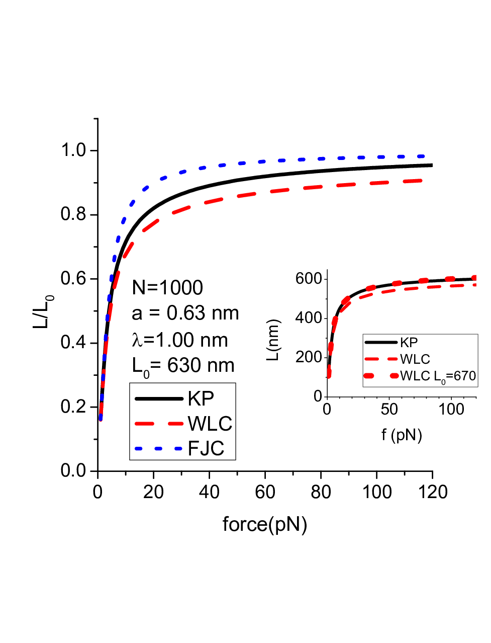
The first model is the Kratky-Porod model with segments, a monomer distance nm and a stiffness constant pN nm, which from (4) implies nm at a temperature 20 C. The second model is a WLC with the same persistence length nm and a contour length nm nm. The third model is a freely-jointed chain (FJC) with a Kuhn length equal to nm and a number of segments .
Fig. 1 displays the force-extension relationship in all three cases. The difference between the KP and WLC cases is fairly significant, of the order of 7% at 70 pN. More importantly however, from a data analysis point of view, use of the WLC enforces a bias towards higher contour length, i.e. monomer distances systematically larger than those mandated by crystallographic data (cf. inset).
III Overstretching of DNA
III.1 The force-extension curve
Fig. 2 displays the DNA force-extension curve as measured in an unconstrained geometry, i.e. where the chain is held fixed at the 3’-3’ opposite ends vanMam2009. The extension is defined relative to the contour length of ds-DNA.
At low force levels, the data can be well described by the WLC model (15), corrected for elastic stretching according to (17) with an elastic (stretch) modulus . The WLC calculation uses a contour length computed with nm and vanMam2009 and a persistence length nm. Only the latter parameter has been adjusted in order to obtain a visual fit to the data; its value is in excellent agreement with the one obtained from earlier force-extension measurements Busta1994.
At forces beyond the 65 pN plateau, the data can be fitted to the KP model force-extension relationship (11) for ss-DNA, corrected for elastic stretching according to (17). I have used the monomer distance nm, as determined by averaging over crystallographic data Murphy2004, the number of bases as given in vanMam2009, and a stretch modulus equal to 8.4 nN GaubNetz2005. Again, the only adjustable parameter used to obtain a visual fit is the local stiffness pN nm, which corresponds to a persistence length nm.

III.2 Thermodynamics (unconstrained geometry)
The plateau in the force-extension curve characterizes the coexistence of the two phases, double-stranded and single-stranded. In this geometry only one of the two single strands of the molten phase is under an applied force. Coexistence occurs when the difference in elastic free energies between single- and double-stranded phases fully compensate the stability free energy of the force-free double helix.
III.2.1 Elastic free energies
The force-dependent parts of the elastic free energies per unit are
| (18) |
and
| (19) |
for the single- and double-stranded cases, respectively, where the second term expresses the elastic stretch energy.
III.2.2 Thermal stability of the double helix
The double helix is generally stable at room temperature. This is usually expressed in terms of the free energy difference per base pair between duplex and molten state
| (20) |
where and denote, respectively, average values for the energy and the entropy of dissociation per base pair and is the melting temperature. A detailed analysis of DNA melting thermodynamics Blake91 suggests that the constant value consistently describes, in a mean-field fashion, the cooperative melting behavior of a large number of samples of varying sequence and composition.The above value has been used in RouzinaBloomfield2001a and will also be used here.
The melting temperature can be calculated from the empirical Marmur-Schildkraut-Doty MarmurDoty1960; SchildkrautDoty1962; DelcourtBlake1998 equation
| (21) |
where is the GC fraction and the salt concentration. At and mM, this leads to C. It should be noted that (20) accounts for all effects involved in the relative stability of ds- and ss-DNA, including entropic polymer contributions, except those explicitly originating in the applied force and described by (18) and (19).
III.2.3 Overstretching threshold
Fig. 3 displays the free energy difference at C. After an initial positive phase at very low forces ( pN), the difference becomes increasingly negative, until, at 64 pN it compensates fully for the room temperature duplex stability (20). This marks the onset of the overstretching transition.

III.3 Thermodynamics (constrained geometry)
In the constrained geometry version of the overstretching experiment all four ends of the 3’5’-5’3’ chain are attached; each single strand is subjected to half the external force. The elastic free energy difference is now . According to the discussion of the previous subsection, overstretching should occur at the force level where the elastic free energy difference fully compensates duplex stability.
The geometry of the constraint however prohibits a full entropy release in the overstretched state. The two strands remain stretched, attached at both ends. Moreover, available conformational space is further restricted by the entanglement brought about by the previous double helical order. An “inherited” structure seems to persist in the overstretched state for which “the most straightforward explanation … is that the new structure, generated during overstretching at 110 pN, consists of two single DNA strands lacking hydrogen bonds between the bases, wrapped around each other with a linking number close to that of relaxed dsDNA”vanMam2009.
The quantity of interest in this context is the entropy difference between the molten state with the geometrical constraint and the one without it, or, in other words, the entropic penalty required to maintain the molten DNA with the inherited plectonemic structure, compared to the reference state of the two free strands. Geometrically constrained force-induced melting does not release the full amount of DNA melting entropy (cf. previous subsection), but a reduced amount . This modifies the relative thermodynamic stability of the double helix (20), which now reads
| (22) |
per base pair, and results in an enhanced stability of the double helix. Overstretching will now occur when the elastic free energy difference compensates (22).
In what follows I will calculate the entropic penalty associated with the plectonemic structure in two different ways, based on the FJC and the KP model, respectively.
III.3.1 FJC estimate of the constraint entropic penalty
The overstretched DNA with the plectonemic structure can be visualized as consisting of the two strands joined at successive points with a spacing , equal to the pitch of the original double helix. In the FJC model this corresponds to segments in each strand, where nm and 2.0 nm, are, respectively, the monomer distance and the Kuhn length of ss-DNA. The total number of such plectonemic joints will be .
Neglecting any effects arising from excluded volume, the probability of two distinct, partially stretched strand pieces, each with elements, starting from a common origin and meeting at a certain point in space within an axial distance and a radial distance , is , where
| (23) |
and
| (24) |
is the vector end-to-end distance probability distribution of the FJC chain.
The conformation with all plectonemic joints has a probability of occurrence , i.e. an entropic penalty per original base pair. Inserting some plausible values for the contact in terms of the inherited double helix, e.g. , results in an estimate of .
III.3.2 KP estimate of the constraint entropic penalty
The calculation is quite analogous with that of the FJC, the only difference now being that and
| (25) |
where is now the vector end-to-end distance distribution function, measured in units of the segment length, computed for the KP model with the stiffness parameter of ss-DNA (cf. section III.1).
Computation of the KP end-to-end distribution function can be performed, for relatively small systems, by direct matrix multiplication and Fourier transform inversion Errami2007; Marko2005. Noting that the Fourier transform of the end-to-end distribution function, , can be formally obtained from (cf. (3) and (5) above) by the substitution , it is possible to write
where the matrix is defined as in (7), with substituted by
| (26) | |||||
where are now spherical Bessel functions, e.g. .
The result of the numerical computation, for , is , leading, for the same values of the parameters and as in the FJC case, to an entropic penalty of per original base pair.
III.3.3 Estimation of the overstretching threshold
Fig. 4 displays the difference in the elastic free energies as a function of the applied force. Also shown is the stability free energy of the double helix, with and without the entropic penalty of the constraint, as given by (22) and (20), respectively. Both effects lead to an enhanced stability of double-stranded DNA, in comparison with the unconstrained geometry. The overstretching force threshold, as estimated from the KP model calculation, with lies at 111 pN, in excellent agreement with experiment vanMam2009.

IV Discussion
The present analysis of the overstretching transition involves a number of material parameters for both ds- and ss-DNA. It is important to keep in mind that only two of these parameters originate in fitting the data of the experimental force-extension curve, namely the persistence lengths. All others are either standard accepted values (e.g. the crystallographically obtained values for the monomer distances and , melting temperature and melting entropy per base pair) or, at the very least (e.g. elastic stretch moduli for ss-DNA and ds-DNA), physically plausible published values. Not surprisingly, the limited space of the remaining free parameters improves the quality of the estimates obtained. Both persistence lengths are consistent with the values obtained by other researchers.
In particular the value nm obtained here for the persistence length of ss-DNA deserves a brief comment. The value lies at the low end of current estimates of 0.75 - 2 nm. Specifically, it compares well with the zero loop origami-based estimates of Roth2018, in accordance with the expectation of loops unwinding under the influence of the external force. On the other hand, it is significantly lower than the SAXS result of LipfertDoniach2012, 1.7 nm at 150 mM NaCl concentration. Although a small part of the discrepancy may be accounted for by the use of the WLC continuum model in LipfertDoniach2012, it is instructive to consider the physics underlying the different experiments. The partially - or fully - stretched state of vanMam2009, is probably far less responsive to the electrostatic repulsion forces acting between segments. As a result, observed persistence lengths may be lower than those observed in force-free solutions and actually approach values corresponding to high salt concentrations, where screening of electrostatic repulsion occurs. Independent sets of measurements LipfertDoniach2012; Chen2012 suggest that there is an intrinsic lower limit of ss-DNA persistence length, of approximately 1 nm and 0.75 nm, respectively, which is approached at high salt concentrations. It is quite possible that experiments performed near the stretched state effectively detect this limit of extreme chain flexibility.
The results of the thermodynamic analysis confirm the conjecture made vanMam2009 about the plectonemic nature of the overstretched state in the constrained geometry. It should be noted that this conclusion does not depend on the detailed choice of geometrical parameters specifying what exactly is considered as a contact. For example, increasing the linear dimension of a contact from to in both the radial and axial directions would result in a decreased entropy penalty by , changing the predicted threshold to pN.
Are there any plausible alternatives to the plectonemic structure of the geometrically constrained molten state? A radically weaker variant would be to consider solely the entropic effect of holding the ends of both strands stretched, without imposing any further structural motifs. Such an alternative calculation results in a much smaller entropic penalty of the order per base pair, roughly ten times less than that of the plectonemic structure, and cannot account for the observed force threshold. In other words, entropic considerations effectively demand some sort of internal structure in the molten state. The “inherited” structure with knots following the pitch of the double helix, as conjectured in vanMam2009 is probably the simplest model satisfying this demand.
I would like to thank Michel Peyrard for reading an earlier version of the manuscript.
I acknowledge support by the project “Advanced Materials and Devices (MIS 5002409)”, implemented under the “Action for the Strategic Development on the Research and Technological Sector”, funded by the Operational Program “Competitiveness, Entrepreneurship and Innovation (NSRF 2014-2020)” and cofinanced by Greece and the European Union (European Regional Development Fund).
References
- (1) I. Rouzina and V. A. Bloomfield, Force-induced melting of the DNA double helix 1. Thermodynamic analysis, Biophysical Journal 80, 882 (2001).
- (2) S. B. Smith, Y. Cui, and C. Bustamante, Overstretching B-DNA: the elastic response of individual double-stranded and single-stranded DNA molecules, Science 271, 795 (1996).
- (3) J. van Mameren, P. Gross, G. Farge, P. Hooijman, M. Modesti, M. Falkenberg, G. J. L. Wuite, and E. J. G. Peterman, Unraveling the structure of DNA during overstretching by using multicolor, single-molecule fluorescence imaging, Proceedings of the National Academy of Sciences 106, 18231 (2009).
- (4) H. Chen, S. P. Meisburger, S. A. Pabit, J. L. Sutton, W. W. Webb, and L. Pollack, Ionic strength-dependent persistence lengths of single-stranded RNA and DNA, Proceedings of the National Academy of Sciences 109, 799 (2012).
- (5) A. Y. L. Sim, J. Lipfert, D. Herschlag, and S. Doniach, Salt dependence of the radius of gyration and flexibility of single-stranded DNA in solution probed by small-angle x-ray scattering, Physical Review E 86, 021901 (2012).
- (6) A. Bosco, J. Camunas-Soler, and F. Ritort, Elastic properties and secondary structure formation of single-stranded DNA at monovalent and divalent salt conditions, Nucleic Acids Research 42, 2064 (2014).
- (7) E. Roth, A. Glick Azaria, O. Girshevitz, A. Bitler, and Y. Garini, Measuring the conformation and persistence length of single-stranded DNA using a DNA origami structure, Nano Letters 18, 6703 (2018).
- (8) M. C. Murphy, I. Rasnik, W. Cheng, T. M. Lohman, and T. Ha, Probing single-stranded DNA conformational flexibility using fluorescence spectroscopy, Biophysical Journal 86, 2530 (2004).
- (9) O. Kratky and G. Porod, Röntgenuntersuchung gelöster Fadenmoleküle, Recueil des Travaux Chimiques des Pays-Bas 68, 1106 (1949).
- (10) T. Hugel, M. Rief, M. Seitz, H. E. Gaub, and R. R. Netz, Highly stretched single polymers: Atomic-force-microscope experiments versus ab-initio theory, Phys. Rev. Lett. 94, 048301 (2005).
- (11) H. Clausen-Schaumann, M. Rief, C. Tolksdorf, and H. E. Gaub, Mechanical stability of single DNA molecules, Biophysical Journal 78, 1997 (2000).
- (12) J. Errami, M. Peyrard, and N. Theodorakopoulos, Modeling DNA beacons at the mesoscopic scale, The European Physical Journal E 23, 397 (2007).
- (13) J. Yan, R. Kawamura, and J. F. Marko, Statistics of loop formation along double helix DNAs, Phys. Rev. E 71, 061905 (2005).
- (14) M. Abramowitz and I. Stegun, Handbook of Mathematical Functions (Dover 1964).
- (15) C. Bustamante, S. B. Smith, J. Liphardt, and D. Smith, Single-molecule studies of DNA mechanics, Current Opinion in Structural Biology 10, 279–285 (2000).
- (16) J. F. Marko and S. Cocco, The micromechanics of DNA, Physics World 16, 37 (2003).
- (17) C. Bustamante, J. F. Marko, E. D. Siggia, and S. B. Smith, Entropic elasticity of lambda phage DNA, Science 265, 1599 (1994).
- (18) S. G. Delcourt and R. D. Blake, Stacking energies in DNA, Journal of Biological Chemistry 266, 15160 (1991).
- (19) J. Marmur and P. Doty, Determination of the base composition of deoxyribonucleic acid from its thermal denaturation temperature, Journal of Molecular Biology 5, 109 (1960).
- (20) C. Schildkraut, J. Marmur, and P. Doty, Determination of the base composition of deoxyribonucleic acid from its buoyant density in CsCl, Journal of Molecular Biology 4, 430 (1962).
- (21) R. D. Blake and S. G. Delcourt, Thermal stability of DNA, Nucleic Acids Research 26, 3323 (1998).