Kinematics of a cascade decay for the precision measurement of binding energy
Abstract
Comparison of a Penning trap and a flat-crystal spectrometer experiments gives a direct test of . The result is for and . The dominant uncertainty is on the -ray measurement in neutron capture reactions, and the secondary -ray has the uncertainty 4.0 eV for . We calculated the Doppler effect of the secondary -ray as eV from the relativistic energy momentum relation of the reaction. This corresponds to the full wave of half maximum of 431.3 eV. The error 4.0 eV comes mainly from the Bragg angle measurement between the centroids of the linewidths which means that only the most probable part of the whole data has been considered. It is necessary to confirm the assumption of the isotropy for the object to measure. We discussed a coincidence measurement as one of the methods to overcome the assumption.
Keywords:
Nuclear Binding Energy, Radiative Capture, Gamma Ray Spectroscopy, Doppler Effect, Angular Correlationpacs
21.10.Dr, 25.40.Lw, 27.30.+t, 29.30.KvNuclear binding energy is the mass difference between initial and final states. The Penning trap in the recent report [1, 2] measures the masses with fractional uncertainty below . Another method is to measure the -ray energy emitted from the transition between initial and final states. The flat-crystal spectrometer in the recent report [3] measures the -ray energy with relative uncertainty below . The comparison of the two methods is a direct test for Einstein’s famous formula , and the result is [4]. In the report the systematic error on the comparison is currently dominated by the uncertainty on the -ray measurements.
The binding energy is the sum of the -ray energy and the recoil energy in traditional -ray measurements and the flat-crystal measurement [3]. This is the result of non-relativistic energy momentum relation. From relativistic energy momentum relation we calculated the Doppler effect of the secondary -ray due to the recoil ( of the intermediate nucleus as eV. This uncertainty is far greater than the error eV of the secondary -ray energy of reported in Ref. [3]. Increasing the number of measurements can reduce the statistical error to eV, but can not reduce to the reported value 4.0 eV which comes chiefly from the measurements of the Bragg angle between the centroids of the spectrum and the lattice spacing of the silicon crystal. Thus, the error bar is shown to be quite out of the diagonal line at the level of 1.2 in Fig. 5 of Ref. [3].
A secondary -ray has angular correlation with respect to the primary -ray in a cascade decay [5, 6]. We calculated the angular correlation function for the decay of . The most probable directions of the secondary -ray are parallel and anti-parallel with the direction of the primary -ray. The Doppler shift of the secondary -ray is maximum at these angles. Therefore, the average value of the two energies of the Doppler shifted -rays at angles and is free from Doppler broadening caused by the recoil of the nucleus. Thus, the profile of the shifted -ray might be even closer to a natural linewidth which can also give the lifetime of the intermediate state of the nucleus. The Institut Laue-Lagnevin (ILL) high-flux reactor provides one of the most intense neutron sources in the world and has a through beam tube [7]. This leads us to calculate the kinematics of the cascade decay for nucleus.
The binding energy for a neutron capture reaction is the mass difference between initial and final states as follows:
| (1) |
where means the mass of the particle in parentheses, is an atomic number, and is the separation energy of a neutron or the binding energy of a neutron which is carried by the -rays and the recoiled nuclei. If a single -ray is emitted dominantly in a neutron capture reaction, the problem has been considered in determining the deuteron binding energy [8]. In this letter we are interested in a cascade decay emitting two -rays successively which is the most probable channel. The excited state of is the case as shown in Fig. 1, and the values in parentheses are the number of -rays emitted per 100 neutron captures [9]. This decay channel can be represented as a Feynman diagram shown in Fig. 2 (a) and can be separated into two parts as Figs. 2 (b), and (c). The equation of the binding energy Eq. (1) is also separated into two parts corresponding to the separated Feynman diagrams, respectively, as follows:
| (2) | |||||
| (3) | |||||
| (4) |
where asterisk means an excited state of the final nucleus.
Fig. 2 (b) represents the neutron capture reaction , and the energy-momentum conservation law is given by
| (5) | |||||
| (6) |
where and . The energy-momentum relations of the final state are for the intermediate state of the silicon nucleus and for the primary photon. It should be noted that the velocity of the center of mass for the neutron and the silicon system is only 114 for the incident neutron flux energy 0.056 eV, while that for the neutron and the proton system is 1.6 as calculated in Ref. [8]. Hence, the Doppler effect of the silicon nucleus due to the incident neutron is negligible ( eV) so that it is not necessary to consider the kinetic energy of the incident neutron [3].
Solving the equations of the energy-momentum conservation laws with respect to the primary photon energy and replacing the initial masses by the binding energy, one can obtain the binding energy of the intermediate nucleus as follows:
| (7) |
which is reduced to the non-relativistic result that is usually used in the literature. Since the incident kinetic energy of the neutron is ignored, there is no Doppler effect in this equation. The last term in the above equation is the kinetic energy of the recoiling intermediate nucleus, and its velocity can be checked as follows:
| (8) |
where the natural unit() is used in every equation throughout this letter, and thus, the velocity in SI unit should be multiplied by the light velocity.
Since the velocity causes the secondary -ray to be Doppler shifted considerably, it is important to take into account the kinetic energy and momentum of the intermediate nucleus in calculation. Hence, the law of the energy-momentum conservation for the process shown in Fig. 2 (c) should be given by
| (9) | |||||
| (10) |
where the energy-momentum relation of the ground state silicon nucleus is , and that of the secondary photon is . To specify the angle between two photons that is of importance in the Doppler effect and angular correlation, it is useful to show the following intermediate step:
| (11) |
where the direction of the primary photon is , and the direction of the recoiling nucleus is . Solving the equation as the same way of the previous energy-momentum law, one can obtain the binding energy of the ground state of the silicon as follows:
| (12) | |||||
where the second term is the kinetic energy due to recoil, the third term is negligible kinetic term, and the last term represents the Doppler effect as shown in Ref. [8]. Higher order terms in expansion include the usual relativistic correction term which depends on the square of velocity. It is negligible and is omitted [11]. This result is calculated in laboratory frame and is consistent with the result that is Lorentz transformed from the calculations in rest frame of .
The total binding energy of the silicon is the sum of Eqs. (7) and (12) as follows:
| (13) | |||||
If one is able to handle the angle in the secondary -ray measurement, the Doppler shifted -ray is detected. Otherwise, all the shifted -rays reach the detector and appear in the spectrum as a broadened lineshape. Thus, the 4934-keV profile is significantly Doppler broadened, which decreases the accuracy with which the Bragg angle can be determined [3].
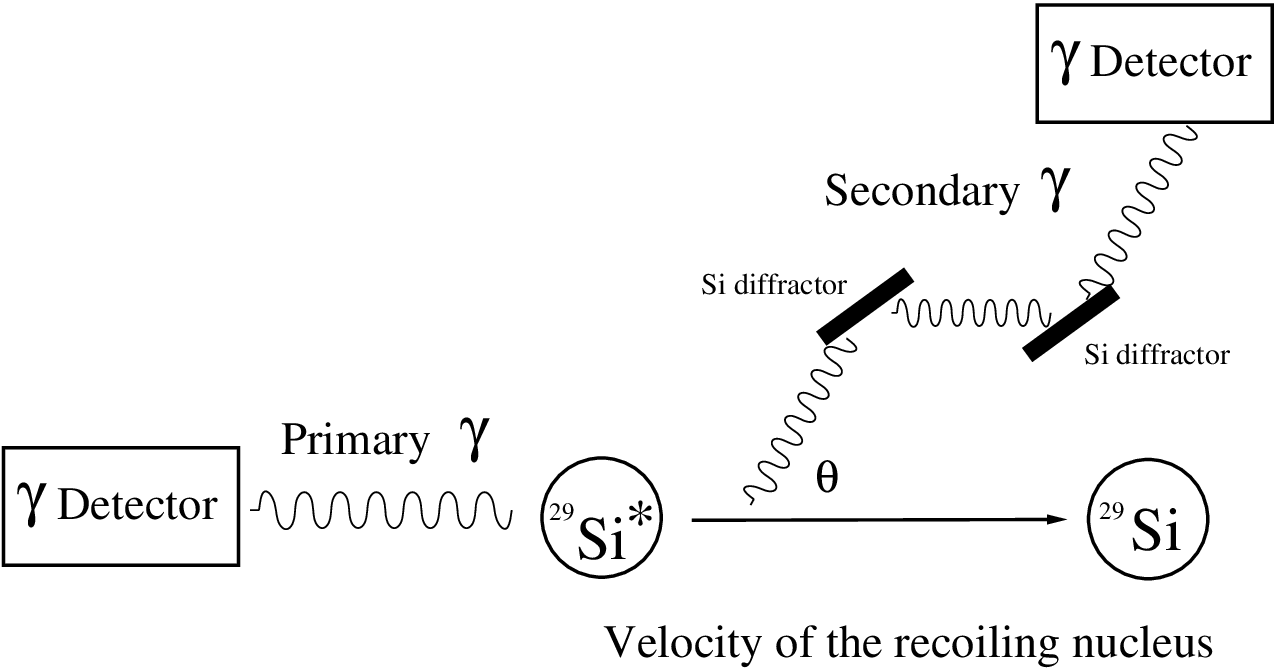
Assuming that the motion of the intermediate nucleus is completely isotropic and the Doppler effect causes nothing in the central value, the authors of Ref. [3] obtained the uncertainty of eV mainly from the measurements of the Bragg angle between the centroids of the diffracted -rays by increasing the number of measurements up to 147 compared to 42 for the primary -ray measurement. If one want to obtain the uncertainty from the linewidths at the level of an uncertainty 4.0 eV, the number of measurements should be more than according to the basic rule of the error propagation: for . The linewidth is too broad compared to the resolution of the flat-crystal spectrometer, but the measurement of Bragg angle between the centroids of the recorded profiles could reduce the uncertainty to 4.0 eV by the assumption. This kind of an assumption about the centroid measurement has a possibility to reduce the uncertainty to less than a natural linewidth, and change the lifetime of the state from which a -ray with a natural linewidth is emitted. The assumption should be confirmed by a proper method. In a coincidence measurement of two successive emitting -rays, the secondary -ray is not broadened but shifted. The magnitude of shifted energy depends on the angle between the primary and secondary -rays. The probability distribution of the secondary -ray with the angle is the angular correlation [5]. Fig. 3 shows a schematic diagram of a coincidence measurement. This is also an diffraction experiment of the secondary -ray which is correlated with the primary -ray and may help to understand quantum physics: one photon is diffracted.
Since the polarizations of the -rays are not observed and only directional correlation is observed, the angular correlation function [5, 6] is simply given by
| (14) |
where is the Legendre polynomial of order . The coefficient for the transition of angular momentum states with emitting photons of the angular momenta and in order is given by
| (15) | |||||
| (16) |
where is a Racah coefficient, and is known as the radiation parameter. The angle of the binding energy in Eq. (13) is related with the angle in the correlation function as , but it is not necessary to distinguish between the angles, because only even power of the Legendre polynomial contributes to the angular correlation function. For a simple example, if a dipole-dipole transition occurs through the nuclear states of , and , the angular correlation function is . If a dipole-dipole transition takes place through the nuclear states of , and as assigned from the -rays measurement in Ref. [10] and shown in Fig. 1, the angular correlation function is
| (17) |
One can see here that the secondary -ray has the most probable distribution at the angles , or . The Doppler shift is maximum at these angles in Eq. (13).
However, the surrounding environment in which the recoiling nucleus is located is not a free space. It is not sure that the shifted -ray at the angle gives exact information on the binding energy. Moreover, the surrounding environment varies from nucleus to nucleus. For example, the velocities of the intermediate state of other nuclei and are greater than that of , but they suffer from the Doppler broadening less than the case of [3]. The reason can be that the lifetime of the intermediate state of and is much longer than that of as shown from experimental data [12, 13]. The velocity of these intermediate nuclei could be attenuated during their long lifetimes, and thus, the Doppler broadening would be considerably reduced. This aspect also reflects in the branching ratio of the decay, that is, the number of -ray per 100 neutron capture in Fig. 1 of Ref. [3]. Since the lifetime of the intermediate nucleus is quite short, it is likely that the velocity of the intermediate nucleus is a little slowed down, and the angular correlation between the two successive -rays is clear.
Let us think twice coincidence measurements at angle and angle . Using Eq. (13), one can obtain the following quantities:
| (18) | |||||
| (19) |
where the first equation is the desired binding energy, and the second equation contains the magnitude of the Doppler shift attenuated by the surrounding environment, namely, attenuation factor [14, 15]. is the velocity of the intermediate nucleus. The average binding energy is free from the major uncertainty due to the Doppler effect of the secondary -ray. Since the primary and secondary -rays come from the electric dipole transitions in view of the spin-parity of the states shown in Fig. 1, a coincidence measurement may rule out uncertainties due to other multipole transitions.
The lifetime of the 4943 keV in old data [12, 13] is 1.16 fs which corresponds to the linewidth of 0.57 eV. The resolution of the flat-crystal spectrometer is this order. Hence, it may be possible to extract the lifetime from the thermal broadened linewidth which can be obtained from the classical Maxwell-Boltzmann distribution by virtue of the modified Lorentzian shape used in Ref. [15]. This can be compared with the Doppler shift attenuation method. If the energy difference Eq. (19) is compared to the calculated value in Eq. (13), one can extract the attenuated velocity of the recoiling nucleus which provides a lifetime in the Doppler shift attenuation method.
In conclusion, the equipment of a flat-crystal spectrometer is an extremely accurate tool for a -ray measurement. The recorded profiles along with interferometer angles have valuable physical facts such as the influence of Si diffractors, a Doppler broadened linewidth, an attenuated linewidth, or a thermal broadened linewidth. For example, the 6111 keV profile of in Fig. 3 of Ref. [3] shows the linewidth of 93.5 eV for arcsec where a scale is 0.05 arcsec and its uncertainty 4.0 eV corresponds to 0.00043 arcsec. Likewise, the average linewidth of the recorded profiles, especially , should have been shown for the justification of the accuracy 4.0 eV in Ref. [3]. It is insufficient to understand the accuracy 4.0 eV from the broadened width 431.3 eV without further information on the first observed data. Usually, centroid measurements have been made in the past when the resolution of a detector is greater than the linewidth, and the assumption for symmetry have been tried to prove [16, 17]. Moreover, the assumption is not for the object to measure but for the detector. An assumption for the object to measure is unusual in any experiment. As a result, the 1.2 discrepancy is an evidence for observing only part of the whole data.
A flat-crystal spectrometer is more suitable to measure sharp -rays or natural linewidths without unnecessary assumptions rather than to measure broadened -rays due to the Doppler effect. A coincidence measurement of two -rays with considering their angular correlation can reduce the uncertainty caused by the Doppler effect in the determination of binding energy. This improved binding energy measurement would lead to one step upgrade, at least 1.2 discrepancy, for the direct test of and relativistically consistent comparison between energy and mass [4]. If the measurement technique for cascade decay is developed, one can confirm one of the postulates of quantum physics and learn the more accurate information about the nuclear structure, for examples, accurate energy level, the lifetime of the nuclear intermediate state, and so on.
Acknowledgements
This work was supported by the National Research Foundation of Korea (Grant No. 2009-0074238).
References
- [1] S. Rainville, J. K. Thompson and D. E. Pritchard, Science 303, 334 (2004).
- [2] W. Shi, M. Redshaw, and E. G. Myers, Phys. Rev. A 72, 022510 (2005).
- [3] M. S. Dewey, E. G. Kessler Jr., R. D. Deslattes, H. G. Börner, M. Jentschel, C. Doll, and P. Mutti, Phys. Rev. C 73, 044303 (2006).
- [4] S. Rainville, et al., Nature(London) 438, 1096 (2005).
- [5] L. C. Biedenharn and M. E. Rose, Rev. Mod. Phys. 25, 729 (1953).
- [6] R. R. Roy, and B. P. Nigam, Nuclear Physics, (John Wiley Sons, inc. New York, 1967).
- [7] E. G. Kessler Jr., M. S. Dewey, R. D. Deslattes, A. Henins, H. G. Börner, M. Jentschel, C. Doll, and H. Lehmann, Nucl. Instrum. Methods Phys. Res. A 457, 187 (2001).
- [8] Y. Ko, M. K. Cheoun, and I. T. Cheon, Phys. Rev. C 59, 3473 (1999).
- [9] M. A. Lone, R. A. Leavitt, and D. A. Harrison, At. Data. Nucl. Data Tables 26, 511 (1981).
- [10] S. Raman, E. T. Jurney, J. W. Starner, and J. E. Lynn, Phys. Rev. C 46, 972 (1992).
- [11] R. Moreh and D. Nemirovsky, Phys. Rev. C 67, 014602 (2003).
- [12] P. M. Endt, Nucl. Phys. A 521, 1 (1990), ibid. A 633, 1 (1998).
- [13] S. J. Skorka, T. W. Retz-Schmidt, H. Schmidt, J. Morgenstern, and D. Evers, Nucl. Phys. 68, 177 (1965).
- [14] K. P. Lieb, H. G. Börner, M. S. Dewey, J. Jolie, S. J. Robinson, S. Ulbig, and Ch. Winter, Phys. Lett. B 215, 50 (1988).
- [15] H. G. Börner and J. Jolie, J. Phys. G 19, 217 (1993).
- [16] R. C. Greenwood and R. E. Chrien, Phys. Rev. C 21, 498 (1980).
- [17] R. C. Greenwood, R. G. Helmer, and R. J. Gehrke, Nucl. Instrum. Methods 159, 465 (1979).