Kinetic limitations of cooperativity based drug delivery systems
Abstract
We study theoretically a novel drug delivery system that utilizes the overexpression of certain proteins in cancerous cells for cell specific chemotherapy. The system consists of dendrimers conjugated with ”keys” (ex: folic acid) which ”key-lock” bind to particular cell membrane proteins (ex: folate receptor). The increased concentration of ”locks” on the surface leads to a longer residence time for the dendrimer and greater incorporation into the cell. Cooperative binding of the nanocomplexes leads to an enhancement of cell specificity. However, both our theory and detailed analysis of in-vitro experiments indicate that the degree of cooperativity is kinetically limited. We demonstrate that cooperativity and hence the specificity to particular cell type can be increased by making the strength of individual bonds weaker, and suggest a particular implementation of this idea. The implications of the work for optimizing the design of drug delivery vehicles are discussed.
Nanoparticle based drug delivery systems have attracted substantial attention for their potential applications in cancer treatment (Hilgenbrink and Low 2005,Lu and Low 2002,Stella et al. 2000,Quintana et al. 2002,Choi et al. 2004). It is hoped that by selectively targeting cancer cells with chemotherapeutic agents one can reduce side effects and improve treatment outcomes relative to other drug delivery systems which do not discriminate between normal and cancerous cells. For example, many epithelial cancer cells are known to overexpress the folate receptor(Toffoli et al. 1997,Ross et al. 1994,Weitman et al. 1992a,Weitman et al. 1992b,Campbell et al. 1991). A nanoparticle with many folic acid ligands will preferentially bind to cancerous cells. A recent studyHong et al. 2007 of a potential drug delivery platform consisting of generation 5 PAMAM dendrimers with different numbers of folic acid found that multivalent interactions have a pronounced effect on the dissociation constant . This enhancement is the signature for cooperativity of the binding, which should lead to a greater specificity to cancerous cells in vivo.
In this letter we present a theoretical study of these key-locking nanodevices (see Fig. 1). We introduce the idea that there are kinetic limitations to cooperativity-based drug delivery systems. In vivo the finite timescale for endocytosis prevents arbitrarily high cooperativity in the drug delivery system. In the first part we provide a detailed analysis of the in vitro experimentsHong et al. 2007. Although enhancement of the association is the signature of greater cooperativity, in this case it is due mostly to non-specific binding of the dendrimers to the surface. Due to the finite time window of the experiments, only indirect support can be offered to the notion of enhanced cooperativity. In the second part we expand the notion of kinetically limited cooperativity to the system in vivo. The equilibrium coverage of nanodevices on the cells is related to the concentration of folate-binding proteins and the strength of the key-lock binding. We quantify the preferential adsorption of nanodevices to the cancerous cells, and discuss how kinetic effects prohibit arbitrarily high cooperativity in the drug delivery system. The implications of the work for designing new drug delivery vehicles with enhanced specificity to cancerous cells are discussed.

We now consider a simple model of the nanodevice system. A dendrimer with a maximum of keys (e.g. folic acids) interacts with locks (e.g. folate-binding proteins) in the cell membrane surface. A simple order of magnitude estimate for can be obtained from the ratio of the surface area of the dendrimer to the surface area of the folic acid. In this way we implicitly take into account the excluded volume effect between the keys. The free energy for the dendrimer connected to the surface by key-lock bridges isLicata and Tkachenko 2006
| (1) |
The dimensionless energy parameter contains information about the binding energy of a single key-lock pair, and the entropy loss associated with localizing a dendrimer on the cell-membrane surface. An estimate of can be obtained from the dissociation constant of free folic acid using the equilibrium relation between the dissociation constant and the free energy change for the formation of a single key-lock bridge, . Here is the localization volume of an ”unbound” key. Below we determine the value from analysis of the in vitro experiments, which was used to determine .
The measured association rate constant of the dendrimer with folic acid is a factor of times greater than of free folic acid. Only a factor of can be attributed to the dendrimer having many folic acids attached to it. Here is the average number of keys attached to the dendrimer. This pronounced enhancement of is the primary evidence for non-specific attraction between the dendrimer and the surface.
| (2) |
The non specific attraction accounts for the Van der Waals attraction to the surface and hydrophobic enhancement. The experimentally measured values are reproduced by a reasonable energy scale (see Fig. 2).
We provide a simple explanation for the experimentally observed dependence of the dissociation rate constant on . The dissociation rate constant of free folic acid provides a characteristic departure time of for those dendrimers attached by a single key-lock bridge. Moreover, the departure time for multiple bridge states increases exponentially in , for two bridges it is . Strictly speaking the relaxation is multiexponential, with time constants for each bridge number. However, the experimental values are well fit by a single exponential. On the timescale of the experiment, we will only see the departure of dendrimers attached by a single bridge.
The experiment measures the departure rate of dendrimers which are connected to the surface by a single bridge, but are unable to form an additional connection. Consider a dendrimer attached to the surface by one key-lock bridge. If the dendrimer has a total of keys, the probability that none of the remaining keys can form bridges is . We now compute the probability that a remaining key is available to form a bridge. In the vicinity of the surface the dendrimer is a disclike structureMecke et al. 2004 with radius . By rotation of the dendrimer about the first bridge, a key located at position searches the annulus of area to find a lock. The probability of encountering a lock in this region is , where the surface density of the locks . By averaging over the key location we obtain the final result
| (3) |
Assuming that during dendrimer preparation the attachment of folic acid to the dendrimer is a Poisson process, the probability of a dendrimer having exactly keys is . The final result is obtained by averaging the probability that no additional bridges can form over this distribution. The factor of counts the number of ways to make the first connection.
| (4) |
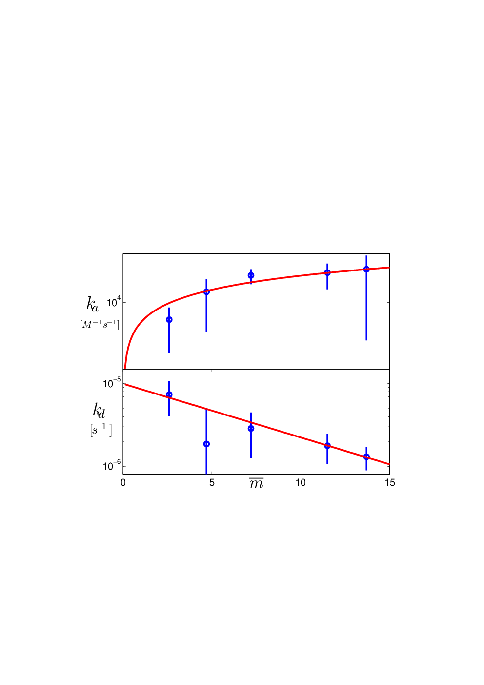
The formula predicts an exponential decay of the effective dissociation rate constant with the average number of folic acids on the dendrimer, which allows for a quantitative comparison to the experiment (see Fig. 2). Using , we can determine the localization length for locks in the experiment from Eq. 3. This estimate for is physically reasonable, and comparable to the bond length of the terminal group on the dendrimer.
Similar to the finite timescale of the experiments in vitroHong et al. 2007, in vivo the endocytosis time provides kinetic limitations to cooperative binding. In equilibrium the concentration of dendrimers on the cell surface is related to the concentration of dendrimers in solution through the association constant . Although it is tempting to use our in vitro results to define the association constant as , this approach is only valid provided there is a single rate for both association and dissociation. Because the dendrimer can form multiple bridges, there are many different rate constants. We present a partition function method which accounts for the multiple rate constants in the problem, and for the possibility that in vivo there is surface diffusion of locks.
To proceed we construct a vector of length , which is a list of the possible sites folic acid can attach to the dendrimer. If a folic acid is present at site we have , and otherwise . The concentration of dendrimers on the cell surface is proportional to the partition function of the system.
| (5) | |||
Here is the surface density of locks on the cell membrane at position , and denotes the total area of the cell membrane. The energy that appears in the Boltzmann weight is the elastic energy penalty required to form multiple bridges. The point is that in solution the dendrimer is roughly spherical, but must flatten to a pancake like shape to form multiple connections with the cell surfaceMecke et al. 2004.
The ensemble averaging is performed by assuming that during nanodevice preparation the attachment of folic acid to the dendrimer is a Poisson process. In this case is given by the success probability that a folic acid attaches to the dendrimer, and the point correlator . In other words, the probability of attachment of a given folic acid to a terminal group on the dendrimer is unaffected by the presence of other folic acids up to an exclusion rule which has already been taken into account. If the interaction potential between locks in the cell membrane is we have . By performing the ensemble averaging we arrive at the result for the equilibrium coverage of dendrimers connected to the cell surface by bridges.
| (6) |
Here is the association constant of free folic acid which has been measured experimentally. Defined in this manner, has a physical interpretation as the Boltzmann weight for the elastic energy of the optimal bridge configuration. The membrane surface can only accomodate a finite number of locks in the vicinity where the dendrimer is attachedHong et al. 2007. As a result for since forming additional key-lock pairs would require deforming the dendrimer into configurations prohibited by elastic stress and steric hindrance. The calculation of the equilibrium coverage above is applicable with and without diffusion of locks in the cell membrane. In the regime of fast diffusion the locks are free to diffusively explore the surface. Their positions are ergodic variables, and the overall ensemble averaged equilibrium coverage counts the Boltzmann weights for different lock configurations. In the regime of slow diffusion, locks are immobilized in the cell membrane. This is the relevant situation when the locks have phase separated into protein rich (lipid rafts) and protein poor phases.
When kinetic effects are taken into account, these two regimes are drastically different. When locks are diffusing, the dendrimer is able to attain the maximum cooperativity . After the dendrimer makes the first connection, it simply waits for locks to diffuse in the vicinity of available keys to make additional connections. In the absence of diffusion, the optimal configuration can only be obtained by multiple binding and unbinding events, the timescale for which is prohibitively long. This is the case for lipid rafts where the locks are immobilized similar to the experiments in vitroHong et al. 2007, and the dendrimer is unable to attain the maximum cooperativity. This is the kinetic origin of limited cooperativity in the drug delivery system.
We now quantify the preferential attachment of nanodevices to the cancerous cells, taking into account kinetic effects. Let denote the concentration of dendrimers attached to the cell by bridges. We can construct a differential equation for by considering linear response to the deviation from thermal equilibrium .
| (7) |
Here is the rate for endocytosisWiwattanapatapee et al. 2000. The dissociation rate constant for breaking all bridges is:
| (8) |
The steady state concentration is the solution to . As a result we obtain the total coverage of dendrimers on the cell surface in the following form:
| (9) |
We now have a means to discuss the preferential attachment of dendrimers to the cancerous cell. The folate binding proteins on the cancerous cell are overexpressed, i.e. if their concentration on the normal cell is , their concentration on the cancerous cell is with . The value of is determined by the biology, and cannot be changed by the experimenter. To quantify the preferential binding of the dendrimer to the cancerous cells we calculate the ratio of coverage on cancerous to normal cells . Values of this ratio greater than indicate the nature of cooperative dendrimer binding (see Fig. 3).


The current experimental scheme uses direct targeting with folic acid (), which does not optimize the coverage on cancerous cells. By decreasing the drug delivery can be tuned to the favorable regime. To do so, consider binding to the cell through an intermediary, perhaps single-stranded DNA (ssDNA). Instead of folic acid, attach many identical sequences of ssDNA to the dendrimer. Then, one also constructs a folic acid-ssDNA complex with the ssDNA sequence complementary to that of the ssDNA attached to the dendrimer. The folic acid will bind very strongly to the folic acid receptors on the cell membrane, leaving the unhybridized ssDNA as a receptor (see Fig. 4). Effectively one has replaced with a new value which can be tuned very precisely by controlling the length and sequence of the DNA. Due to the large degree of overexpressionToffoli et al. 1997, this change substantially increases the ratio of dendrimers on cancerous to normal cells. As indicated in Fig. 3, with there is a fold improvement over direct targeting with folic acid!
In this work we presented a theoretical study of a cell-specific, targeted drug delivery system. A simple ”key-lock” model was proposed to determine the effective dissociation rate and association rate constants of the dendrimers as a function of the average number of folic acids, which permits a direct comparison to the experimental results. The equilibrium coverage of dendrimers on the cell surface was calculated, and the differences between in vitro experiments and in vivo studies were discussed. The degree of cooperativity of the drug delivery system is kinetically limited. We quantified the notion of preferential selection of dendrimers to cancerous cells, and demonstrated that the selectivity can be enhanced by decreasing the strength of individual bonds. A particular implementation of this idea using ssDNA was discussed.
Acknowledgements.
This work was supported by the ACS Petroleum Research Fund (PRF Grant No. 44181-AC10). We acknowledge B. Orr, M. Holl, P. Leroueil, and C. Kelly for valuable discussions.References
- Hilgenbrink and Low (2005) A. R. Hilgenbrink and P. S. Low, J. Pharm. Sci. 94, 2135 (2005).
- Lu and Low (2002) Y. J. Lu and P. S. Low, Adv. Drug Deliv. Rev. 54, 675 (2002).
- Stella et al. (2000) B. Stella, S. Arpicco, M. T. Peracchia, D. Desmaele, J. Hoebeke, M. Renoir, J. D’Angelo, L. Cattel, and P. Couvreur, J. Pharm. Sci. 89, 1452 (2000).
- Quintana et al. (2002) A. Quintana, E. Raczka, L. Piehler, I. Lett, A. Myc, I. Majoros, A. K. Patri, T. Thomas, J. Mule, and J. R. B. Jr., Pharm. Res. 19, 1310 (2002).
- Choi et al. (2004) Y. Choi, A. Mecke, B. G. Orr, and J. R. B. Jr., Nanoletters 4, 391 (2004).
- Toffoli et al. (1997) G. Toffoli, C. Cernigoi, A. Russo, A. Gallo, M. Bagnoli, and M. Boiocchi, Int. J. Cancer (Pred. Oncol.) 74, 193 (1997).
- Ross et al. (1994) J. F. Ross, P. K. Chaudhuri, and M. Ratnam, Cancer 73, 2432 (1994).
- Weitman et al. (1992a) S. D. Weitman, A. G. Weinberg, L. R. Coney, V. R. Zurawski, D. S. Jennings, and B. A. Kamen, Cancer Res. 52, 6708 (1992a).
- Weitman et al. (1992b) S. D. Weitman, R. H. Lark, L. R. Coney, D. W. Fort, V. Frasca, V. R. Zurawski, and B. A. Kamen, Cancer Res. 51, 3396 (1992b).
- Campbell et al. (1991) I. G. Campbell, T. A. Jones, W. D. Foulkes, and J. Trowsdale, Cancer Res. 51, 5329 (1991).
- Hong et al. (2007) S. Hong, P. R. Leroueil, I. J. Majoros, B. G. Orr, J. R. B. Jr., and M. M. B. Holl, Chemistry and Biology 14, 107 (2007).
- Licata and Tkachenko (2006) N. A. Licata and A. V. Tkachenko, Phys. Rev. E 74, 041408 (2006).
- Mecke et al. (2004) A. Mecke, I. Lee, J. R. B. Jr., M. M. B. Holl, and B. G. Orr, Eur. Phys. J. E 14, 7 (2004).
- Wiwattanapatapee et al. (2000) R. Wiwattanapatapee, B. Carreño-Gómez, N. Malik, and R. Duncan, Pharmaceutical Research 17, 991 (2000).